What is the treatment for tinea capitis?
Treatment for tinea capitis in adults includes oral antifungal medication as well. You'll need to take the medication for at least six weeks to make sure the infection clears completely. Oral antifungal medications for adults include terbinafine and itraconazole.Mar 3, 2022
What is the treatment for tinea capitis in children?
Oral griseofulvin remains the treatment of choice for tinea capitis in children.May 15, 2008
How can you prevent the spread of tinea capitis?
How can I prevent the spread of tinea capitis?Use antifungal shampoo as directed. Use a clean towel each time you wash your hair. ... Do not share personal items. ... Wash items in hot water. ... Keep your skin, hair, and nails clean and dry. ... Have infected pets treated by a veterinarian.
How do you treat tinea capitis at home?
Here are six simple ways to treat ringworm.Apply a topical antifungal. Most cases of ringworm can be treated at home. ... Let it breathe. ... Wash bedding daily. ... Change wet underwear and socks. ... Use an antifungal shampoo. ... Take a prescription antifungal.
What is tinea capitis in childhood?
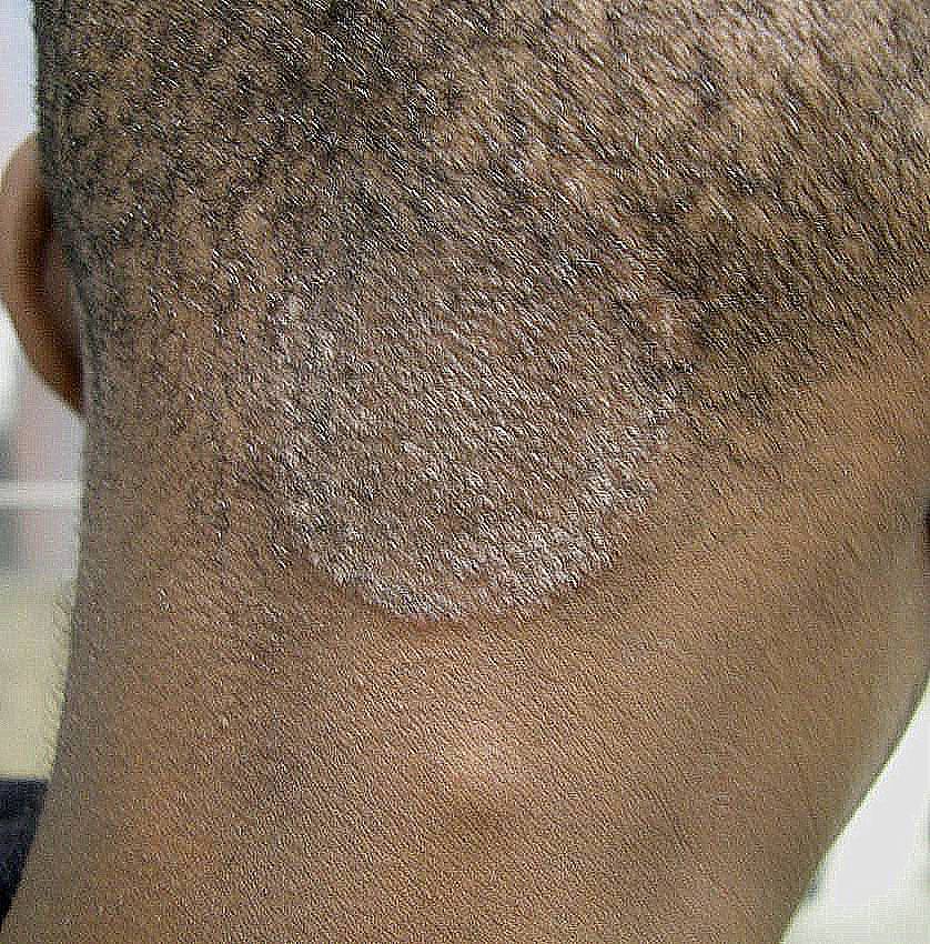
Tinea capitis, also known as ringworm, is a fungal infection of the scalp. It is the most common fungal infection in children under 12 years of age. Ringworm is caused by several kinds of fungus, some of which may also infect dogs and cats.
What is tinea capitis caused by?
Tinea capitis is a disease caused by superficial fungal infection of the skin of the scalp, eyebrows, and eyelashes, with a propensity for attacking hair shafts and follicles (see the image below). The disease is considered to be a form of superficial mycosis or dermatophytosis.Feb 21, 2020
When can a child with tinea capitis return to school?
Child care or School: Until treatment has been started or if the lesion cannot be covered. If on the scalp, until 24 hours after treatment has been started.
How do you treat a fungal infection on the scalp?
One of the most common medications used to treat fungal infections of the scalp is griseofulvin, an oral antifungal. You may need to use griseofulvin or another medication for four to eight weeks to properly treat the infection. Other medications used to treat scalp fungus include itraconazole, fluconazole and others.Feb 24, 2021
How do you treat a fungal scalp infection?
Treatment of yeast on scalp. Most scalp yeast infections can be treated with topical over-the-counter (OTC) treatments. These come in the form of ointments, shampoos, or foams. Research shows that antifungal drugs known as azoles, such as fluconazole (Diflucan), are very successful, as well as allylamines.
How long do fungal spores stay in hair?
Considering that fungal spores can persist in the hair follicle for a year or more, the relatively low rate of recurrence, even at the beginning of the griseofulvin era, is surprising. Although it has since been forgotten, it seems that patients develop immunity to fungal infections. Kligman [cited in 8] reported inoculation studies in human subjects in which 3–4 months after successful primary infection, not one of the patients developed re‐infection; the scalp proved to be resistant. In veterinary medicine, a special vaccine with inactivated M. canis as the antigen has been tested for effectiveness and is available on the market under the name Biocan M Plus ® or Biofel M Plus ® [ 11 ].
What is the most common dermatophytosis in Europe?
Tinea capitis continues to be the most common pediatric dermatophytosis in Europe. In the United States, tinea capitis, which is usually caused by Trichophyton tonsurans, has been called a modern‐day epidemic [ 1 ]. In Africa, an estimated 8% of children are affected (international figures [ 2 ]). In view of this situation, we present a critical review and assessment of current therapy options. The modern antifungals terbinafine, itraconazole, and fluconazole have unfortunately not been able to fulfill hopes of shortening treatment in children with tinea capitis compared with the more established drug, griseofulvin.
How do azoles and allylamines work?
Azoles and allylamines primarily target the fungal cell membrane by inhibiting ergosterol biosynthesis. This means that they are fully effective only against proliferating fungi which synthesize ergos‐terol but not against fungal spores or mycelia in the resting phase. Still, studies have shown that terbinafine, at 1000 times more than minimum inhibitory concentrations, can fully eradicate arthrospores and resting mycelia of both T. mentagrophytes and T. rubrum [ 7 ]. Griseofulvin acts by inhibiting mitosis as well as interrupting intracellular transport and causes curling of fugal spores. Griseofulvin has only fungistatic properties.
Where are antifungals accumulated?
Systemic antifungals are accumulated in the keratin in the skin. The active ingredients are absorbed by the hair through the hair papilla into the hair shaft. The hair above the skin is composed of dead keratinized cells, and hence does not absorb the agent. There is therefore no active transport of the antifungal from the hair papilla to the distal portions of the hair.
Where do microspores invade?
Infection begins in the scalp epidermis in the stratum corneum and in the follicle orifice. The hyphae grow on the surface of the hair and then deep into the follicle. The intrafollicular hyphae convert into arthrospores. While one portion of the hyphae invade the interior of the hair, growing down into the keratogenous zone, the other portion attacks by destroying the follicle with ectothrix spore chains. Growing hairs that are enclosed in this sheath of spores break off, sometimes only a few millimeters above the scalp. The majority of infectious particles (arthrospores) are not located in the hair, however, but in the hair follicle – which is the root of the problem for drug therapies.
When was griseofulvin introduced?
The introduction of griseofulvin as antifungal treatment in 1958 virtually eradicated favus and Microsporum infections in Central Europe. Between 1955 and 1966 T. schönleinii was detected 48 times in East Germany and M. audouinii 199 times; between 1967 and 1971 these two species were not isolated at all [ 12 ]. Of the 38,738 patients with dermatophyte infection (confirmed on culture), only 18 had tinea capitis, caused by M. canis, or 3.6 reports per year. This analysis includes all mycological laboratories in East Germany and is thus representative [ 12 ]. Elsner and colleagues [ 13] reported for the Würzburg area between 1976 and 1985 that M. audouinii was no longer detected although there was one report of a T. Schoenleinii isolate. These epidemio‐logical data demonstrate the effectiveness of griseofulvin in pediatric tinea capitis. In 1962, Rieth [ 14] proposed a 3–12 week‐long griseofulvin regimen for microsporia. Based on the literature, Götz [ 15] reported treatment durations of 18 to 40 days and for M. canis 6 weeks, or occasionally, 14 weeks. According to Götz, “persistence occurs because, while most of the hair is soon free of fungus, surviving spores can remain for some time in the follicle.” The suggested pediatric dosage of griseofulvin proposed by Rieth was 20 to 40 mg/kg per day.
Is itraconazole safe for children?
Itraconazole is available as a capsule or suspension (Sempera ® liquid). It is not approved for use in children. International studies have reported on the effectiveness of the following dosages. In Germany and Austria, these may only be given on an individual basis, and with all legal consequences in mind: in capsule form 5 mg/kg once daily with the main meal for 4–8 weeks (or longer), or as a continuous dose or pulse therapy (5 mg/kg per day for one week, followed by a 3‐week‐long treatment‐free interval of 2 to 4 months) for tinea capitis. Capsules may also be divided, depending on the patient's weight. The recommended dosage for the suspension is 3 to 5 mg/kg for 4 weeks [ 20 ]. Important note: Sempera ® liquid is not approved for use in Germany or Austria for the treatment of dermatophyte infections or for use in children. Laboratory tests should be performed before initiating treatment (blood differential, liver, and kidney function as well as potassium levels).
Author Post
A qualified & experienced Trichologist (Grad. from International Association of Trichology, Australia), Dr. Sharmishtha Deshpande brings along with her an experience of more than 25 years and has successfully treated patients suffering from all kinds of Hair & Scalp problems.
Dr. Sharmishtha Deshpande
A qualified & experienced Trichologist (Grad. from International Association of Trichology, Australia), Dr. Sharmishtha Deshpande brings along with her an experience of more than 25 years and has successfully treated patients suffering from all kinds of Hair & Scalp problems.
How old is the most affected by tinea capitis?
Tinea capitis is overwhelmingly a scourge of childhood; the predominant age range affected is between 3 and 7 years of age. 17, 18, 19, 20, 21, 22, 23, 24, 25, 26 The incidence of tinea capitis may also vary by sex, depending on the causative organism. When the etiologic agent is M audouinii , the ratio of infected boys to infected girls is as high as 5:1. 27 With M canis, the ratio varies considerably, but the infection rate in boys is usually higher. 28 Although tinea capitis is predominantly a disease of prepubescent children, adults do become infected from time to time. Thus Trichophyton infections of the scalp affect girls and boys equally, whereas in adults, women are infected more frequently than men. 27
What causes tinea capitis?
Tinea capitis is commonly caused by approximately 6 dermatophytes. Epidermophyton floccosum and Trichophyton concentricum , however, do not invade scalp hair and Trichophyton rubrum , which is the most common dermatophyte isolated worldwide, is not a frequent cause of tinea capitis. *Produces ectothrix infections.
When was griseofulvin first used?
Griseofulvin was originally isolated in 1939 as a metabolite that accumulated in cultures of the mold Penicillium griseofulvum , 57 but it was not until 1958 that the compound was reported to be effective in the treatment of experimental fungal infections in animals and in human dermatophyte infections. 58 Since then, it has become the drug of choice for treating dermatophyte infections in infants and children. 59
What is the term for a parasitic infestation of the skin?
In an extensive historical review, Sabouraud 1 cited Horace as stating that in Roman times, the word tinea indicated the insect whose larvae feed on clothes and books. He also cited Galen that tinea subsequently came to mean any verminous or parasitic infestation of the skin. By the mid 16th century, the term tinea was used to describe all diseases of the hairy scalp. The term ringworm came into use at about the same time and referred to any skin disease in which the lesions were arranged in rings. 2
How can tinea capitis be transmitted?
21 Tinea capitis can be transmitted via infected persons, fallen infected hair s, and select animal vectors. Spread of tinea capitis by fomites (contaminated barbershop instruments, hairbrushes, combs, and shared hats) is also common. T tonsurans has been isolated from combs, hairbrushes, bedding, and clothing. 29 M audouinii has been isolated from infected hair, clothing, furniture in the home, and from the back of seats in movie theaters. 30 These data suggest that infectious fungal particles are viable in fomites for many months. T violaceum and T schoenleinii can be transmitted from person to person by sharing clothing or towels. Infection due to these dermatophytes has also been perpetuated within families from one generation to another through a low-grade infection of the scalp or by asymptomatic carriers. 30
Is Griseofulvin safe for children?
Known side effects include headaches and gastrointestinal disturbances, but overall griseofulvin is generally safe and well tolerated. According to the package insert, oral griseofulvin was found to be embryotoxic and teratogenic to pregnant rats and, therefore, should not be prescribed to pregnant patients or women comtemplating pregnancy.
What is an asymptomatic carrier?
We define an asymptomatic carrier as a person with no signs or symptoms of tinea capitis but from whom positive scalp cultures can be isolated. 20, 25, 33 Mackenzie, Burrows, and Wallby 34 were early in recognizing this state in tinea capitis. They described many of its features during their study of an outbreak of tinea capitis caused by T tonsurans in a boarding school in Northern Ireland. Positive fungal cultures were obtained from the hairbrushes and clothing of several children who were “not ostensibly infected.” They also noted “exceptional difficulty in detecting infection” in some cases in which 8 or 9 examinations were needed before establishing a diagnosis of tinea capitis. They concluded: “Perhaps the greatest single factor which has prevented complete elimination of the pathogen is the inability to detect infection, particularly that of the scalp. In our opinion, it is almost impossible to detect what is virtually a carrier phase…” 34 (page 1058)
What is the treatment for tinea capitis?
Tinea capitis is a common fungal infection of the hair of the scalp affecting predominately prepubertal children. In the US, griseofulvin has been considered a first-line therapy agent for tinea capitis since the 1960s. However, it has been falling out of favor due to significant treatment failure, high cost, and long duration of treatment. Other antifungal agents have been researched as an alternative to griseofulvin. This paper will review the relevant pharmacologic properties, dosing, cost, efficacy, and adverse events profile for griseofulvin, terbinafine, itraconazole, fluconazole, and some adjuvant therapy options such as selenium sulfide shampoos and topical ketoconazole.
What is tinea capitis?
Tinea capitis describes a dermatophyte infection of scalp and hair that predominately occurs in children. The diagnostic workup includes microscopic examination, culture and/or molecular tests. Treatment is guided by the specific organism involved and should consist of systemic agents as well as adjuvant topical treatment. The aim of the present update of the interdisciplinary German S1 guidelines is to provide dermatologists, pediatricians and general practitioners with a decision tool for selecting and implementing appropriate diagnostic and therapeutic measures in patients with tinea capitis. The guidelines were developed based on current international guidelines, in particular the 2010 European Society for Pediatric Dermatology guidelines and the 2014 British Association of Dermatologists guidelines, as well as on a review of the literature conducted by the guideline committee. This multidisciplinary committee consists of representatives from the German Society of Dermatology (DDG), the German‐Speaking Mycological Society (DMykG), the German Society for Hygiene and Microbiology (DGHM), the German Society of Pediatric and Adolescent Medicine (DGKJ) and the German Society for Pediatric Infectious Diseases (DGPI). The Division of Evidence‐based Medicine (dEBM) provided methodological assistance. The guidelines were approved by the participating medical societies following a comprehensive internal and external review.
What is the fungus that causes tinea capitis?
Objectives: Trichophyton violaceum is an anthropophilic dermatophyte that is endemic to parts of Africa and Asia and is sporadic in Europe. T. violaceum mainly causes tinea capitis in both children and adolescents. Although the infections caused by T. violaceum are of considerable medical importance, its antifungal susceptibility profile remains poorly examined. Methods: In this study, we tested the in vitro antifungal susceptibility of a set of clinical T. violaceum isolates obtained from tinea capitis patients, using the CLSI broth microdilution method. We tested eight antifungals and used isolates collected from Western China (21), Eastern China (12), the Middle East (1), Europe (20), South Africa (7) and Canada (1). Results: The geometric means of the MICs of the antifungals for all isolates were as follows (in increasing order): posaconazole, 0.021 mg/L; terbinafine, 0.023 mg/L; voriconazole, 0.062 mg/L; amphotericin B, 0.20 mg/L; itraconazole, 0.34 mg/L; caspofungin, 0.56 mg/L; fluconazole, 4.23 mg/L; and flucytosine, 8.46 mg/L. No statistically significant differences in the susceptibility profiles of T. violaceum were detected within the geographical regions tested. Conclusions: Posaconazole, terbinafine and voriconazole were shown to be the most potent antifungal agents against T. violaceum isolates obtained from tinea capitis patients worldwide. These results might help clinicians in developing appropriate therapies that have a high probability of successfully treating tinea capitis due to T. violaceum.
How do fungal infections affect humans?
Invasive fungal infections can cause significant morbidity and mortality in humans and different animal species, worldwide. Antifungal therapy remains a central component of protecting human and vertebrate animals against fungal infections. Depending on the strategy chosen, topical and/or systemic drugs can be used based on the clinical picture of the host and mycological identification of the etiologic agent. For effective treatment, it is important to correctly identify the causative agents at the species level, which will enable administration of suitable therapeutics and initiation of appropriate therapeutic modalities. In addition, the management of fungal infections in animals usually includes systemic or topical treatment of the animal and environmental decontamination if necessary. Only a few products are licensed for animals, and, as a consequence, off-label use of the drugs approved for use in humans is quite common.
What is dermatophytosis? What are its causes?
These infections are caused by the keratinophilic fungi Trichophyton spp., Microsporum spp. , and Epidermophyton, which have been recovered from both symptomatic and asymptomatic individuals. Although dermatophytosis is generally not a life-threatening condition, these types of infections are among the most common infections worldwide, and their incidence has continued to increase consistently in recent years. Area covered: This article provides an overview of the general characteristics of dermatophytes, including their taxonomy and epidemiology, as well as the different clinical forms and laboratory diagnostics of dermatophytosis. We further classify the topical and systemic antifungal compounds currently used to treat dermatophyte infections. Expert Commentary: Antifungal therapy is a central component of patient management for dermatophytosis, and depending on the strategy chosen, topical and/or systemic drugs can be used. However, for effective treatment, it is important to correctly determine the causal agents at the species level, which will enable administration of suitable therapeutics and initiation of appropriate management strategies.
Is tinea capitis asymptomatic?
Tinea capitis has a high incidence with a global changing pathogen distribution, making this condition a public health concern around the world. As the infection is initially asymptomatic, it is easily spread. Moreover, it is present in many fomites, including hairbrushes, pillows, and bedding. Prompt recognition and treatment is necessary for kerion, an inflammatory subtype characterized by tender boggy plaques with purulent drainage. Kerion is usually associated with infection by zoophilic dermatophytes, although other sources have been described. Treatment for this severe form of dermatophytic infection can be challenging. In addition to the use of topical treatments, oral administration of griseofulvin, terbinafine, itraconazole, or fluconazole is often required. Griseofulvin, the first-line treatment, may not completely eradicate pathogen colonization of the host and may contribute to reinfection and prevalence of infective but asymptomatic carriers. This review highlights new agents that are being evaluated for the treatment of kerion and typical tinea capitis, enhanced diagnostic criteria, and a grading system for kerion evaluation.
What is Senna Didymobotrya?
Senna didymobotrya is important for its medicinal benefits among most communities in treating a wide range of ailments. Materials and methods Plants were collected from a cluster in Siaya, Nandi and Nakuru counties (Kenya). Stem bark, root bark, leaves, flowers and immature pods were obtained; air-dried and ground into fine powder. Methanol was used to extract the plant extracts. The extracts were reconstituted in water and incorporated into growth media to obtain 0%, 2.5%, 5%, 7.5% and 10%. Bioassays were carried out on T. tonsurans (ATCC 28942) and C. albicans (14053). The growth of cultures on the plates was measured over a period of sixteen days. The area under disease progress stairs was determined and subjected to ANOVA and comparison of means using LSD. Results Results indicated that the growth of C. albicans was not significantly affected by the plant extracts. Growth of T. tonsurans was completely inhibited by immature pods extract at 10%, the leaves and flowers extracts inhibited the growth at 7.5%. The stem and root bark extracts inhibited growth at low dosages of 2.5- 5 %. Conclusion There is need to carry out research on root and stem barks to identify the active phytochemicals that contribute to their high efficacies. On species conservation, harvesting of roots may lead to depletion of S. didymobotrya.
