There is a variety of non-surgical treatments that could help relieve the symptoms of accessory navicular bone syndrome:
- Rest—avoid strenuous activity
- Immobilize—a cast or removable walking boot forces rest and allows the inflammation to diminish
- Ice—reduces swelling. Do not put ice directly on the skin.
- Medicate—over-the-counter medication such as ibuprofen (NSAIDs), may...
How to stop accessory navicular pain?
3 Things to Know About Navicular Bone Pain
- PRP and Stem Cell Injections. Precise, ultrasound and x-ray guided injections can treat Navicular Bone pain due to arthritis, ligament or tendon injuries, and low back dysfunction.
- 3 Things to Know About Navicular Bone Pain. ...
- In Conclusion. ...
Does bone spur removal surgery relieve pain?
Surgery, such as a laminectomy, is designed to relieve the pain and neurological symptoms by removing the bone spurs and thickened ligaments causing painful nerve compression. The majority of patients who undergo surgery for bone spurs experience good results, often gaining years of relief and improved quality of life.
Can boots help with navicular pain?
So, yes, hoof boots with pads can help , as they will allow the horse to feel comfortable landing heel first, and thus eliminate a prime cause of navicular-toe first landing, due to heel pain. There are many causes for heal pain, including high heels with a non functioning frog, or a frog that has thrush.
Can Neulasta cause bone pain?
Bone pain. Bone pain is a common side effect of Neulasta. In clinical studies, 31% of people who took Neulasta reported bone pain compared with 26% of people who took a placebo (treatment with no ...
How do you fix navicular bone?
Surgery for Accessory Navicular Syndrome If this is the case, surgery may be recommended to correct the deformity. Surgery typically involves removing the accessory bone, repairing the posterior tibial tendon, and restructuring the foot back to a normal appearance.
How long does it take for a navicular bone to heal?
It will take about 6 weeks for most people to heal. The goals of treatment are to manage pain and support the bone as it heals. This may include: Medicine to ease pain and swelling.
Can you cure navicular?
Navicular disease can be treated but rarely cured. Corrective trimming and shoeing is important to ensure level foot fall and foot balance. Often a rolled toe egg bar shoe is used to encourage early break over at the toe and good heel support.
Can you walk with a fractured navicular?
In most cases a navicular fracture can be treated conservatively by implementing a cast that immobilises the injured foot for around 6 weeks while the fracture heals. During this period it will be necessary to use crutches to walk and to avoid much weight bearing.
Does a navicular fracture need surgery?
Navicular stress fractures are relatively common, most effectively treated by either non‐weightbearing cast immobilisation or surgical fixation, followed by a graded return to sport. These treatments appear equally effective over the short term.
Is a navicular fracture serious?
Navicular fractures and other foot and ankle issues can become much worse without proper treatment, resulting in far more serious issues that require much more extensive treatment and recovery and can even result in permanent disability.
What does navicular lameness look like?
Horses with navicular usually have a history of subtle onset of lameness. The horse may just look stiff early on in the course of disease and stumble frequently. The lameness may seem inconsistent and switch from one (front) leg to another. Putting the horse on a circle or a hard surface can make it worse.
What are the most common signs of navicular syndrome?
Lameness is the classic sign of navicular syndrome. This can appear suddenly, but a more common pattern is mild lameness that becomes progressively worse over time. A horse with navicular syndrome feels pain in the heels of the front feet, and its movements reflect attempts to keep pressure off this area.
What does navicular disease look like?
Clinical signs of navicular disease include a short, choppy stride with lameness that worsens when the horse is worked in a circle, as when longeing. Frequent stumbling may occur at all gaits, even the walk, or when horses are asked to step over short obstacles such as ground poles.
How painful is a navicular fracture?
Symptoms. Symptoms of a navicular stress fracture usually involve a dull, aching pain in the ankle or at the middle or top of the foot. In the early stages, pain often occurs only with activity. In the later stages, pain may be constant.
How common is a navicular fracture?
A navicular fracture is rare but can be seen, especially in athletes.
How do I know if I have a navicular stress fracture?
What Are the Symptoms of a Navicular Stress Fracture? Your child will have vague, aching pain along the inner side of the foot near the arch. It may come on slowly over time and get worse during and following physical activity. Sprinting, jumping and pushing-off are movements that aggravate the pain.
How long does it take for a navicular fracture to heal?
If you still have pain with pressure on top of the navicular after 6 weeks, you will spend more weeks non-weightbearing. In a small percentage of patients, the navicular stress fracture will not heal and may require surgery.
What is a stress fracture in the navicular bone?
Navicular Stress Fracture Treatment and Prevention. Stress fractures of the navicular bone are a relatively common foot injury – especially in athletes. Navicular stress fractures are most likely to occur in athletes who participate in sports with a lot of jumping and direction change, such as basketball, soccer, high jumpers and sprinters.
Where is the navicular stress fracture?
A navicular stress fracture, figure 1, is an injury to the bone on the inside of the foot between the ankle bone and the midfoot. People with navicular injury typically have vague pain in the mid portion of the foot – just in front of the ankle. Initially, the pain is worse during and just after athletic activity and reduces with a period of rest.
Can navicular stress fractures heal?
If not treated correctly navicular stress fractures tend not to heal. It is critical to eliminate pressure on the bone to allow it to heal. If you suspect you may have a navicular injury make an immediate appointment to see us in Seattle or a podiatrist in your area.
What is the best treatment for a navicular bone injury?
Precise, ultrasound and x-ray guided injections can treat Navicular Bone pain due to arthritis, ligament or tendon injuries, and low back dysfunction. The Centeno-Schultz Clinic are experts in the treatment of foot, ankle, and leg injuries. Board-certified, fellowship-trained physicians provide multiple treatment options including PRP and the use of your own stem cells. These procedures require extensive training and can not be performed by your Primary Care Physician (PCP) or orthopedist. To watch a PRP injection into the foot, see the video below.
What causes navicular bone pain?
3 Things to Know About Navicular Bone Pain 1 Common causes of navicular bone pain are fracture and arthritis 2 Important but less appreciated causes include ligament injury, irritation of low back nerves, and Accessory Navicular Bone. 3 PRP and stem cells are effective, nonsurgical, natural treatment options for Navicular Bone pain.
What is the treatment for a ruptured ligament?
Ligament Instability: Rest, elevation, and immobilization are first-line treatment options. Surgery is necessary for ruptures where both ends of the ligament are physically separated. Low Back Nerve Injury: Treatment is dependent upon the severity of the specific condition and amount of nerve dysfunction.
Where are the accessory navicular bones located?
They are typically located on the inside aspect of the Navicular Bone. Accessory Navicular bones are classified into three groups based upon shape and position (7). Accessory Navicular syndrome occurs when the extra piece of bone causes pain. It typically occurs after a trauma or from shoes rubbing against the extra bone.
Why does cartilage help with osteoarthritis?
Cartilage protects the joint and enables the joint to move smoothly. The most common causes of Navicular osteoarthritis are degeneration and trauma (4). Pain is aggravated by prolonged standing and walking is common.
What is the treatment for osteoarthritis?
Osteoarthritis: Conservative care involves activity modification, weight loss, orthotics, walking boot, and oral medications including fish oil and turmeric.
Where is the Navicular Bone?
In Conclusion. The Navicular Bone is a small C shaped bone located on the inside portion of the midfoot. It provides important support of the foot and arch during movement. Fracture and arthritis are common causes of pain.
What is the name of the drug that is used to treat navicular syndrome?
That’s when the Food and Drug Administration (FDA) approved two new medications for the treatment of navicular syndrome, specifically targeting bony changes seen in many cases. Both drugs, sold under the names Tildren and Osphos, belong to a medication class known as bisphosphonates.
How does osteoblast work?
At times of bone stress, such as during extreme physical activity or after a traumatic injury, this process is accelerated, with osteoclasts working faster to tear down the damaged bone. Generally, osteoblasts can keep up the increased pace, laying down new, stronger bone quickly, but the process isn’t instantaneous.
Can navicular syndrome be treated with shoeing?
An infuriatingly vague diagnosis, the term “navicular” was often applied to cases of otherwise unexplained heel pain. Therapeutic trimming and shoeing could help preserve soundness for a few years, as could non-steroidal anti-inflammatory drugs ...
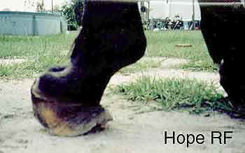
What is the navicular bone on a horse?
Navicular Bone Horse: Causes, Diagnosis and Treatment. The navicular bone is a small fat bone that lies across the back of the coffin bone of horse hoof. Navicular bone horse is attached to the pedal bone by a strong ligament and the pastern bone by a suspensory ligament. There are several soft tissue structures are associated with navicular bone.
How long does a horse need to rest after a fracture?
The horse is given 2 months box rest followed by a gradually increasing, controlled exercise program. Whatever method of conservative treatment is used, healing will be slow and by the fibrous union because of the difficulty in immobilizing the fracture.
Can horses develop navicular bone problems?
Horses of all foot shape can develop navicular bone problems. Balanced ration, correct shoeing and maintaining good stable management can reduce the incidence of the condition. Though the prognosis is poor in the case of navicular bone problems, careful management can reduce the sufferings of your horse. As a horse owner, you must take care of the ...
Can a palmar digital nerve block cause lameness?
A palmar digital nerve block will often improve the lameness, however, a negative response to the nerve block does not rule out a fracture of the navicular bone. The diagnosis can be confirmed by radiography.
Can a tripartite navicular bone be lame?
Although rare, congenital bipartite and tripartite navicular bones have been reported. These may not cause lameness and can be distinguished from fractures radiographically by their smooth rounded edges, and the overall symmetry of the bone.
How to diagnose accessory navicular syndrome?
To diagnose accessory navicular syndrome, the Doctor at American Foot will ask about symptoms and conduct a full evaluation of foot structure, joint and muscle strength, and range of motion. He or she may press the bony prominence to gauge the degree of pain. X-rays are used to confirm the diagnosis. An MRI or other advanced imaging tests may be used to further evaluate the condition.
Where is the extra bone on the foot?
Some members of the population—7-19%—are gifted with an extra bone located on the inner side of the foot just above the arch. This bone, the accessory navicular, os tibial externum, is present at birth.
Kidner Procedure
If these non-operative measures fail, the option of surgical treatment can be performed. The terms used to describe the procedure include removal or excision of the bone that never fused normally in development and/or removal of the prominence of bone of the navicular.
Before and After X-Rays of Os Navicular
This post-op x-rays images above demonstrate the resection and re-creation of a smooth and flat inner border of the foot. The first x-ray on the left is taken before the procedure. The second x-ray, on the right, is taken after successful completion of the procedure.
