Can Amyloodinium be prevented?
Amyloodinium is a common and deadly disease, but if proper quarantine procedures are followed coupled with early recognition and treatment, you can hopefully avoid the damage caused by this disease. Faifield, T.A. Commonsense Guide to Fish Health.
Can Amyloodinium cause serology?
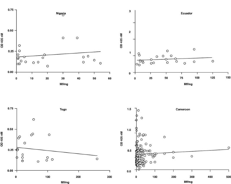
Serology. Amyloodinium-infected fish can develop a specific, antibody-mediated response to the parasite. This has been described by Smith et al. (1992) in blue tilapia (Oreochromis aureus) and by Cobb et al. (1998a) in the tomato clown fish (Amphiprion frenatus).
What are the stages of Amyloodinium ocellatum?
The disease is more common among tropical fish and takes on a dusty, brownish to gold coloration on a fish’s body. The 4 main stages of Amyloodinium ocellatum life cycle are as follows: Trophont Feeding Stage – An active feeding protozoan attaches itself to the skin or gills of the fish.
How Amyloodinium infection affects the fish food industry?
Infection of Amyloodinium has also affected the fish food industry very much. Amyloodinium has two flagella like a whip which is used to move around. There are different stages in the life cycle of Amyloodinium.

What is the feeding stage of a trophont?
Trophont Feeding Stage – An active feeding protozoan attaches itself to the skin or gills of the fish. It takes the form of a nodule or epithelium which looks like white spots on fish.
What causes coral disease?
Amyloodinium ocellatum aka Marine Velvet, or Coral Disease, is caused by parasites called dinoflagellates. These parasites are found in both freshwater (Oodinium), and saltwater (Amyloodinium). The disease is more common among tropical fish and takes on a dusty, brownish to gold coloration on a fish’s body.
How is amyloodinium transmitted?
Amyloodinium is transmitted through direct contact with live dinospores. Thus, the infection can be spread via water contaminated with live dinospores, including aerosolized droplets (Roberts-Thompson et al., 2006). Dinospores range in size from 12 to 15 ?m in diameter (Brown, 1931; Nigrelli, 1936; Landsberg et al., 1994). It is also likely that the parasite may be transmitted by fomites (nets, hands, shoes, equipment, etc.) that have contacted contaminated water. However, there is no evidence that dinospores are “sticky” like some other protozoan parasites.#N#Birds, wildlife or other terrestrial animals that might move between culture systems (including dogs that swim in different areas on a farm) might also transmit parasites via infected water or even by moving an infected fish into a non-infected area. Dead fish can also be a reservoir for Amyloodinium in that trophonts can drop off into the sediment and divide to form tomonts, or can even divide on the dead fish. For this reason, it is advisable to remove dead fish as quickly as possible from the system. If possible, the aquarium bottom should also be routinely siphoned to remove tomonts, helping to reduce the number of potentially infective dinospores (J. Landsberg, personal communication).#N#Minimizing the movement of parasites around a culture facility is a critical element in a preventive health program. This is discussed in more detail below, but the goal of foot baths, net rinses, and physical separation is ultimately to prevent the movement of these tiny, infectious, parasitic stages to areas where uninfected fish may be housed.
How does Amyloodinium affect aquaculture?
Amyloodinium can devastate aquacultural businesses, since outbreaks typically have an acute onset, spread very rapidly, and cause extensive mortalities . Farms that raise “high risk” species such as red drum and clownfish are strongly advised to implement strong biosecurity protocols and educate workers about the importance of keeping this organism out of the culture systems. Once established on a farm that raises sensitive species of fish, the parasite may prevent the business from being financially successful.
What are the three stages of life?
The three life stages are the trophont (the adult stage that feeds on the fish), the tomont (detached from the fish, this stage divides to form dinospores), and dinospores (the freeswimming stage that searches for and infects a host).
What is the most important parasite in cultured fish?
Amyloodinium ocellatum, an Important Parasite of Cultured Marine Fish. This factsheet by the Southern Regional Aquaculture Center (SRAC) gives information on amyloodinium ocellatum, an important parasite of cultured marine fish. Amyloodinium ocellatum was described by Brown (1931) and is one of the most important pathogenic parasites affecting ...
How do you know if a fish has amyloodiniosis?
If the primary site of infection is gill, which seems to be the most common, the primary clinical signs will be respiratory. These may include increased respiratory rate (rapid gilling and movement of the opercula), “piping,” and gathering at the surface or in areas with higher dissolved oxygen concentrations, as well as reduced appetite. If the primary site of infection is skin, infected fish sometimes develop a white or brown coloration (“velvet”) or cloudy appearance, which is most visible when viewed with indirect lighting such as a flashlight (Levy et al., 2007). Such fish may display signs of “flashing” or rubbing on tank walls, the substrate, or other structures in their environment. Again, feeding behavior likely will be poor and some fish may appear emaciated. Fish with Amyloodinium infection alone do not typically have ulcers, white spots , or fuzzy lesions, but the skin can seem “hazy” in appearance. If the infection is confined to the gill, the “velvet” appearance will not be present.
Is piscinoodinium a parasite?
A morphologically similar parasite, Piscinoodinium, infects freshwater fish but is much less common and less pathogenic than A. oscellatum. While the infection of feral fish with A. oscellatum is not unusual, heavy mortality in wild populations is rare. Serious outbreaks have occurred in cultured red drum (Sciaenops ocellatus), ...
Can epizootics be found in cultured fish?
Epizootics have been reported in feral and cultured fish, as well as in home and public aquaria. Rapid spread of the parasite and high mortality are common in cultured fish if the organism is not recognized and treated early in the course of an outbreak.
What is the life cycle of amyloodinium?
The life cycle of amyloodinium is complex compared with those of many other fish parasites. Many single-celled parasites that infect the external surface of fish divide by binary fission. This simply means that an adult organism divides in half and produces two. Those two divide, resulting in four, and so forth. There are two important exceptions to this generalization: the organisms that cause "white spot disease" (Icthyopthirius multifiliis in freshwater fish and Cryptocaryon irritans in marine fish, described in IFAS Extension Circular 920), and the organisms that cause "velvet" (Amyloodinium ocellatum in marine fish and Oodinium spp. in freshwater fish).
How does amyloodinium spread?
A common example is introduction of the infection into facilities that use natural seawater. Tomonts or infective dinospores can be introduced directly with incoming seawater, becoming a source of infection for fish in the system. Obviously, introducing fish infected with trophonts into a culture system will serve as a source of infection as soon as the throphonts detach and begin the reproductive process. It is also possible to introduce the infection through food items. The authors are aware of one amyloodinium outbreak that was traced directly to fish caught in the wild and fed to animals in a closed, recirculating system.
What is the organism that infects the gills and skin of both marine and brackish water fish
Amyloodinium ocellatum is a dinoflagellate that infects the gills and skin of both marine and brackish water fishes. The organism may be more closely related to a toxic algae than to the protozoans with which it has been grouped in the past. A similar organism, Oodinium spp., is found in freshwater fish. The disease caused by these organisms has been referred to as "velvet," "rust" and "gold dust disease" because of the shiny sheen the parasite imparts to heavily infected fish.
How to tell if a fish has amyloodinium?
Often, the first indication of an amyloodinium infection is dead or dying fish. Amyloodinium should always be considered as a possible cause of mortality when a disease outbreak involving marine or brackish water fish occurs. Behavioral signs may include a decrease in or complete lack of feeding activity, flashing (rubbing against objects in the tank or on the bottom substrate) and coughing (backflushing water across the gills). The skin of heavily infected fish may have a dull gold or brown sheen. Closer examination of the skin may reveal scale loss and patchy accumulation of mucus.
Is amyloodinium safe for aquarists?
Although a wide variety of chemical treatments have been used to control amyloodinium outbreaks over the years, none have proven completely effective or safe for target animals.
How many stages does Oodinium have?
Similar to Cryptocaryon, Oodinium has 3 stages in its life-cycle: The infective Dinospore, which is free-swimming; The attached Trophont, which is found on external surfaces in contact with environmental water; And the mature cyst/ dividing Tomont.
Why do oodinium dinospores attack fish?
The gills (similar to Brooklynella) are where Oodinium Dinospores attack first due to the soft tissue that is easy to penetrate. The Dinospore attaches a filament into the host fish for feeding, becoming a Trophont.
How long does it take for Brooklynella to die?
In tanks without properly installed UV Sterilizers the progression of Oodinium or Brooklynella was as quick as 48 hours from first symptom to death, while in tanks with properly installed UVs, this progression was often as long as 10-14 days!
What is the parasitic dinoflagellate that kills saltwater fish?
Oodinium is a parasitic dinoflagellate which can infect and kill many species of saltwater fish. Similar to Crytptocaryon in mode of transfer (Marine Ich and other external fish parasites), this Dinoflagellate is generally more dangerous in the confines of an aquarium, especially a small overcrowded tank due to rapid re-infection.
What is marine oodinium?
MARINE OODINIUM (Amyloodinium Ocellatum) a species of dinoflagellates; Also occasionally known as Coral Fish Disease or Saltwater Velvet. Updated 9/28/18. Although closely related to freshwater velvet (Piscinoodinium pillulare), these two external parasites differ in that the marine variety ...
Is oodinium the same as Brooklynella?
Oodinium is also sometimes mistaken for Brooklynella (& Vice Versa), as symptoms and disease progression are similar (and thankfully so is treatment, so do not stress on a certain diagnosis). In fact the only relatively easy to discern difference is the heavy amount of slime that is produced by Brooklynella usually starting near the gills.
Is copper sulfate effective in marine oodinium?
Unfortunately, Copper sulfate is not as effective in Marine Oodinium , as compared to freshwater Velvet. Partly due to the differences in the dinoflagellate. One difference is that Marine Oodinium does NOT contain Chloroplasts. I have however had slightly better results with " Ionic Copper ".
Description of Causative Organism
Life Cycle
Potential Impact on Aquaculture Businesses
Potential Impact on Zoos and Aquaria
Diagnosis
- Disease Presentation
Morbidity and mortality from amyloodiniosis can be severe, sometimes with rapid onset over a period of a few days. Affected fish may die suddenly, showing few clinical signs, but in most cases behavioral and physical changes will be observed before death. If the primary site of infec… - Preliminary Diagnosis
Clinical diagnosis of amyloodiniosis is straightforward, although less experienced examiners have confused A. ocellatum and Cryptocaryon irritans. Both are ectoparasites that are easily detected by microscopic examination of infected tissue. Gill or skin biopsies (Figs. 2 and 3) reveal the par…
Treatment Strategies
Treatment Options For Ornamental Aquarium Fish
Role of The Rearing System in Treatment Protocols
Preventing Infection
Summary