Conclusions: The diagnosis of an orbital hematoma should be made as quickly as possible to permit adequate early therapy. Decrease of vision or blindness caused by orbital hematoma may be improved through a lateral canthotomy as emergency measure and subsequently by draining the hematoma to relieve compression of the optic nerve. Publication types
What are the treatment options for orbital hemorrhage?
If the orbital hemorrhage occurs in the preseptal space, the clinician has the option of observing the hematoma or draining it. If the preseptal hematoma is expanding rapidly, then making a small drainage incision would be acceptable therapy.
How serious is orbital hematoma after sinus surgery?
Orbital hematoma is an uncommon but serious complication of sinus surgery. Appropriate perioperative attention may minimize risk, but early diagnosis and appropriate management are crucial to preventing vision loss.
What is included in patient education about orbital hemorrhage before surgery?
Before eyelid or orbital surgery, the patient should be counseled regarding the rare but serious risk of orbital hemorrhage. If an orbital hemorrhage has no identifiable cause, then the orbit should be imaged for possible vascular tumors and inflammation.
What is orbital hemorrhage?
Orbital hemorrhage is bleeding within the orbit that can quickly cause vision loss if not addressed in a timely manner. Vision loss occurs due to a compartment syndrome within the orbital walls that compromises the optic nerve and its blood supply.

What is an orbital hematoma?
Orbital hematoma is defined as a collection of blood inside the orbit, and the major adverse sequelae that develop arise because the orbit is a bony cone with tight fascial attachments holding the globe at its anterior edge.
How do you manage retrobulbar hemorrhage?
The majority of retrobulbar hemorrhages can be managed conservatively with digital ocular massage or intravenous acetazolamide or mannitol. However, further surgical intervention is indicated when vision is at risk.
What is Extraconal hematoma?
Extraconal hematoma has a classic appearance of a confined lentiform hyperdense hematoma, analagous to an intracranial extradural hemorrhage. It is usually located in the superior half of the orbit and almost always adjacent to a fracture of the bony orbit.
How is retrobulbar hematoma diagnosed?
Signs of retrobulbar hemorrhage include expanding proptosis, ophthalmoplegia, increased intraocular pressure, loss of pupillary reflexes, and optic disc or retinal pallor [3, 4]. Both Computed Tomography (CT) scan and Magnetic Resonance Imaging (MRI) may be performed in the diagnosis.
What is retrobulbar injection?
Retrobulbar anesthesia is a type of regional anesthetic nerve block in the retrobulbar space, located behind the globe of the eye. The technique was first described in 1884 by Herman Knapp.
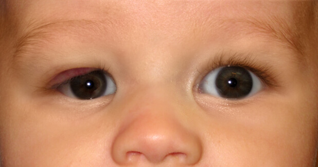
What is a retrobulbar hematoma?
Retrobulbar hematoma is the phenomenon of blood collecting in the retrobulbar space behind the globe. Although it is not common, it is a serious condition that can lead to blindness.
How do you do lateral Canthotomy and Cantholysis?
Use iris scissors to cut from the lateral canthus to the rim of the orbit, about 1 to 2 cm (canthotomy). Cut the inferior and sometimes both crus of the lateral canthal ligament (cantholysis). Most experts recommend starting with the inferior crus. Lift the lateral portion of lower eyelid.
What is orbital blowout fracture?
A blowout fracture is a break in the floor or inner wall of the orbit or eye socket. A crack in the very thin bone that makes up these walls can pinch muscles and other structures around the eye, keeping the eyeball from moving properly. Getting hit with a baseball or a fist often causes a blowout fracture.
What causes periorbital hematoma?
Racoon eye or periorbital ecchymosis is caused by blood tracking into periorbital tissues, which is frequently observed after head trauma but is also observed in systemic diseases, such as amyloidosis, neuroblastoma, and surgical interventions.
How do you get hyphema?
Hyphema is the medical term for bleeding inside your eye. Specifically, hyphema causes blood to pool behind your cornea (the outermost layer of your eye) and your iris (the colored part of your eye). It's usually caused by something hitting your eye. Sports injuries are the most common cause of hyphema.
What is retrobulbar pain?
What Is It? Retrobulbar neuritis is a form of optic neuritis in which the optic nerve, which is at the back of the eye, becomes inflamed. The inflamed area is between the back of the eye and the brain.
What causes retrobulbar optic neuritis?
Retrobulbar optic neuritis (RON) is mainly caused by multiple sclerosis, a common demyelinating disease. The cardinal signs of RON are the loss including visual acuity or/and contrast sensitivity, periocular pain induced with ocular movements, RAPD and CVD.
What causes vitreous haemorrhage?
A vitreous haemorrhage is usually due to a blood vessel within the retina breaking, and bleeding into the vitreous cavity. Common causes of bleeding are a result of the development of fragile new blood vessels on the retina due to either diabetes or blockages in the retinal veins (Retinal Vein Occlusions).
How do you do lateral Canthotomy and Cantholysis?
Use iris scissors to cut from the lateral canthus to the rim of the orbit, about 1 to 2 cm (canthotomy). Cut the inferior and sometimes both crus of the lateral canthal ligament (cantholysis). Most experts recommend starting with the inferior crus. Lift the lateral portion of lower eyelid.
What is orbital hematoma?
Orbital hematoma is defined as a collection of blood inside the orbit, and the major adverse sequelae that develop arise because the orbit is a bony cone with tight fascial attachments holding the globe at its anterior edge. Therefore, the occurrence of orbital bleeding and hematoma formation can cause the pressure in the globe to increase rapidly, ...
Why should a microdebrider not be used in medial orbital wall dehiscence?
In the setting of medial orbital wall dehiscence or fat exposure, the microdebrider should not be used, because rapid debridement of soft tissue (fat, muscle, nerve) can occur ( Fig. 5 ).
What bones are in the orbit?
Seven bones contribute to the orbit: maxillary, lacrimal, ethmoid, frontal, zygomatic, sphenoid, and palatine ( Fig. 1 ). The thickness of these bones along the orbit is variable, but the medial and inferior walls are the thinnest, less than 1 mm in certain places. Natural dehiscences are uncommon in the medial orbital wall, also known as the lamina papyracea . The orbit is surrounded by bony walls on all sides except anteriorly, where fascial planes hold the globe in position. The globe is supported by the extraocular muscles, which are suspended by several ligaments to the bony orbit. Increased intraorbital pressure therefore causes proptosis, but the amount of globe protrusion is limited by these tendons and ligaments. As pressures approach mean arterial pressure, decreased arterial perfusion of the retina and optic nerve occur, and venous outflow may also be impaired. To release the globe anteriorly for temporary pressure relief, division of the medial or lateral canthal connections is required.
How long does it take to get blind from a retinal artery ischemia?
Animal models of central retinal artery ischemia suggest that irreversible blindness may occur within 100 minutes.
Is orbital hematoma traumatic?
Orbital hematoma is best grouped into categorie s of spontaneous, traumatic, or iatrogenic. The iatrogenic category is a feared complication of endoscopic sinus surgery (ESS), blepharoplasty, or reconstructive trauma surgery. Awareness, diagnosis, and management of this complication is essential for every head and neck surgeon when operating ...
Is orbital hematoma a complication of sinus surgery?
Orbital hematoma is an uncommon but serious complication of sinus surgery. Appropriate perioperative attention may minimize risk, but early diagnosis and appropriate management are crucial to preventing vision loss. The surgeon, postoperative care staff, and patient must be aware of the signs and symptoms.
Can an anterior ethmoid artery be damaged?
The posterior ethmoid artery is rarely encountered, but the anterior ethmoid artery is reliably encountered posterior to the frontal recess, and can often be on a pedicle that is easily damaged. Use of through-cutting forceps or a curette along the skull base in these regions can directly injure these arteries.
What is the blood supply to the orbit?
The orbit has a rich blood supply. Arterial supply. The blood supply to the orbit is primarily from the ophthalmic artery via the internal carotid artery. Major branches of the ophthalmic artery are branches to the extraocular muscles, central retinal artery, and posterior ciliary arteries.
What to do if hemorrhage is limited?
Patient management. If hemorrhage is limited, not progressive, and does not threaten optic nerve function, close observation can be appropriate. If vision is threatened and there is progressive tense proptosis, perform urgent canthotomy and cantholysis of the inferior crus of the lateral canthal tendon.