What is ECG monitoring and why is it important?
The initial extracorporeal shockwave lithotripsy (ESWL) machine, the Dornier H3, uses fluoroscopy for stone localization and treatment monitoring. Although this imaging method has the benefit of being familiar to urologists, it has some drawbacks, such as difficulty in imaging some types of stones and the radiation exposure of the operator and ...
What is an ESWL procedure?
Oct 04, 2017 · The goal of continuous ECG monitoring is to aid in: (a) immediate recognition of sudden cardiac arrest to improve time to defibrillation; (b) recognize deteriorating conditions such as early after-depolarizations or nonsustained arrhythmias that may lead to a life-threatening arrhythmia; (c) facilitate management of arrhythmias; and (d ...
What is the role of ECG monitoring in the treatment of bradycardia?
ECG monitoring is indicated for patients with newly diagnosed critical left main coronary artery disease or its equivalent (eg, proximal left anterior descending and circumflex disease) who are candidates for urgent revascularization. Monitoring should continue uninterrupted while these patients await intervention.
What is the purpose of the ECG practice standards 2004?
Apr 19, 2010 · Electrocardiograms. The electrocardiogram (ECG) is a laboratory test which has long been used to diagnose a variety of heart conditions and (in adults) to screen for overall risk of heart disease ...
Does lithotripsy affect the heart?
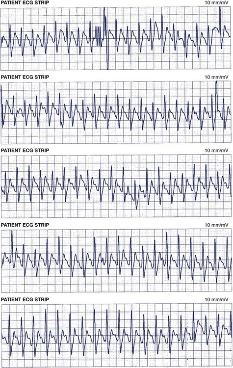
It is well known that, during extracorporeal shock wave lithotripsy (ESWL) for kidney stones, shock waves delivered by a spark-gap generator frequently elicit cardiac arrhythmias.
Which of the following is contraindicated for ESWL procedure?
Contraindications. The formal contraindications for ESWL are: pregnancy, untreated urinary tract infection/urosepsis, decompensated coagulopathy, uncontrolled arrhythmia, and abdominal aortic aneurysm >4.0 cm.
What is the difference between ESWL and lithotripsy?
Shock Wave Lithotripsy (SWL) is the most common treatment for kidney stones in the U.S. Shock waves from outside the body are targeted at a kidney stone causing the stone to fragment. The stones are broken into tiny pieces. lt is sometimes called ESWL: Extracorporeal Shock Wave Lithotripsy®.
What are the complications of ESWL?
ESWL has been reported to have a low complication rate and a low mortality rate. Most of the complications include transient gross hematuria, flank pain, and urinary tract infection that can usually be treated with conservative management.
Which stones are resistant to ESWL?
Cystine and brushite calculi are the most resistant to ESWL, followed by calcium oxalate monohydrate, struvite, calcium oxalate dihydrate, and uric acid stones. Stone composition affects the resistance to fragmentation and the type of fragments produced.
When is ESWL indicated?
ESWL is the main noninvasive treatment for kidney stones. It works well for people with smaller stones that can be easily seen with an X-ray. ESWL is not recommended for people with chronic kidney infection, because some fragments may not pass and bacteria will not be completely eliminated from the kidney.
Is ESWL considered surgery?
An outpatient surgical procedure for kidney stone disease. ESWL, or extracorporeal shockwave lithotripsy, is a very common, non-invasive method for treating stones in the kidney or ureter, the tube which drains the urine from the kidney to the bladder.
How effective is ESWL?
The overall success rate of ESWL was 97.3%. No child had postoperative urinary infection or ureteral obstruction. Conclusions: ESWL is an efficient and safe modality for the treatment of pediatric ureteral stones.
Does ESWL use radiation?
Radiation dose to personnel working in the extracorporeal shock wave lithotripsy suite averaged less than 2 mrem. (0.02 mSv.) per case, making it a procedure that is safe in regard to radiation exposure.
Is ESWL safe?
Conclusions: ESWL is a safe and efficient first-line therapy for treatment of isolated small lower pole kidney stones with acceptable stone-free rates, low morbidity, few complications and a low stone recurrence rate.
Why is there blood in urine after lithotripsy?
Your Recovery This treatment uses sound waves to break kidney stones into tiny pieces. These pieces can then pass out of the body in the urine. You may have a small amount of blood in your urine after this treatment.
What size kidney stone requires lithotripsy?
Most kidney stones that develop are small enough to pass without intervention. However, in about 20 percent of cases, the stone is greater than 2 centimeters (about one inch) and may require treatment.
How does ESWL work?
ESWL involves the administration of a series of shock waves generated by a machine called a lithotripter. The shock waves are focused by x-ray onto the kidney stone and travel into the bodythrough skin and tissue, reaching the stone where they break it into small fragments. For several weeks following treatment, ...
Is ESWL a non-invasive procedure?
Patients who once required major surgery to remove their stones could be treated with ESWL, and not even require an incision. As such , ESWL is the only non-invasive treatment for kidney stones, meaning no incision or internal telescopic device is required.
What is ESWL in kidneys?
As such, ESWL is the only non-invasive treatment for kidney stones, meaning no incision or internal telescopic device is required.
What is ESWL in medical terms?
ESWL involves the administration of a series of shock waves generated by a machine called a lithotripter. The shock waves are focused by x-ray onto the kidney stone and travel into the bodythrough skin and tissue, reaching the stone where they break it into small fragments.
Is ESWL an outpatient procedure?
The Procedure. Because ESWL is a completely non-invasive therapy, most ESWL treatments are performed on an outpatient basis. Although the use of anesthesia does depend on patient and physician preference, recent data suggest that the results of ESWL may be improved with the administration of a mild anesthetic.
When was shock wave lithotripsy introduced?
The introduction of extracorporeal shock wave lithotripsy (ESWL) in the early 1980s revolutionized the treatment of patients with kidney stone disease. Patients who once required major surgery to remove their stones could be treated with ESWL, and not even require an incision.
How is ESWL performed?
ESWL can be performed either under general anesthesia or under intravenous sedation. Once the patient is anesthetized, the lithotripter machine is positioned in contact with the patient’s flank on the side of the stone. A series of up to 2,000 shock waves are delivered to fragment the stone.
Can ESWL be used for kidney stones?
ESWL is well suited to patients with small kidney stones (generally < 1cm) that can be easily seen by x-ray. Certain stones within the upper portion of the ureter may be treated with ESWL as well. Those that are further down the ureter are often approached by ureteroscopically.
Who is the lead scientist for ESWL?
The Department of Urology at the University of Florida was one of six sites within the United States to investigate the efficacy of ESWL lead by Dr. Birdwell Finlayson, a world renowned expert in stone disease. ESWL accomplishes stone fragmentation by utilizing shock waves generated by a sophisticated spark plug electrode housed within ...
Can you have a urine infection prior to ESWL?
It is very important that your urine remain free of infection prior to ESWL. Therefore if you suspect that you may have a urinary tract infection (burning on urination, blood in the urine, urinary frequency and urgency, fevers), please notify your surgeon immediate so that proper cultures and treatment may be provided.
How long does an ESWL procedure last?
ESWL procedures typically last approximately one hour. ESWL can be performed either under general anesthesia or under intravenous sedation.
How long does ESWL surgery take?
ESWL procedures typically last approximately one hour. ESWL can be performed either under general anesthesia or under intravenous sedation. Once the patient is anesthetized, the lithotripter machine is positioned in contact with the patient’s flank on the side of the stone. A series of up to 2,000 shock waves are delivered to fragment the stone. ESWL is performed under Xray guidance to accurately target the stone in efforts to maximize stone fragmentation while minimizing adjacent organ injury. On occasions, a ureteral stent may be required to dilate the ureter, avoid stone obstruction, and facilitate stone passage down to the bladder.
Is ESWL safe?
Although ESWL has proven to be very safe over decades of use and experience, there are potential risks that patients must be aware of which include: Bleeding and Transfusion: A small amount of bleeding will occur as a result of ESWL and often manifests by visible blood in the urine following the procedure.
What is ECG monitoring?
Ambulatory electrocardiographic (ECG) monitoring is used to help doctors diagnose intermittent cardiac arrhythmias that occur only infrequently and unpredictably. Such arrhythmias often produce sudden symptoms, but typically are no longer present by the time a person gets to a doctor.
Is ambulatory ECG monitoring helpful?
Often, ambulatory monitoring is the most expeditious approach to a diagnosis. Less often, ambulatory ECG monitoring is also helpful in evaluating the effectiveness of the treatment of a cardiac arrhythmia , or in assessing a person’s prognosis who has various types of underlying heart (or other types of) disease.
Why do we use ambulatory ECG?
Interpreting Results. The main reason for using ambulatory ECG monitoring is to see whether a person’s unexplained symptoms are due to a cardiac arrhythmia —or not. When interpreting the results of this kind of monitoring, it is critical to keep in mind two things.
What are the different types of ECG monitors?
These include Holter monitors, event monitors, patch monitors, and implantable monitors. In addition, consumer devices are now available that can perform some of the functions of ambulatory ECG monitors.
What is a Holter monitor?
A Holter monitor (named after its inventor, a biophysicist), consists of several “leads” (wires) attached to the skin and plugged into a small, battery-operated recording device that is worn around the neck.
How long does a Holter monitor last?
The Holter monitor is worn continuously for a fixed, relatively short, period of time (usually for 24 or 48 hours), and records each and every heartbeat during that time. The recorder is then analyzed to look for any cardiac arrhythmias that may have occurred during the recording period.
What is the report that your doctor receives after an arrhythmia event is transmitted?
The report that your doctor receives after an arrhythmia event is transmitted consists of the ECG tracing itself, an interpretation of the ECG by a technician, and a report of any symptoms you reported as being associated with the event.
What is ECG monitoring?
ECG monitoring is recommended for patients with major trauma, acute respiratory failure, sepsis, shock, acute pulmonary embolus, major noncardiac surgery (especially in older adult patients with a history of coronary artery disease or coronary risk factors), renal failure with electrolyte abnormalities (eg, hyperkalemia), drug overdose (especially from known arrhythmogen ics, eg, digitalis, tricyclic antidepressants, phenothiazines, antiar rhythmics), and other illnesses. It is estimated that ≈1 in 5 patients admitted to intensive care will develop significant arrhythmias, most commonly atrial fibrillation or ventricular tachycardia. 34 Clinically significant arrhythmias have been reported in a variety of surgical populations requiring intensive care, for example, patients undergoing major noncardiothoracic surgery, 35 colorectal surgery, 36 and pulmonary surgery. 37 ECG monitoring should be continued until patients are weaned from mechanical ventilation and are hemodynamically stable.
How long should ECG be monitored after heart surgery?
ECG monitoring should be performed after uncomplicated cardiac surgery for a minimum of 48 to 72 hours. For patients at high risk for developing postoperative atrial fibrillation, monitoring should continue until hospital discharge. Risk factors for the development of postoperative atrial fibrillation include advanced age, history of atrial fibrillation, presence of valvular disease, and preoperative β-blocker withdrawal. 15,16 Creswell et al 17 reported that the incidence of postoperative atrial fibrillation in a sample of >4000 patients is 32% after coronary artery bypass surgery, 64% after combined bypass and mitral valve replacement surgery, 49% after combined bypass and aortic valve replacement, and 11% after heart transplantation. The incidence of postoperative atrial fibrillation in minimally invasive coronary bypass procedures is not significantly different than it is with traditional techniques. 18–20
What is class 1 ECG?
Class I includes all patients at significant risk of an immediate, life-threatening arrhythmia. If a patient is required to leave the monitored unit for diagnostic or therapeutic procedures, then cardiac monitoring should be continued with a portable, battery-operated monitor-defibrillator used by a healthcare provider who is skilled in ECG interpretation and defibrillation. These patients are divided into 16 subcategories.
How long should I monitor for angioplasty?
Monitoring in patients who have undergone uncomplicated, nonurgent percutaneous coronary interventions (ie, not for acute MI) should begin immediately postintervention, but it need not continue after 6 to 8 hours if patients received a stent. Patients who undergo coronary angioplasty without stenting should be monitored for 12 to 24 hours because of the higher incidence of abrupt closure.
Can you monitor ECG after groin puncture?
ECG monitoring may be indicated immediately after the procedure , however, because vasovagal reactions causing symptomatic bradycardia are not uncommon in this setting.
Should ST monitoring be discontinued?
ST monitoring should not be discontinued in patients who have experienced recurrent chest pain or anginal symptoms or who have had a second elevation in cardiac enzymes indicating infarct extension until they have experienced a 24-hour-long ST event–free period. If the patient has recurrent symptoms of ischemia after ST monitoring is discontinued, then ST monitoring should be restarted. A potential benefit of ST monitoring in the postacute MI period is to assess a patient’s readiness for early mobilization and discharge from the hospital. The absence of ischemic events with increasing physical activity in the hospital provides justification for the efficacy of the antianginal regimen and for early discharge of the patient.
What is ST monitoring after cardiac surgery?
The potential benefits of ST monitoring after cardiac surgery are to (1) distinguish incisional from ischemic chest pain, (2) assess graft patency and detect reocclusion, and (3) determine whether postoperative cardiac complications (eg, arrhythmias, heart failure) have an ischemic basis. It is important to point out that experience with ST monitoring after cardiac surgery is limited. Moreover, few if any clinical studies exist to guide clinicians in distinguishing the gradual diffuse ST-T–wave changes that are frequently observed after pericardiotomy from changes that are indicative of acute myocardial ischemia.
What is an ECG test?
The electrocardiogram (ECG) is a laboratory test which has long been used to diagnose a variety of heart conditions and (in adults) to screen for overall risk of heart disease. This simple, painless, and relatively inexpensive test records the heart’s electrical activity.
Is ECG used for heart disease screening?
However, the accuracy of the ECG as a screening tool for heart disease in children prior to medication use is controversial. There has long been interest in using the ECG to screen for heart disease in young athletes, as part of the “preparticipation” evaluation. In that experience in the United States, overdiagnosis of serious conditions by ECG ...
Our Surgeons
Prior to The Procedure
Preparation For Procedure
The Procedure
Potential Risks and Complications
What to Expect After Surgery
When to Call Your Doctor
Frequently Asked Questions
- What is the advantage of ESWL as compared to other stone treatments?
1. The primary advantage of ESWL is that it is completely non-invasive in that there are no skin incisions required or invasive instrumentations used to treat the stone. Instead, the energy from shock waves is used to fragment the stone into smaller pieces that will then pass down the uret… - Are there disadvantages?
1. Unfortunately not all stones will fragment successfully with ESWL as the fragility of the stone is dependent on many factors including size, location, and stone composition. Therefore on theoretical disadvantage to ESWL as compared to more invasive stone procedures is that more …
Standard vs. Ambulatory
When It's Used
Best Types
Interpreting Results
- The main reason for using ambulatory ECG monitoring is to see whether a person’s unexplained symptoms are due to a cardiac arrhythmia—or not. When interpreting the results of this kind of monitoring, it is critical to keep in mind two things. First, many cardiac arrhythmias are actually benign and may not produce any symptoms at all. Second, all th...