What are the treatment options for fibular hemimelia?
The treatment of fibular hemimelia is highly dependent on the potential of the foot to be reconstructed, and should be held primarily in mind when deciding on amputation versus limb salvage.
What is the fibular developmental field in hemimelia?
(Graham, 1983) The so-called fibular developmental field is thought to influence the development of lateral foot rays, the ACL, patella, proximal femur, acetabulum and pubic bone, which would explain some of the spectrum of deformities observed in patients with fibular hemimelia. (Lewin, 1986)
Can fibular hemimelia be passed down?
A person with fibular hemimelia has a 1 in 40,000 chance of having a child with fibular hemimelia. Since this genetic mutation is spontaneous, there is no increased risk of fibular hemimelia being passed down to the next generation. What causes fibular hemimelia? The exact cause of fibular hemimelia is unknown.
What are the signs and symptoms of fibular hemimelia?
The severity of fibular hemimelia has a wide spectrum. For example, one patient may have a predicted 5-cm (2-inch) mild leg length discrepancy (LLD), five toes/rays present on the foot, and mild instability of the ankle.
How is fibular hemimelia treated?
Treatment for Severe Fibular Hemimelia Doctors might amputate (do surgery to remove) part of the foot or leg so the child can wear a prosthesis. Orthotists then fit the child with a prosthetic lower leg. New prosthetics let kids who have had an amputation run, climb, and jump like other kids. Most kids can play sports.
How many people in the world have fibular hemimelia?
Fibular hemimelia (FH) is a very rare disorder, occurring in only 1 in 40,000 births. Bilateral fibular hemimelia (affecting both legs) is even rarer. It is currently unknown why fibular hemimelia occurs.
When is fibular hemimelia diagnosed?
Fibular hemimelia (FH) is a congenital longitudinal limb deficiency characterized by complete or partial absence of the fibula. Typically, it has been diagnosed at birth, when the neonate is seen to have lower limb shortening and a foot with missing toes.
Who suffered from fibular hemimelia?
Fibular hemimelia often causes severe knee instability due to deficiencies of the ligaments. Severe forms of fibula hemimelia can result in a malformed ankle with limited motion and stability. Fusion or absence of two or more toes are also common.
Can a person walk without fibula bone?
The fibula is a long, thin bone of the outer leg alongside the shinbone. It is sometimes used to harvest bone that can be used in certain reconstructive surgeries of bone. The fibula can be removed without impacting the individual's ability to walk or bear weight.
Can leg length discrepancy be corrected?
If a child has stopped growing, orthopedists can sometimes correct a leg length discrepancy by shortening the longer leg. This is done by removing a piece of bone from the longer leg. Limb lengthening surgery also can be done.
How do you treat fibula pain?
Ice is used to relieve the pain and reduce swelling. If no surgery is needed, crutches are used for mobility and a brace, cast, or walking boot is recommended while healing takes place. Once the area has healed, individuals can stretch and strengthen weakened joints with the help of a physical therapist.
What causes Hemimelia?
In many cases, the cause of fibular hemimelia is unknown. Studies have shown that the condition can be related to genetic abnormalities. However, these seem to occur randomly and are not passed down from parents to children.
Can you live without a tibia?
The underlying cause is generally unknown. Although most isolated cases occur sporadically in people with no family history of the condition, absence of the tibia can rarely affect more than one family member.
What is an Epiphysiodesis surgery?
Epiphysiodesis is the surgical ablation of a physis to stop its future growth, generally used to correct a leg length discrepancy. Prediction of leg length discrepancy at skeletal maturity can be difficult and multiple methods have been developed to provide an estimate.
What happens if one leg is shorter than the other?
Structural LLD occurs when either the thigh bone (femur) or the shin bone (tibia) is shorter in one leg than in the other. The condition typically presents at birth, but it can also happen as a child grows. Some potential causes of structural LLD include: Bone injuries: Bone breaks can slow down bone growth in one leg.
What does Hemimelia mean?
hemimelia, the absence of any of the long bones, resulting in a shortened limb; From: Dictionary of Toxicology (Third Edition), 2015.
What is a fibular hemimelia?
Fibular hemimelia is a congenital (at birth) limb deficiency where the fibular bone is partially or completely missing in the lower leg. This shortens the affected leg; there is also usually a lower leg deformity or bow and an abnormally positioned foot with missing toes. Although most of the limb abnormalities are concentrated in ...
How many chances are there of having a child with Fibular Hemimelia?
A person with fibular hemimelia has a 1 in 40,000 chance of having a child with fibular hemimelia. Since this genetic mutation is spontaneous, there is no increased risk of fibular hemimelia being passed down to the next generation.
What is ankle valgus?
This is termed ankle valgus. When severe ankle valgus is present, this gives the appearance that the patient is walking on the inner side of the ankle. The ankle position is a combination of the above stated factors that are all related to fibular hemimelia. The foot in fibular hemimelia is affected in various ways.
What is the tibia bone?
The tibia (shin bone) is always shorter on the affected side with an abnormally positioned ankle and/or foot. The tibia usually has a dimple on the front side of the leg marking the bow or deformity in the tibia. Photos of a patient with fibular hemimelia that show the variable appearances of the foot and ankle.
What bone is the ankle bone fused to?
In fibular hemimelia, the ankle bone (talus) is usually fused or coalesced to the heel bone (calcaneus). This results in the absence of the subtalar joint. The subtalar joint is the joint between the ankle bone and heel bone that allows the foot and ankle to rock side to side.
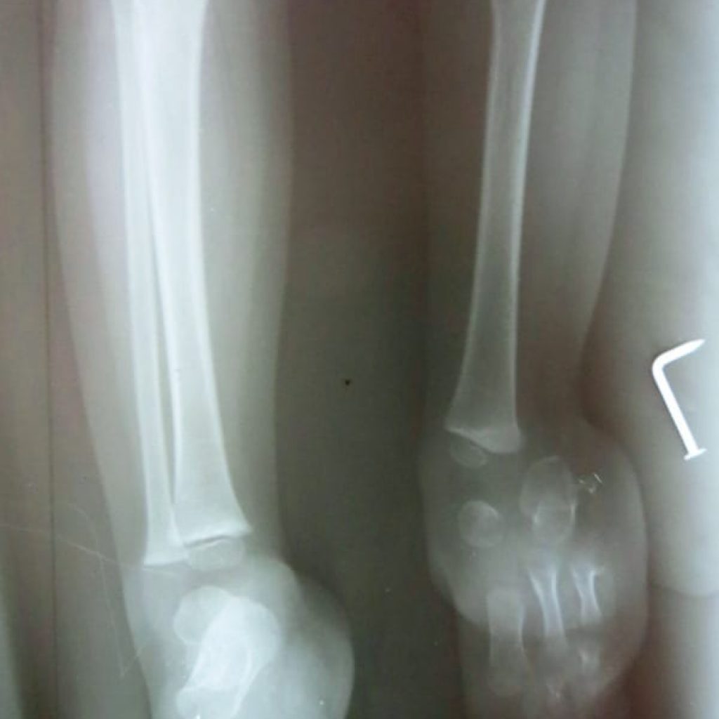
What is the fibula on X-rays?
When the fibula is totally absent on the X-rays, there is always a fibrous remnant connected to the calcaneus (heel bone) that is very tight and contracted. This fibrous fibula remnant is termed the fibular anlage.
What classification system is used for hemimelia?
There are five different fibular hemimelia classification systems used in North America. The earliest one is the Coventry and Johnson classification system. The most commonly used system is the Achterman and Kalamchi classification. The other systems are the Stanitski classification system, ...
What is the treatment for fibular hemimelia?
Most cases are sporadic and the etiology of fibular hemimelia remains unclear. Treatment often includes orthotics and prosthetics, with or without surgical intervention. The decision to amputate versus reconstruct the extremity depends on the extent of foot deformity, the LLD, and the desires of the individual patient and family.
What is fibular hemimelia?
The treatment of fibular hemimelia aims to achieve a functional limb nearly equal in length to the normal limb at maturity , with a plantigrade, stable and flexible foot . The treatment of fibular hemimelia is highly dependent on the potential of the foot to be reconstructed, and should be held primarily in mind when deciding on amputation versus limb salvage. Treatment of fibular hemimelia should be highly individualized to the patient and undertaken in experienced centers with access to a multidisciplinary management team including a pediatrician, physical therapists and social workers.
What is Genu Valgum?
Genu valgum in fibular hemimelia is thought to be related to the hypoplasia of the lateral femoral condyle, the tibial angulation and the resultant increase in compressive forces across the lateral half of the physis, culminating in stunted growth in this area.
What are the clinical features of fibular hemimelia?
The most common clinical features of fibular hemimelia are limb-length discrepancy (LLD) and foot deformities. The ability to reconstruct the foot and the extent of the LLD will ultimately determine the treatment decision of amputation versus limb salvage. In the presence of significant upper extremity deformity amputation should be avoided.
What is the shortened tibia?
The shortened tibia often includes an anterior or anteromedial bow, thought to arise due to the tethering effect of the rudimentary fibular anlage which clinically presents as a taut posterior band in the fibular area. There may be a skin dimple at the apex of the bow.
Is fibular hemimelia autosomal dominant?
Sporadic appearance is most common, but chromosomal anomalies with autosomal dominant, autosomal recessive and x-linked transmission have been reported. Theories exist that intimate an exogenous vascular or mechanical interference with limb bud function in utero. (Graham, 1983) The so-called fibular developmental field is thought to influence the development of lateral foot rays, the ACL, patella, proximal femur, acetabulum and pubic bone, which would explain some of the spectrum of deformities observed in patients with fibular hemimelia. (Lewin, 1986)
What causes a deformity in the limb?
The deformity is probably due to disruptions during the critical period of embryonic limb development, between 4th and 7th week of gestation. Vascular dysgenesis, viral infections, trauma and environmental influences have been suggested as possible causes.
Is the foot in an equinovalgus position?
The foot is generally in an equinovalgus position . As there is limited growing potential within the affected bone, the extent of the deformity tends to increase with growth. Occasionally, fibular hemimelia is associated with congenital shortening of the femur (femur-fibula-ulna complex; see this term).
Is the right fibula affected more often than the left?
Unilateral involvement occurs in two-thirds of cases, with the right fibula being affected more often than the left. Agenesis of both fibulae is rare. Fibular hemimelia may vary from partial absence of the fibula (10% of cases) with relatively normal-appearing limbs, to absence of the fibula with marked shortening of the femur, curved tibia, ...