
- Mechanism and Epidemiology. The talus is a strong bone since it bears the entire body weight. ...
- General Treatment. Talus fractures are typically very painful. The first step in treatment is to place you in a splint to keep the foot and ankle from moving, which can ...
- Post-operative Care. After surgery, you will be placed into a splint or a cast. This is to keep your foot still and allow your bones and soft tissues to heal.
- Long Term. Talus fractures are a difficult injury. Some people experience continued pain, stiffness, and swelling even after their bones heal.
- More Information
How long for fractured talus to heal?
- raise your ankle if possible
- gently hold an ice pack (or a bag of frozen peas) wrapped in a towel on your ankle for 15 to 20 minutes every 2 to 3 hours
- stop any bleeding – put pressure on the wound using a clean cloth or dressing
- if your ankle is not at an odd angle, wrap it loosely in a bandage to help support it
What are the different ways to treat a bone fracture?
Treatments for non-unions and delayed unions include:
- Ultrasound therapy: A medical professional will apply low intensity ultrasound to the affected area. This may help fractures heal.
- Bone graft: If the fracture does not heal, a surgeon will transplant a natural or synthetic bone to stimulate the broken bone.
- Stem cell therapy: Stem cell-derived therapies may assist in the healing of bone fractures.
What are the signs of a fractured talus bone?
- Bruising
- Difficulty walking
- Pain
- Swelling
Is a fractured talus the same as a broken ankle?
The talus is the small bone between the heel bone (the calcaneus), and the tibia and fibula. A broken ankle is very painful. You might hear the bone break at the time of injury. It may sound like a snapping or grinding noise. Other symptoms include: A broken ankle isn’t the same as a sprained ankle.
Do all talus fractures need surgery?
Most of the time fractures involving the talus require surgery. However, if the fracture is in a good alignment and seems stable, you might be treated without surgery using a splint or cast. If the bones are shifted out of place, surgery is usually needed to reset the bones.
How long is recovery from talus surgery?
Recovery can be prolonged. No weight or walking on the leg will be allowed for 8-12 weeks. Once the bone is healed, exercise and physical therapy is started to maximize the function of the ankle. The patient should expect some swelling about the foot for several months after the procedure.
Can talus bone heal itself?
The fractured ends of the bone are still basically lined up properly. The break can usually heal without surgery.
Can a fractured talus heal without surgery?
If the parts of the bone are aligned, your doctor can put you in a cast and give you crutches so you don't put weight on your foot. The bone can heal without surgery. Physical therapy might be needed after the bone has healed. If the parts of the bone have moved out of place, surgery is required.
Can a talus bone be replaced?
The talus spacer is made for each patient individually, modeled from computed tomography (CT) imaging, and is fitted to a patient's specific anatomy. During the replacement surgery, the patient's talus bone is removed and replaced with the implant, which is made from cobalt chromium alloy.
Why does my talus hurt when walking?
Osteochondral Lesions of the Talus (OLT) A sudden injury like a sprain can damage cartilage on your talus (heel bone) or cause fractures, blisters or sores in the bone underneath. You might notice a catch in your ankle, or it could lock up or still hurt months after a treated injury, which could be an OLT.
What happens if talus bone dies?
Avascular necrosis of the talus can be quite devastating and lead to total loss of the ankle joint with arthritis, deformity and pain. The development of AVN is determined to a large extent by the type of the talus fracture.
How do you realign talus?
2:225:51How to SELF Adjust Your Ankles - YouTubeYouTubeStart of suggested clipEnd of suggested clipPress into the talus. And then bring the foot into dorsiflexion. So you're bringing it back up whileMorePress into the talus. And then bring the foot into dorsiflexion. So you're bringing it back up while you maintain that pressure into the ankle. And just hold it for a little bit.
Is a talus fracture serious?
A talus fracture can be a severe injury. A talus fracture can result in a significant loss of motion and function of your ankle and foot joints. This can affect your ability to walk and bear weight on your foot.
How do I know if my talus is broken?
A fractured talus usually causes:Extremely severe pain.An inability to put weight on the foot.Swelling and tenderness.
Why do talus fractures require surgery?
Because the talus is important for ankle movement, a fracture often results in substantial loss of motion and function. A talus fracture that does not heal properly can lead to complications, including a limp, arthritis, and chronic pain. For this reason, most talus fractures require surgery.
Where to go for talus fracture?
Most people with talus fractures will go to an urgent care center or emergency room for initial treatment because of the severity of their symptoms.
What is the talus in the foot?
Anatomy. The talus is the bone that makes up the lower part of the ankle joint (the tibia and fibula make up the upper part). The ankle joint allows your foot to move up and down. The talus also sits above the heel bone (calcaneus). Together, the talus and calcaneus form the subtalar joint.
How to classify talus fracture?
Talus fractures can be classified by how much the pieces of bone have moved out of their normal position.
What joint is the talus in?
Together, the talus and calcaneus form the subtalar joint. This joint allows your foot to move inward and outward, which is important for walking on uneven ground. The talus is the main connector between the foot and leg, helping to transfer weight and pressure forces across the ankle joint.
Where is the talus bone located?
The talus bone sits between the bones of the lower leg and the calcaneus (heel bone).
What is the procedure for repositioning bone fragments?
Open reduction and internal fixation. During this operation, the bone fragments are first repositioned (reduced) into their normal alignment. They are then held together with special screws or metal plates and screws.
What is the term for a fractured talus?
Another common place for the talus to break is on the outside part of the bone called the "lateral process, " which can happen when the foot is forced in an outward direction. This is sometimes called a "snowboarder fracture" because it often occurs when snowboarding.
Where is the talus fractured?
The most common area the talus fractures is in the mid portion in an area called the neck. The neck is located between the "body" of the talus, which is by the ankle joint, and the "head" of the talus, which is by the foot. Figure 3: Side view x-ray showing fracture of the talar neck (shown by white arrow). Another common place for the talus ...
How to heal a swollen ankle?
In this case, your surgeon may choose to keep you in a splint until the swelling goes down. You may be placed in an external fixator to hold the bones in place. This also helps take the pressure off of the skin and other soft tissues. With an external fixator, large pins are inserted into certain bones of your foot and ankle. These pins are visible outside of your skin, with special bars used to connect the pins to each other. This is usually temporary for a couple of weeks until the swelling goes down.
What is the talus in the foot?
The talus is a bone in your foot that forms part of the ankle joint. It sits between your shin bone (tibia), and your heel bone (calcaneus). The joint between the talus and the calcaneus is called the subtalar joint. This joint allows for side to side motion of the foot and helps us when we walk on uneven ground. The blood supply to the talus comes from several small arteries. Fractures of the bone often damage the blood supply to the bone and can affect healing. The talus is kept in place by strong ligaments (which connect bone to bone). The ligaments can also be injured and cause the talus to become unstable in the ankle joint.
Why do surgeons need more x-rays after talus fracture?
Over time, your surgeon will likely want more x-rays, not only to check the alignment of the fracture, but also at the quality of the bone. Because the blood supply to the bone is delicate, it can easily be disrupted by the fracture. If there is no blood supply to the bone, the bone cells may die. This is called avascular necrosis, and it is a common complication after a talus fracture. If this happens more surgery may be needed.
What to do after foot surgery?
After surgery, you will be placed into a splint or a cast. This is to keep your foot still and allow your bones and soft tissues to heal. Whether you had surgery or not, you will usually not be allowed to put any weight on your foot. If you put too much weight on your foot before the bones heal, the fracture may move.
Why do you put pins in your foot?
You may be placed in an external fixator to hold the bones in place. This also helps take the pressure off of the skin and other soft tissues. With an external fixator, large pins are inserted into certain bones of your foot and ankle.
What is the procedure for a fractured talus?
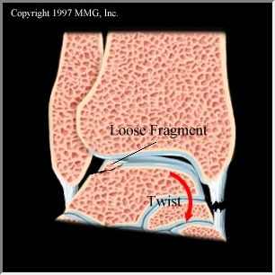
M ost fractures of the talus do require surgery to reset the bone and help minimize later complications. The orthopaedic surgeon realigns the broken bone with metal screws placed inside the bone. Any small fragments of bone discovered during this procedure will be removed and bone grafts will be used to help restore the shape of the joint.
How long does it take for a talus to heal after surgery?
After surgery, the patient will then be placed in a cast for approximately eight to twelve weeks. The patient will not be allowed to put any weight on the foot for at least two to three months. During healing, the physician may request X-rays or an MRI to be done. This will reveal if the talus bone has a good blood supply.
How long does it take to cast a broken bone?
After surgery, the patient will then be placed in a cast for approximately eight to twelve weeks.
Can a talus bone be MRI?
During healing, the physician may request X-rays or an MRI to be done. This will reveal if the talus bone has a good blood supply. Even if the bones heal well, arthritis may still develop. Since most of the talus is covered with cartilage, bones are allowed to move smoothly against each other.
How to treat osteochondral cysts?
There are multiple treatment options available for osteochondral lesions with associated subchondral cysts and one should determine the best treatment option with the assistance of MRI, CT and intraoperative findings. Utilize retrograde drilling as the primary treatment option when there is intact cartilage. When treating larger lesions, the utilization of allograft and autograft may be necessary. Newer allograft and matrix treatment methods provide promising results, and additional research will help assist and guide future treatment protocols.
What should be done after immobilization?
Patients should have physical therapy following immobilization to gradually increase range of motion, weightbearing and activity. After the failure of conservative care, a combination of MRI and CT findings along with intraoperative arthroscopic evaluation are necessary to determine the best treatment option.
What Are The Most Effective Treatment Options?
Non-surgical treatment consists of non-weightbearing and immobilization for approximately six weeks. One can utilize non-steroidal anti-inflammatory drugs (NSAIDs) and other anti-inflammatory modalities during this time period. Patients should have physical therapy following immobilization to gradually increase range of motion, weightbearing and activity.
What is arthroscopic treatment?
Arthroscopy is a key component in treating osteochondral and cystic lesions of the talus. When possible, utilize arthroscopic treatment as this allows for direct visualization of the pathology while avoiding an arthrotomy and potential complication. A wide variety of treatment strategies exist for osteochondral defects of the talus and these treatments and techniques have continued to evolve over the last 15 years.
How to treat cystic lesions?
One can treat large, chronic, cystic lesions with OATS, mosaicplasty and autogenous bone grafting. Perform these techniques following excision and debridement of the lesion and the underlying subchondral bone. Then harvest these osteochondral grafts and utilize them to restore mechanical, structural and biochemical properties of the initial hyaline cartilage.
What is osteochondral lesion?
An osteochondral lesion is an injury or fracture to the chondral surface or underlying subchondral bone. These lesions occur in the talus and are generally secondary to single or multiple traumatic events. Traumatic events can cause multiple mechanisms of injury including shearing, fracture, avulsion and compaction.
What causes pain in the subchondral bone?
Over time, osteolysis and subchondral cyst formation occur. As cystic changes occur, the increase in fluid pressure results in increasing pain due to the innervation of the subchondral bone.
What is the treatment for Gustilo IIIC?
The choice of appropriate treatment remains controversial and may include amputation, primary tibiocalcaneal arthrodesis [6,8], talar body prosthesis [9] or total ankle arthroplasty [7,10].
What are the drawbacks of tibiocalcaneal arthrodesis?
There are two possible drawbacks of our treatment. First, the recovery period would probably have been shorter if our patient had allowed the tibiocalcaneal arthrodesis soon after his initial clinical status had stabilized. We believe, however, that our decision to perform a primary reconstruction by the use of an urgent free flap transfer after radical debridement, blood vessel reconstruction, and temporary Kirschner wire fixation was appropriate because it avoided primary below-knee amputation and provided primary wound healing. Second, an early arthrodesis would also have prevented later injuries such as the decompensation of the tibiocalcaneal joint 20 years after the initial treatment.
How to avoid amputation of foot?
To avoid amputation, our primary treatment plan was first to perform a microsurgical reimplantation of the foot and use temporal fixation to avoid a pending infection that could be caused by a more complex treatment. This would be followed by a late tibiocalcaneal arthrodesis, which would provide viability of the microvascular flap and stabilize the general condition of our patient. After the initial treatment and successful healing of the soft tissues, his condition was stable. He was without pain and his injured leg could carry his full body weight. His tibiocalcaneal joint was fully functional. At this stage, during his hospitalization after his treatment, he was advised to complete the treatment by tibiocalcaneal arthrodesis. However, he has consistently refused any further treatment, referring to his fully functional condition and the absence of pain. We strongly suggested treatment by tibiocalcaneal arthrodesis after his hospitalization, during his rehabilitation and at several follow-up examinations, but we could not convince him, since he was satisfied with the outcome of the treatment and afraid of any potential complications from additional treatment.
Can a talar prosthesis be used for a Gustilo IIIc injury?
A case with a total ankle arthroplasty and a custom-built talar prosthesis resulted in a better outcome than in our case [10]. However, our patient suffered a more serious Gustilo IIIc injury. The primary use of a talar prosthesis for this type of injury is controversial because of possible high infection and complication rates [7]. In addition, implantation of a metal talar body prosthesis for an extruded and lost talus is a rarely performed approach [7,9,10]. Primary below-knee amputation as a salvage procedure was another option for our treatment, but we decided against it since our patient was a teenager at the time of his injury.
Is tibiocalcaneal arthrodesis performed?
Due to his explicit request, the tibiocalcaneal arthrodesis was not performed, resulting in an uncommon treatment that led to the self-formation of a new type of tibiocalcaneal joint and a surprisingly successful long-term result. His foot was functional for 20 years after the initial treatment, when minor injuries caused by a jump from a height of 0.7m led to decompensation of pseudarthrosis, which was treated with a standard arthrodesis procedure.
Can you reposition an extruded talus?
On the other hand, there are various appropriate choices for successful treatment when an extruded talus is not lost. Wound lavage and debridement before reduction of the extruded talus should be performed immediately after the injury [2,3,11-13]. Repositioning of the extruded talus is important for restitution of ankle biomechanics and represents a worthy alternative to tibiocalcaneal arthrodesis despite the fact that complication rates of avascular necrosis and infection remain high [2-4,7,12,14,15]. Primary reimplantation of a totally extruded talus is recommended even for contamination or articulare damage, while osteonecrosis of the talus does not necessarily lead to an unsatisfactory result [2,3,5-7,12]. A tibiocalcaneal arthrodesis after a totally extruded talus is recommended as a second stage or as a salvage procedure with remarkably satisfactory results [7,11]. Several authors also report the successful reimplantation of a talus after complete open extrusion after Gustilo IIIa and IIIb injury [2]. A successful reimplantation was also reported for a case where the talus was lost and retrieved several hours after the accident [6].
Can a talus be amputated?
We have presented a case with the most extreme variant of a totally extruded and lost talus accompanied with a semi-amputated foot. A review of the literature revealed only a few reports of similar cases, but none involving an adolescent in combination with foot reimplantation. In the first of these reports, the talus was lost and the foot was so severely damaged that the leg was amputated below the knee [18].
What to do if you have a hip joint?
Apart from physical therapy, make efforts to minimize your daily physical activity movement as much as possible. You may need the aid of crutches or a walker if the affected joint is your hip, knee or ankle. Consider crutches an invitation to stay off your legs.
What is the disease that causes bone to collapse?
Learn more... Avascular Necrosis (AVN) is a disease that occurs from temporary or permanent poor blood supply to the bones, leading to death of the bone tissues. This process can make breaks in the affected bone that eventually causes bone collapse.
How to treat AVN?
Another way to treat AVN is to limit your alcohol intake, which can worsen your condition. You'll also want to keep your cholesterol levels as low as possible, which will help strengthen your heart and blood. Acupressure therapy can also help treat AVN by pressing on certain areas of the body to help you relax. While you can do acupressure yourself, you can also see a professional for a fully relaxing experience. If possible, see a physiotherapist, who can show you certain exercises to maintain or enhance your joint movement. To learn how to use medication to treat AVN, keep reading!
Can you put your bones in line with electrical stimulation?
If surgery puts your bones in line, electrical stimulation sets the ball in motion. However, it's not right for everyone, so ask your doctor if it's a feasible option.
Diagnosis
During the physical exam, your doctor might feel around your joint to pinpoint your pain. Your doctor might also order X-rays or other imaging tests to view your joints and bones.
Treatment
If your bone spurs cause pain, your doctor might recommend over-the-counter pain relievers, such as acetaminophen (Tylenol, others), ibuprofen (Advil, Motrin IB, others) or naproxen sodium (Aleve, others).
Preparing for your appointment
You'll likely first see your family doctor, who might refer you to a doctor who specializes in the diagnosis and treatment of joint disorders (rheumatologist).

Basic Anatomy
Mechanism and Epidemiology
- The talus is a strong bone since it bears the entire body weight. To break the talus bone, it usually takes a lot of energy such as falling off of a ladder, a roof, or from a car accident. The most common area the talus fractures is in the mid portion in an area called the neck. The neck is located between the "body" of the talus, which is by the ankle joint, and the "head" of the talus, w…
General Treatment
- Talus fractures are typically very painful. The first step in treatment is to place you in a splint to keep the foot and ankle from moving, which can help with the pain. You may also need a CT scan of the bone. The CT scan will help your surgeon to decide if surgery is needed and to see if any other bones in the foot are broken. Most of the time fr...
Post-Operative Care
- After surgery, you will be placed into a splint or a cast. This is to keep your foot still and allow your bones and soft tissues to heal. Whether you had surgery or not, you will usually not be allowed to put any weight on your foot. If you put too much weight on your foot before the bones heal, the fracture may move. Once the bone heals, you can gradually increase the amount of weight you p…
Long Term
- Talus fractures are a difficult injury. Some people experience continued pain, stiffness, and swelling even after their bones heal. This often improves over time, but you may always notice a difference compared to your other side. Because talus fractures often occur in high-energy situations such as falls from height, an injury to the cartilage often occurs. When this happens, y…
More Information