Resistance exercise and whole body vibration can prevent fatty infiltration in skeletal muscle and also improve muscle strength. Therapeutic strategies to prevent myosteatosis may improve muscle function and reduce fall risk in the elderly, potentially impacting the incidence of bone fracture.
How to prevent fatty infiltration of muscles with aging?
One of the most effective countermeasures against fatty infiltration of muscle with aging is physical activity and regular exercise.
How can fatty infiltration of the liver be treated?
Fatty infiltration of the liver, if caught at early stages could be treated by practicing a healthy diet and with the help of exercise. How can I prevent fatty infiltration of the liver? The best way to prevent fatty infiltration of the liver is through proper diet and exercise.
What is fatty infiltration in skeletal muscle?
Fatty Infiltration in Skeletal Muscle: Cellular and Molecular Mechanisms. There are several stem cell populations in skeletal muscle, the most well defined being muscle satellite cells (SCs), which lie below the basil lamina of muscle fibers and contribute to myogenesis during the process of muscle regeneration.
How do you measure fatty infiltration in muscle?
Fatty infiltration was analyzed by comparing muscle with minimal fatty infiltration to muscle with moderate and severe amount of fatty infiltration, respectively, with chi square of the maximum likelihood test for qualitative variables. Moderate and severe amounts of fatty muscle infiltration were also compared to each other.
Can you reverse fatty infiltration of muscle?
Fatty infiltration is irreversible and progressive if left untreated. Slight reversal of muscle atrophy has been noted after repair in some studies.
What causes fatty infiltration in muscles?
The main causes of fatty infiltration are muscular disuse and spinal injury, similar to the causes of atrophy (Elliott et al., 2006; Hodges et al., 2006).
How does fatty infiltration develop?
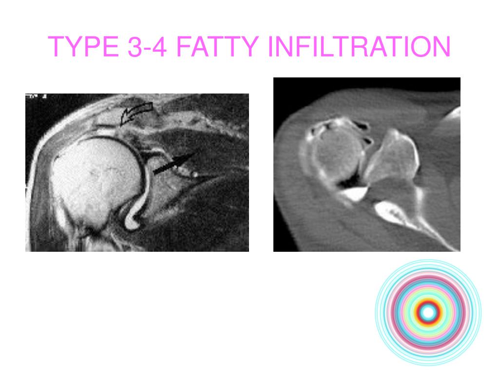
More specifically, a full-thickness tear of a rotator cuff tendon can result in retraction of the muscle belly and its tendon. Retraction of the muscle belly may lead to a change in the pennation angle between the muscle fibers and the subsequent development of fatty infiltration [9].
What is fatty degeneration of muscle?
Summary. Fatty degeneration is a degenerative condition of the tendon-muscle unit of rotator cuff muscles, characterized by atrophy of muscle fibers, fibrosis, and fatty accumulation within and around the muscles.
What happens when a muscle atrophies?
If you have atrophied muscles, you'll see a decrease in your muscle mass and strength. With muscle atrophy, your muscles look smaller than normal. Muscle atrophy can occur due to malnutrition, age, genetics, a lack of physical activity or certain medical conditions.
How do you burn intramuscular fats?
The quickest way to get rid of intramuscular fat is with low-level aerobic activity. Staying under 75% of max heart rate will keep you burning predominately fat, and exercising while on a ketogenic diet is an even better way to do it; nutritional ketosis increases exercise-induced intramuscular fat oxidation 20-fold.
Can fat form under muscle?
Subcutaneous fat is present underneath the skin and on top of abdominal muscles. 2. Visceral fat is stored within the abdominal cavity (underneath the abdominal muscles) and surrounds your organs. As a quick rule of thumb: if the fat is visible or pinchable it is most likely subcutaneous fat.
What does fatty infiltration mean?
Medical Definition of fatty infiltration : infiltration of the tissue of an organ with excess amounts of fat.
Does fat attached to muscle?
It serves as a passageway for nerves and blood vessels between your skin and your muscles. It insulates your body, helping it regulate temperature. It attaches the dermis to the muscles and bones with its special connecting tissue.
What causes fatty infiltration in the rotator cuff?
Rotator cuff pathology is not only a tendon pathology, but also a muscle pathology. Tendon tear can lead to the onset of progressive and irreversible fatty infiltration (FI) of the muscle belly as well as the belly of the adjacent muscles.
What causes fatty atrophy?
The causes of fat atrophy are varied. Fat atrophy can be associated with Parry-Romberg syndrome and linear scleroderma, or exist as an isolated condition with unknown causes. Loss of fat tissue may also be associated with reactions to medication, corticosteroid injections, systemic illness, or as a result of trauma.
Can supraspinatus atrophy be reversed?
Gerber et al. showed that the muscular atrophy in cases of massive rotator cuff tear was at least stopped and might be reversed in successfully repaired supraspinatus musculotendinous units at least within two years.
Introduction
Osteoporosis affects ~10 million people in the U.S. and results in over 1.5 million bone fractures per year. Hip fractures are a major cause of morbidity and mortality among the elderly: ~40% of those suffering a hip fracture will end up in a nursing home and 20% will never walk again.
Factors Contributing to Bone Marrow Adipogenesis
Bone cell populations are heterogeneous and include cells of both hematopoietic (e.g., megakaryocytes and osteoclasts) and mesenchymal (e.g., osteoblasts and AC) origin. Aging is accompanied by an accumulation of AC as well as increase in ACs size within the bone marrow cavity ( 5 ).
Fatty Infiltration in Skeletal Muscle: Cellular and Molecular Mechanisms
Aging in humans is accompanied by a loss of subcutaneous fat but an accumulation of AC and lipids in non-adipose depots, such as bone marrow, liver, and skeletal muscle ( 2 ). Fatty infiltration of skeletal muscle (myosteatosis) has, in particular, been recognized as an important component of aging and frailty ( 21 – 26 ).
Functional Consequences of Fatty Infiltration in Muscle
Protein synthesis enhances muscle hypertrophy and the maintenance of muscle strength, whereas impaired protein synthesis contributes to muscle atrophy.
Discussion: Targeting Adipogenesis and Lipid Accumulation in Muscle to Prevent Fracture
One of the most effective countermeasures against fatty infiltration of muscle with aging is physical activity and regular exercise.
Author Contributions
MH wrote the initial draft and prepared the manuscript illustrations. MM-L contributed additional narrative material on bone marrow adipogenesis and edited the manuscript. DF contributed narrative material on exercise and whole body vibration and on myosteatosis. DF also edited the manuscript.
Conflict of Interest Statement
The authors declare that the research was conducted in the absence of any commercial or financial relationships that could be construed as a potential conflict of interest.
Abstract
Fatty infiltration (FI) and muscle atrophy are common findings following RC tears. Fatty degeneration varies among rotator cuff muscles. The pathophysiology is complex, and is characterized by increased fibrosis. Age is reported to correlate with degeneration and atrophy.
About this chapter
Hantes M., Komnos G. (2020) Fatty Infiltration and Muscle Atrophy. What It Means and What Happens After Repair?. In: Sampaio Gomes N., Kovačič L., Martetschläger F., Milano G. (eds) Massive and Irreparable Rotator Cuff Tears. Springer, Berlin, Heidelberg. https://doi.org/10.1007/978-3-662-61162-3_10
What is intramuscular fat?
Intramuscular fat is the visible fat found within a muscle. Intermuscular is considered to be an ectopic fat depot similar to visceral adipose tissue (VAT) found in the abdomen. IMAT is most commonly measured via computed tomography (CT) or magnetic resonance imaging (MRI).
What is the difference between intramuscular and intermuscular fat?
Intermuscular fat is generally considered to be any fat (including the fat between muscle groups and within a muscle) found beneath the fascia of a muscle and is the widest definition for fat beneath the fascia of a muscle. Intramuscular fat is the visible fat found within a muscle.
What is IMAT in a muscle?
Many studies have used slightly different definitions of IMAT (i.e., adipose tissue in a muscle, adipose tissue between muscles, or adipose tissue under the fascia of the thigh), and conclusions drawn across studies should be interpreted within this context. 3. IMAT and Metabolism.
What is IMAT in medical terms?
Intermuscular adipose tissue (IMAT) is a significant predictor of both muscle function and mobility function in older adults and across a wide variety of comorbid conditions such as stroke, spinal cord injury, diabetes, and COPD. IMAT is also implicated in metabolic dysfunction such as insulin resistance.
What is IMAT in skeletal muscle?
IMAT has been referred to in the literature by a variety of names and definitions including myostasis, intermuscular fat, intramuscular fat, and low density lean tissue. Intermuscular fat is typically the broadest definition of fatty infiltration in the muscle referring to storage of lipids in adipocytes underneath the deep fascia of muscle. This includes the visible storage of lipids in adipocytes located between the muscle fibers (also termed intramuscular fat) and also between muscle groups (literally intermuscular) [ 46#N#D. C. Karampinos, T. Baum, L. Nardo et al., “Characterization of the regional distribution of skeletal muscle adipose tissue in type 2 diabetes using chemical shift-based water/fat separation,” Journal of Magnetic Resonance Imaging, vol. 35, no. 4, pp. 899–907, 2012. View at: Publisher Site | Google Scholar#N#See in References#N#] (See Figure 1 ). While not frequently isolated as a separate fat depot by itself, there also exists a smaller group of lipids stored within the muscle cells themselves known as intramyocellular lipids or IMCL; IMCL has been reviewed extensively elsewhere [ 47#N#P. M. Coen and B. H. Goodpaster, “Role of intramyocelluar lipids in human health,” Trends in Endocrinology and Metabolism, vol. 23, no. 8, pp. 391–398, 2012. View at: Google Scholar#N#See in References#N#]. Increased levels of IMCL are found both in obese insulin resistant individuals and in highly trained endurance athletes; these paradoxical findings have led to the conclusion that lipids stored within muscle cells are not always harmful to the cell [ 47#N#P. M. Coen and B. H. Goodpaster, “Role of intramyocelluar lipids in human health,” Trends in Endocrinology and Metabolism, vol. 23, no. 8, pp. 391–398, 2012. View at: Google Scholar#N#See in References#N#]. For the remainder of this review, the term IMAT will refer to any measure of fat beneath the deep fascia of the thigh, not including studies that have used methods that independently quantify IMCLs (i.e., histochemical or spectroscopic methods).
What is the role of structural composition in muscle strength?
Muscle’s structural composition is an important factor underlying muscle strength and physical function in older adults. There is an increasing amount of research to support the clear disassociation between the loss of muscle lean tissue mass and strength with aging. This disassociation implies that factors in addition to lean muscle mass are responsible for the decreases in strength and function seen with aging. Intermuscular adipose tissue (IMAT) is a significant predictor of both muscle function and mobility function in older adults and across a wide variety of comorbid conditions such as stroke, spinal cord injury, diabetes, and COPD. IMAT is also implicated in metabolic dysfunction such as insulin resistance. The purpose of this narrative review is to provide a review of the implications of increased IMAT levels in metabolic, muscle, and mobility function. Potential treatment options to mitigate increasing levels of IMAT will also be discussed.
How to decrease IMAT?
Exercise and weight loss may act to directly decrease IMAT, improve factors associated with increased IMAT such as obesity and inactivity, and improve metabolic, muscle, and mobility dysfunction. Multiple studies have examined the effects of diet, exercise, or a combination of diet and exercise on IMAT [ 20.
What is a fatty infiltration?
Fatty infiltration (FI) is an important prognosis factor in the anatomical and functional outcomes of rotator cuff repairs. The objective of this study was to analyze the natural history of muscle FI and better evaluate its onset and aggravation time frame.
What is the FI of a rotator cuff?
FI of the rotator cuff muscles is closely related to patient age, the extent of the tendon rupture, and the time to onset. The more extensive the tear and the older the patient, the more rapidly one should make a decision for surgical repair. Other factors such as the extent of tendon and muscle retraction seem to play an important role but could not be evaluated in this study.
Can rotator cuff tears be surgically treated?
Nevertheless, all rotator cuff tears do not warrant surgical treatment and most of them, notably those occurring after 55 years of age, should first be treated medically (rest, adaptation of daily and occupational movements, rehabilitation, NSAIDs, antalgics, physical therapy, infiltrations, etc.).
Abstract
In some patients nonoperative treatment of a rotator cuff tear is sufficient, while in others it is only the first stage of treatment prior to surgery.
Introduction
Pathology of the rotator cuff influences both the tendon and corresponding muscle. More specifically, a full-thickness tear of a rotator cuff tendon can result in retraction of the muscle belly and its tendon.
Patients and Methods
We retrospectively reviewed 2500 patients with rotator cuff tears who underwent open or arthroscopic tendon repair, arthroscopic débridement, or a reverse shoulder arthroplasty between 1988 and 2005. We excluded 812 patients with previous surgery on the affected shoulder or incomplete imaging studies.
Results
The appearance of fatty infiltration of the supraspinatus was related to increasing patient age, delay between onset of symptoms and diagnosis, and the number of involved tendons (p < 0.001 for all three variables).
Discussion
Understanding the relationship of fatty infiltration and rotator cuff muscle atrophy and the clinical and temporal factors that influence this process may help physicians improve outcomes or provide patients with better prognostic information.
Introduction
Factors Contributing to Bone Marrow Adipogenesis
- Bone cell populations are heterogeneous and include cells of both hematopoietic (e.g., megakaryocytes and osteoclasts) and mesenchymal (e.g., osteoblasts and AC) origin. Aging is accompanied by an accumulation of AC as well as increase in ACs size within the bone marrow cavity (5). Adipose tissue represents ~20% of bone marrow tissue before the third decade in life …
Fatty Infiltration in Skeletal Muscle: Cellular and Molecular Mechanisms
- Aging in humans is accompanied by a loss of subcutaneous fat but an accumulation of AC and lipids in non-adipose depots, such as bone marrow, liver, and skeletal muscle (2). Fatty infiltration of skeletal muscle (myosteatosis) has, in particular, been recognized as an important component of aging and frailty (21–26). Lipid accumulation in muscles of the lower limb is also associated …
Functional Consequences of Fatty Infiltration in Muscle
- Protein synthesis enhances muscle hypertrophy and the maintenance of muscle strength, whereas impaired protein synthesis contributes to muscle atrophy. Insulin is an anabolic factor for skeletal muscle, and accumulation of muscle ACs and IMC lipid decreases insulin sensitivity, impairing the capacity for normal protein synthesis in skeletal muscle ...
Author Contributions
- MH wrote the initial draft and prepared the manuscript illustrations. MM-L contributed additional narrative material on bone marrow adipogenesis and edited the manuscript. DF contributed narrative material on exercise and whole body vibration and on myosteatosis. DF also edited the manuscript.
Conflict of Interest Statement
- The authors declare that the research was conducted in the absence of any commercial or financial relationships that could be construed as a potential conflict of interest.
Acknowledgments
- The authors are grateful to E. Scheller and W. Cawthorn for the opportunity to prepare this contribution. Funding to MH is provided by the National Institute on Aging (NIA AG036675) and funding to MM-L is provided by the American Diabetes Association (1-16-JDF-062). The authors thank Donna Kumiski in the Electron Microscopy and Histology Core Facility for her assistance …