What are the types of orthodontic x-rays?
- Cephalometric radiograph. Cephalometric x-ray, also known as lateral ceph, is one of the essential types of dental x-ray used in orthodontics.
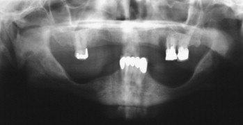
- Panoramic x-ray. Panoramic x-rays are a two-dimensional extraoral radiograph that displays information about both the jaws, upper and lower teeth, alveolar bone, and temporomandibular joint.
- CBCT. ...
Which radiographs are best for orthodontics?
The radiographic examination comprised a panoramic radiograph, a lateral cephalogram and six intraoral anterior periapical radiographs. Initially, only records from the clinical examination were used for diagnosis and treatment planning. If required, the dentist could choose any of the radiographs to accomplish the task.
What is the importance of X-rays in orthodontics?
The cephalometric radiograph is an invaluable adjunct to the clinical examination and models of the dentition, which together form the basis for orthodontic diagnosis and treatment planning. However, the user should be aware of the various errors which can detract from the reliability of this technique. Cephalometric landmarks are identified and tracings of the radiograph are made.
Why are panoramic radiographs used in orthodontics?
May 27, 2020 · Based on the central question the Taskforce established 7 clinical questions (CQs), one per type of radiograph, on the contribution to orthodontic diagnosis, treatment planning and post-treatment outcome evaluation of CQ#1 an orthopantomogram (OPT), CQ#2 a lateral cephalogram (LC), CQ#3 a hand-wrist (HW) radiograph, CQ#4 a peri-apical (PA), CQ#5 a …
Can orthodontic treatment be carried out without X-rays?
Jun 06, 2021 · The respondents' usage of radiographic techniques. Figure 1 summarizes the use of radiographic techniques during the different stages of orthodontic treatment. The most frequently used techniques during all stages were panoramic, lateral, …
What radiograph is most commonly taken on an orthodontic patient?
The bitewing, lateral cephalometric and panoramic radiographs were the most frequently ordered radiographs for both young and older patients. Radiographic and clinical examinations were judged to be of equal importance to more than fifty percent of the dentists.
Why are radiographs used in orthodontics?
Position of Teeth An orthodontist uses x-rays to look for any malformed, missing or extra teeth. Abscesses, cysts and some tumours that are hidden, may be seen on x-rays.Nov 25, 2019
What are the two radiographs that are needed for orthodontic treatment planning?
Traditionally, dental models, facial and intra-oral photographs and a set of two-dimensional radiographs are used for orthodontic diagnosis and treatment planning. As evidence is lacking, the discussion is ongoing which specific records are needed for the process of making an orthodontic treatment plan.Nov 12, 2013
What radiograph is used for orthodontic treatment group of answer choices?
Lateral Cephalometric Radiographs (LCR) are a common decision-making aid in orthodontic treatment planning and are routinely used in clinical practice.Apr 16, 2018
What is a panoramic radiograph used for?
Panoramic radiography, also called panoramic x-ray, is a two-dimensional (2-D) dental x-ray examination that captures the entire mouth in a single image, including the teeth, upper and lower jaws, surrounding structures and tissues.
What is cephalometric radiography?
Cephalometric x-rays (also called ceph x-rays or radiographs) show a side view of your head, exposing teeth, jaw, and surrounding structures. This technology is considered safe and often useful or necessary to help professionals evaluate and assist patients.
What is a lateral cephalometric radiograph?
A lateral cephalometric radiograph (LCR) is a standardised, reproducible radiograph used primarily for orthodontic diagnosis and treatment planning. It is taken from a distance of 1.5m with the head at a right angle to the X-ray beam at a distance of 30cm, (although this has been found to vary slightly).Mar 3, 2015
What charts and records would the dentist require for orthodontic treatment?
Radiographs, photographs, study models and 3D study models Records such as radiographs, photographs, visual recordings and study models, either as hard copies or in digital format, complement the written record. To be of value, these records must be of good quality.
What diagnostics records are indicated for an orthodontic consultation?
If it is determined that orthodontic treatment is indicated, you will be scheduled for diagnostic records. These records usually consist of study models, diagnostic photographs and orthodontic x-rays.
What is occlusal radiography?
An occlusal radiograph is a radiograph designed to be placed between the occlusal surfaces of the teeth with the central beam directed at 90o or at 50 -60o to the plane of the film depending on what is required to be viewed.
What extraoral radiograph is used to best see the maxillary sinus?
Bregma–menton viewiv. Bregma–menton view. This projection is primarily used to demonstrate the walls of the maxillary sinus (especially in the posterior areas), the orbits, the zygomatic arches and the nasal septum.Apr 11, 2016
What is a Cephalogram used for?
The lateral cephalogram shows facial structure, bone, and soft tissue. Your radiologist or dental practitioner can use these images to study the relation of your teeth to your jaw, assess problems in alignment or growth patterns, and prepare treatment.Sep 28, 2015
Abstract
The aim was to investigate the objective and choice of different radiographic examinations used in orthodontic treatment of children and adolescents, using a web-based questionnaire directed toward specialists in orthodontics. The questionnaire was distributed to 255 orthodontists in Sweden.
INTRODUCTION
Many common dental radiographic examination techniques can be used for the assessment of teeth and jaws when planning and conducting orthodontic treatment. The risk associated with repeated exposures to radiation at an early stage in life is not negligible.
MATERIAL AND METHODS
Ethical approval for the study was obtained from the Regional Ethical Review Board, Gothenburg, Sweden (Dnr 847-18).
RESULTS
The questionnaires were collected between June and October 2017 and were followed by telephone interviews in October 2018. The response rate to the questionnaire was 57% (144 out of 255).
DISCUSSION
The findings from the questionnaire showed that panoramic radiography together with lateral and intra-oral radiography were preferred for treatment planning while intra-oral radiographs of the incisors were preferred during treatment and for follow-up.
Acknowledgements
The authors wish to thank all orthodontists participating in the questionnaire study and follow-up interviews, Per Ekman for statistical support, and Vincent Collins for language assistance.
AUTHOR CONTRIBUTIONS
Conceptualization: Christina Stervik, Agneta Lith, Annika Ekestubbe; Methodology: Christina Stervik, Agneta Lith, Annika Ekestubbe; Software: Christina Stervik, Per Ekman; Validation: Christina Stervik, Agneta Lith, Anna Westerlund, Annika Ekestubbe; Formal analysis: Christina Stervik, Agneta Lith, Anna Westerlund, Annika Ekestubbe, Per Ekman; Investigation: Christina Stervik; Resources: Christina Stervik; Data Curation: Christina Stervik, Per Ekman; Writing - original draft preparation: Christina Stervik, Agneta Lith, Annika Ekestubbe; Writing - review and editing: Christina Stervik, Agneta Lith, Anna Westerlund, Annika Ekestubbe; Visualization: Christina Stervik, Agneta Lith, Annika Ekestubbe; Supervision: Annika Ekestubbe; Project administration: Christina Stervik; Funding acquisition: Christina Stervik..
How many chances of developing cancer in Orthodontic patients?
ICRP currently estimate that there is a 1 in 20,000 chance of developing a fatal cancer for every 1 mSv of effective dose of radiation.
What is an IRMER operator?
IRMER Operators are responsible for conducting any practical aspect of a medical exposure including exposing the radiograph or processing the image. tIRMER Operators and IRMER Practitioners must have received adequate training. tIRMER Operators and IRMER Practitioners must undertake continuing education.
What is stochastic effect?
Stochastic effects are the chance or random effects, governed by the laws of probability, that patients may develop from any dose of radiation. In dental X-ray imaging, the risk of heritable (genetic) effects is considered negligible, so the stochastic effect of concern is the risk of cancer induction.
Is ionising radiation dangerous?
As a result, the use of ionising radiation in clinical practice is governed by law criminal law in the UK . Ionising radiations, including X-rays, are dangerous and have the potential to cause damage to human tissue, including fatal malignant change. Radiographic examinations put patients at risk.
What are the different types of dental x-rays?
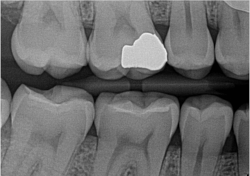
There are 4 common types of dental x-rays: Bitewings. Periapicals. OPG / Panoramic X-ray. CT-Scan. 1. Bitewings are x-rays taken of your back teeth biting together on the left and right sides. These are used to check for tooth decay in between teeth, and bone levels around teeth. They do not show the roots of the teeth.
Why is it important to have an x-ray of your teeth?
First of all it’s important to understand dental x-rays are a useful and important part of dental examination, to ensure the correct diagnosis when there are some signs/symptoms or indications.
What is a periapical x-ray?
Periapicals are x-rays taken of individual teeth – can be 1-2 teeth next to each other, and they show the full length of the tooth – including the root of the tooth. Periapicals are mainly used to check the root / nerve health of a tooth. They are great for detail. 3.
Why is it important to keep x-rays?
X-rays are essential as a diagnostic tool and if you keep a record of them you can avoid having new ones taken too frequently. «.
What is the purpose of X-rays?
X-rays are small doses of radiation that are able to pass through the body then expose an image on a computer or film to show a view of hard tissues of the body. X-rays are great to see bones and calcified objects like teeth. They are not useful to see softer items like cartilage, muscle, tissue.
Can you have orthodontic treatment without x-rays?
The British Orthodontic Society states that for adults with healthy teeth, orthodontic treatment can be carried out without orthodontic x-rays .#N#“In patients with a healthy definition and supporting structures, orthodontic treatment may be carried out without the need for radiographs.”
Can you diagnose tooth decay with an OPG?
You can’t accurately diagnose early tooth decay on an OPG. They are great for general survey. 4. CT Scan – these are a 3D view of the upper and lower jaws. These are great when doing implant surgery and you want to use 3D planning software. They are however high in radiation and so should only be used when needed.
