Procedures
Pneumothorax Recovery. It usually takes 1 or 2 weeks to recover from pneumothorax. But you have to wait for your doctor to say you’re OK. Until then: Go back to your routine a little bit at a time.
How long does it take to recover from a pneumothorax?
Treatment
- Observation. If only a small portion of your lung is collapsed, your doctor may simply monitor your condition with a series of chest X-rays until the excess air is completely ...
- Needle aspiration or chest tube insertion. ...
- Nonsurgical repair. ...
- Surgery. ...
- Ongoing care. ...
Which medications are used in the treatment of pneumothorax?
Occasionally, the pneumothorax is small enough to go away on its own — under observation. Otherwise, the air in the pleural space must be removed and the lung reinflated. This is typically done by surgically inserting a chest tube into the space, and sucking out the air.
Can pneumothorax go away on its own?
Which medications are used in the treatment of pneumothorax?
- Anestacon) Lidocaine hydrochloride is…
- Fentanyl citrate (Sublimaze) The onset of analgesia with fentanyl citrate is immediate…
- Oramorph SR) Morphine is the drug…
- Midazolam. Benzodiazepine used for sedation component… Facebook Tumblr Reddit LinkedIn WhatsApp Messenger Telegram
What is the standard treatment for a pneumothorax?

What is the most common treatment for a pneumothorax?
Treatment for a pneumothorax usually involves inserting a needle or chest tube between the ribs to remove the excess air. However, a small pneumothorax may heal on its own.
How do doctors fix a pneumothorax?
If you have a large pneumothorax, a chest tube will be placed between the ribs into the space around the lungs to help drain the air and allow the lung to re-expand. The chest tube may be left in place for several days and you may need to stay in the hospital.
What is the first line treatment for pneumothorax?
Contou et al recommended that clinicians consider drainage via a small-bore catheter as a first-line treatment for pneumothorax of any cause.
Does pneumothorax need treatment?
Treatment. A small pneumothorax may go away on its own over time. You may only need oxygen treatment and rest. The provider may use a needle to allow the air to escape from around the lung so it can expand more fully.
What are 3 signs and symptoms of a pneumothorax?
What are the Symptoms of Pneumothorax?Sharp, stabbing chest pain that worsens when trying to breath in.Shortness of breath.Bluish skin caused by a lack of oxygen.Fatigue.Rapid breathing and heartbeat.A dry, hacking cough.
How long does it take to recover from a pneumothorax?
Pneumothorax Recovery It usually takes 1 or 2 weeks to recover from pneumothorax.
How do you fix a collapsed lung at home?
How can you care for yourself at home?Get plenty of rest and sleep. ... Hold a pillow against your chest when you cough or take deep breaths. ... Take pain medicines exactly as directed. ... If your doctor prescribed antibiotics, take them as directed.More items...
Is pneumothorax a collapsed lung?
A collapsed lung occurs when air gets inside the chest cavity (outside the lung) and creates pressure against the lung. Also known as pneumothorax, collapsed lung is a rare condition that may cause chest pain and make it hard to breathe. A collapsed lung requires immediate medical care.
What percent pneumothorax needs a chest tube?
Large (> 25% or apex to cupula distance > 3 cm) pneumothorax requires chest tube placement.
Is pneumothorax a medical emergency?
Pneumothorax is common and life-threatening clinical condition which may require emergency treatment in Emergency Medicine Departments.
How is pneumothorax diagnosed?
Diagnosis of a pneumothorax is typically done via a chest X-ray, which takes images to detect the presence of air in the pleural space (area round the lungs). A CT scan and thoracic ultrasound can also be used to help diagnose a pneumothorax.
What are the complications of pneumothorax?
The complications of pneumothorax include effusion, hemorrhage, empyema; respiratory failure, pneumomediastinum, arrhythmias and instable hemodynamics need to be handled accordingly. Treatment complications refer to major pain, subcutaneous emphysema, bleeding and infection, rare re-expansion pulmonary edema.
What is a Pneumothorax?
A pneumothorax is commonly known as a collapsed lung. Normally, the outer surface of the lung sits next to the inner surface of the chest wall. The lung and the chest wall are covered by thin membranes called pleura.
What to do if your lung is not expanding?
If you have had a serious chest injury or treatment does not expand your lung, your doctor will discuss the possibility of surgery to repair the lung and pleura. Sometimes damaged or scarred portions of lung are removed to allow the collapsed lung to heal. Traditional surgery requires the chest to be opened.
What is lung surgery?
Surgery that removes blebs or areas of scarring or attaches the lung permanently to the chest wall
What causes a collapsed lung?
A collapsed lung is most often caused by an injury to the lungs or chest wall, such as: A penetrating injury, such as a knife or gunshot wound. Blunt trauma from a fall or car accident. Medical procedures, such as removal of fluid from the pleural cavi ty with a needle (thoracentesis) or a lung biopsy.
How to remove air from pleural cavity?
Removal of the air from the pleural cavity by inserting a hollow plastic tube, called a chest tube, between the ribs and attaching it to a suction device. If you need to be admitted to the hospital for treatment, you will likely receive oxygen by nasal prongs or a mask for the first couple days.
What is it called when air can only move into the chest cavity?
If air can only move into the chest cavity, but not out, pressure builds up inside the pocket of trapped air. This is called a tension pneumothorax. A tension pneumothorax can cause complete collapse of the nearby lung and can push the heart and major blood vessels to the other side of the chest.
What is it called when a lung collapses?
Occasionally, a lung will collapse without any direct injury to the lung or chest. This is called a spontaneous pneumothorax. This is more likely to happen in people with lung disease.
What is the goal of pneumothorax treatment?
The goal of treatment is to relieve the pressure on the lung and allow it to re-inflate. The type of treatment selected will depend on the cause and severity of the collapse, and on the patient’s overall health. For a minor pneumothorax, your doctor may simply keep an eye on you, as the lung may re-inflate on its own, ...
What are the Symptoms of Pneumothorax?
Symptoms normally come on almost immediately and commonly begin with chest pain. Other signals that the problem may be a collapsed lung are:
How to remove air from a collapsed lung?
During a needle aspiration, a needle attached to a syringe is inserted between the ribs into the air-filled space that is pressing on the collapsed lung and is used to suction out the excess air. A chest tube involves a similar insertion that involves a one-way valve device that continuously removes air until the lung re-inflates. The tube may need to stay in for a few hours, or even a few days, to ensure that the lung does not collapse again.
How long does a chest tube stay in?
The tube may need to stay in for a few hours, or even a few days, to ensure that the lung does not collapse again.
What is the next step in a lung leak?
In cases that involve an accident, or repeated collapsed lungs, the next step is a non-surgical repair of the leak. This can be done in several ways and is sometimes called pleurodesis.
Can you go to the hospital after a lung collapse?
Avoiding air travel for the first week after a collapse is often suggested. Diving puts patients at high risk, so most doctors suggest permanently avoiding it . Also, people who smoke are at increased risk of a pneumothorax, so quitting is highly recommended.
How to prevent a collapsed lung?
Anyone can take steps to reduce your chances of collapsed lung: Stop smoking. Avoid or limit activities with drastic changes in air pressure ( scuba diving and flying).
How to prevent lung from collapsing again?
Your provider makes an incision and inserts a tube. Then your provider uses chemicals (such as doxycycline or talc) to attach the lung to the chest cavity, eliminating extra space in the chest cavity.
What is a collapsed lung?
A collapsed lung occurs when air enters the pleural space, the area between the chest wall and the lung. Air in the pleural space can build up and press against the lung, causing it to collapse partially or fully. Also called a deflated lung or pneumothorax, a collapsed lung needs immediate medical care.
What is the condition where the lung collapses?
Endometrial tissue lines the uterus. With endometriosis, it grows outside the uterus and attaches to an area inside the chest. The endometrial tissue forms cysts that bleed into the pleural space, causing the lung to collapse.
What is the condition where the chest is hard to breathe?
A collapsed lung occurs when air gets inside the chest cavity (outside the lung) and creates pressure against the lung. Also known as pneumothorax, collapsed lung is a rare condition that may cause chest pain and make it hard to breathe. A collapsed lung requires immediate medical care.
Why do you put a tube in your chest?
Chest tube drainage: If you have a larger pneumothorax, your provider may put a hollow tube in your chest to reduce the air in the pleural space. As the air pressure decreases, the lung re-expands and heals. You may have this tube in place for a couple of days or longer.
How long do you stay in the hospital after a lung transplant?
After treatment, you may be in the hospital for a couple of days or longer. This allows your provider to check your progress and give you oxygen, if necessary. You will make an appointment for follow-up visits. You should contact your provider if symptoms of collapsed lung return.
What are the principles of pneumothorax?
The treatment principles of pneumothorax include five principles: air elimination, reducing air leakage, healing the pleural fistula, promoting re-expand and preventing future recurrences, but treating underlying diseases, preventing and dealing complications are also important. The core of treatment is based on different etiology and pathogenesis. If patients did not get promptly and completely treatment that were combined with pneumothorax or underlying diseases, respiratory failure would be most dangerous. Therefore maintaining necessary respiratory function and stable hemodynamics are the first step of treatment (1-8).
How to treat pneumothorax with needle aspiration?
Air elimination is the first and most important treatment of pneumothorax through needle aspiration and catheter drainage. Their improvements are based on the developments of the material and operation on special positions/circumstances. Needle aspiration is required to exhaust air completely at the first time. In order to approach the target, aspiration should be done in any position of the chest cavity even in some traditional dangerous positions such as the supraclavicular fossa and subclavicular region, guiding by Ultrasound or CT scanning. Additionally, new damage caused by the needle must be avoided during the re-expansion. The current approach is to use cannula or vine remaining needlleiv, even connected to one-way Heimlich valve device to enhance the efficacy and are convenient for patients to activities. In the future there may have sensitive detection method or use the non-metallic needle. Secondary pneumothorax patients especially with pre-existing structuredness lung diseases such as chronic obstructive pulmonary disease (COPD) or tuberculosis, needle aspiration treatment success rate is generally low (9-15).
How long should a pneumothorax tube be clamped?
The ACCP consensus group suggests that the tube should be clamped for approximately 4 hours in primary spontaneous pneumothorax and 5-12 hours in secondary spontaneous pneumothorax. The time is so short but there has some doubts about why BTS 2010 guideline was not refer to clamped observation (15-25).
How to reduce air leakage?
The first step is to find the lung lobe or segment interlinked pleural fistula by using blocking balloon to observe bubbles overflow in the drainage bottle, pressure change or not which can use Chartis system. Without structuredness lung disease or thoracic surgery or trauma, upper lobe is the most likely position of breach and may be treated preliminarily. And CT scanning is needed to infer the possible segment by the image of bulla and adhering band.
Is pneumothorax a recurrent disease?
Except traumatic or iatrogenic factors, pneumothorax usually is also a recurrent disease, especially caused by structure dness lung diseases which named secondary pneumothorax. Some studies revealed its recurrence rate 70% or more. Therefore, to prevent recurrences is other important aspect of the treatment principles.
Is pneumothorax a primary or secondary condition?
In any case it is a dangerous situation that requires immediate attention and treatment. Pneumothorax can be divided in primary and secondary.
Is a small bore chest drain a good replacement for a large bore chest drain?
But if failed, needle aspiration should not be repeated and small-bore (<14 F) chest drains may be a good replacement. On the other side, small-bore chest drains should be the first choice, especially for the patients with pre-existing structuredness lung diseases.
How old is too old to have pneumothorax?
The type of pneumothorax caused by ruptured air blisters is most likely to occur in people between 20 and 40 years old, especially if the person is very tall and underweight.
What happens when a lung collapses?
In a collapsed lung, air from the lung leaks into the chest cavity. The example shown is a complete left pneumothorax.
How to tell if you have a collapsed lung?
On some occasions, a collapsed lung can be a life-threatening event. Treatment for a pneumothorax usually involves inserting a needle or chest tube between the ribs to remove the excess air. However, a small pneumothorax may heal on its own.
Do pneumothorax run in families?
Genetics. Certain types of pneumothorax appear to run in families.
Can pneumothorax be mechanical?
Mechanical ventilation. A severe type of pneumothorax can occur in people who need mechanical assistance to breathe. The ventilator can create an imbalance of air pressure within the chest. The lung may collapse completely.
How to treat pneumothorax without symptoms?
If the pneumothorax is small and without symptoms, pneumothorax treatment is conservative, and generally involves serial chest x-rays to ensure that the air pocket is not getting larger. If the pneumothorax increases in size, or is large to begin with, treatment involves putting a small tube into the chest cavity to drain the air, ...
Why do you need antibiotics for pneumothorax?
Intravenous antibiotics should be commenced at the same time to prevent infection and resultant sputum retention, which may delay re-expansion of the collapsed lung. A recurrent pneumothorax requires more aggressive management, and hence a partial pleurectomy may be performed if the patient is fit to undergo surgery.
What is Pneumothorax?
A Pneumothorax (pl. pneumothoraces) is a collection of free air in the chest outside the lung i.e in the pleural space that separates the lung from the chest wall.
What are the symptoms of tension pneumothorax?
Signs and symptoms of Tension Pneumothorax include surgical emphysema, absent breath sounds on the affected side, mediastinal shift and tracheal deviation to the opposite side and acute respiratory distress. When an intercostal drain is inserted into the pleural space, the air is released under pressure.
What happens if you have a pneumothorax?
These increasing pressures if not corrected, can invert the diaphragm, cause subcutaneous emphysema and ultimately a cardiorespiratory arrest. Signs and symptoms of Tension Pneumothorax include surgical emphysema, absent breath sounds on the affected side, mediastinal shift and tracheal deviation to the opposite side and acute respiratory distress. When an intercostal drain is inserted into the pleural space, the air is released under pressure.
How long does a pneumothorax tube stay in place?
The tube is kept in place until the lung has time to heal and the collection of air has resolved. The size of the pneumothorax, in addition to symptoms, other injuries, and the need for other surgical procedures which would necessitate general anaesthesia all effect the pneumothorax treatment.
How far away from the cupola is a pneumothorax?
A larger pneumothorax (> 3 cm apex-to-cupola distance) will require an intercostal drain and physiotherapy should be withheld until the drain has been inserted.
How is pneumothorax treated?
The treatment of pneumothorax depends on a number of factors, and may vary from discharge with early follow-up to immediate needle decompression or insertion of a chest tube. Treatment also depends on the physician that is going to handle the patient; pulmonary physicians usually perform medical thoracoscopy (minimally invasive) one port, while thoracic surgeons use a surgery suite and two ports. In some cases patient preference is requested.
How to treat tension pneumothorax?
Tension pneumothorax is usually treated with urgent needle decompression. There are several cases where “silent lung” is observed and needle decompression may be required before transport to the hospital upon the site of the accident, and can be performed by an emergency medical technician or other trained professional. The needle or cannula is left in place until a chest tube can be inserted. If tension pneumothorax leads to cardiac arrest, needle decompression is performed as part of resuscitation as it may restore cardiac output.
How long after a pneumothorax can you go to work?
If pneumothorax occurs in a smoker, it may be advisable for someone to remain off work for up to a week after a spontaneous pneumothorax (1-10). For those who have undergone pleurodesis it may take up to two to three weeks off work to recover. Air travel is discouraged for up to seven days after complete resolution of a pneumothorax if recurrence does not occur. Underwater diving is considered unsafe after an episode of pneumothorax unless a preventative procedure has been performed (11-20). Currently professional guidelines suggest that pleurectomy should be performed on both lungs and that lung function tests and CT scan normalize before diving is resumed. Aircraft pilots may also require assessment for surgery (12,21-28).
What is pneumothorax in medical terms?
Pneumothorax is an urgent situation that has to be treated immediately upon diagnosis. Pneumothorax is divided to primary and secondary. A primary pneumothorax is considered the one that occurs without an apparent cause and in the absence of significant lung disease. On the other hand secondary pneumothorax occurs in the presence of existing lung pathology. There is the case where an amount of air in the chest increases markedly and a one-way valve is formed leading to a tension pneumothorax. Unless reversed by effective treatment, this situation can progress and cause death. Pneumothorax can be caused by physical trauma to the chest or as a complication of medical or surgical intervention (biopsy). Symptoms typically include chest pain and shortness of breath. Diagnosis of a pneumothorax requires a chest X-ray or computed tomography (CT) scan. Small spontaneous pneumothoraces typically resolve without treatment and require only monitoring. In our current special issue we will present the definition, diagnosis and treatment of pneumothorax from different experts in the field, different countries and present different methods of treatment.
What is a pneumothorax?
Traumatic pneumothorax occurs when the chest wall is pierced, such as when a stab wound or gunshot wound allows air to enter the pleural space. Traumatic pneumothoraces have been found to occur in up to half of all cases of chest trauma, with only rib fractures being more common in this group. The pneumothorax can be occult in half of these cases, but may enlarge—particularly if mechanical ventilation is required. This type of pneumothorax has also been observed to patients already receiving mechanical ventilation for some other reason.
What causes secondary spontaneous pneumothorax?
Secondary spontaneous pneumothorax occurs due to underlying chest diseases. Most commonly they are observed in patients with chronic obstructive pulmonary disease (COPD), which accounts for approximately 70% of cases. Other known lung diseases that may increase the incidence for pneumothorax are; tuberculosis, necrotizing pneumonia, pneumonocystis carini, lung cancer, sarcoma involving the lung, sarcoidosis, endometriosis, cystic fibrosis, acute severe asthma, idiopathic pulmonary fibrosis, Rheumatoid arthritis, ankylosing spondylitis, polymyositis and dermatomyositis, systemic sclerosis, Marfan’s syndrome and Ehlers-Danlos syndrome, histiocytosis X and lymphangioleiomyomatosis (LAM). Secondary spontaneous pneumothoraces (SSPs), by definition, occurs in individuals with significant underlying lung disease. The following symptoms are usually observed; hypoxemia and hypercapnia in more severe cases. The sudden onset of breathlessness in patients with known underlying lung diseases such as; COPD, cystic fibrosis, or other serious lung diseases should therefore prompt investigations to identify the possibility of a pneumothorax.
What are the two types of pneumothorax?
Keywords: Pneumothorax, medical thoracoscopy, spontaneous, secondary. Primary spontaneous. Spontaneous pneumothoraces are divided into two types: primary, which occurs in the absence of known lung disease, and secondary, which occurs in someone with underlying lung disease.
How is pneumothorax treated?
The treatment of pneumothorax depends on a number of factors and may vary from discharge with early follow-up to immediate needle decompression or insertion of a chest tube. Treatment is determined by the severity of symptoms and indicators of acute illness, the presence of underlying lung disease, the estimated size of the pneumothorax on X-ray, and – in some instances – on the personal preference of the person involved.
How to measure pneumothorax?
the volume of air in the pleural space) can be determined with a reasonable degree of accuracy by measuring the distance between the chest wall and the lung. This is relevant to treatment, as smaller pneumothoraces may be managed differently. An air rim of 2 cm means that the pneumothorax occupies about 50% of the hemithorax. British professional guidelines have traditionally stated that the measurement should be performed at the level of the hilum (where blood vessels and airways enter the lung) with 2 cm as the cutoff, while American guidelines state that the measurement should be done at the apex (top) of the lung with 3 cm differentiating between a "small" and a "large" pneumothorax. The latter method may overestimate the size of a pneumothorax if it is located mainly at the apex, which is a common occurrence. The various methods correlate poorly but are the best easily available ways of estimating pneumothorax size. CT scanning (see below) can provide a more accurate determination of the size of the pneumothorax, but its routine use in this setting is not recommended.
What is the name of the abnormal collection of air in the pleural space between the lung and the chest wall?
A pneumothorax is an abnormal collection of air in the pleural space between the lung and the chest wall. Symptoms typically include sudden onset of sharp, one-sided chest pain and shortness of breath. In a minority of cases, a one-way valve is formed by an area of damaged tissue, and the amount of air in the space between chest wall ...
How many cases of pneumothorax are there in a year?
The surgical treatments usually involve pleurodesis (in which the layers of pleura are induced to stick together) or pleurectomy (the surgical removal of pleural membranes). About 17–23 cases of pneumothorax occur per 100,000 people per year. They are more common in men than women.
Why is tension pneumothorax difficult to spot?
Tension pneumothorax may also occur in someone who is receiving mechanical ventilation, in which case it may be difficult to spot as the person is typically receiving sedation; it is often noted because of a sudden deterioration in condition . Recent studies have shown that the development of tension features may not always be as rapid as previously thought. Deviation of the trachea to one side and the presence of raised jugular venous pressure (distended neck veins) are not reliable as clinical signs.
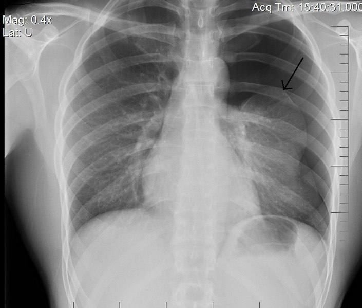
What is the arrow in a pneumothorax?
A large right-sided spontaneous pneumothorax (left in the image). An arrow indicates the edge of the collapsed lung. A pneumothorax is an abnormal collection of air in the pleural space between the lung and the chest wall. Symptoms typically include sudden onset of sharp, one-sided chest pain and shortness of breath.
Which side of the chest is the pneumothorax on?
Chest X-ray showing the features of pneumothorax on the left side of the person (right in image) A plain chest radiograph, ideally with the X-ray beams being projected from the back (posteroanterior, or "PA"), and during maximal inspiration (holding one's breath), is the most appropriate first investigation.

What Is A Pneumothorax?
Symptoms
Diagnosis
Expected Duration

Specialist to consult
Prevention
Treatment
When to Call A Professional
- Your doctor may suspect you have a collapsed lung if you suddenly develop shortness of breath or chest pain, especially if you have had trauma to the chest. He or she will ask about your symptoms, your medical history and your smoking habits. Your doctor will examine you, focusing on your general appearance, your vital signs (temperature, pulse, breath rate, blood pressure), an…
Prognosis
- Once the cause of a collapsed lung is treated, it usually will return to normal within 48 to 72 hours. Recovering from a collapsed lung may take up to several weeks.
Further Information
- Most cases of collapsed lung cannot be prevented. Quitting smoking can reduce your risk of developing the types of lung disease associated with this problem. Wearing your seat belt in the car and avoiding other activities that put you at risk of chest injuries can help you to avoid a collapsed lung caused by trauma.