Treatments include the use of antibiotics, systemic immunosuppressive drugs and surgical repair of the scleral defect with a variety of graft materials.
How do you treat scleral thinning?
Scleral Thinning. Topical antimetabolites such as mitomycin-C or triethylene thiophosphamide have been routinely used as an adjunctive therapy after pterygium removal surgery to decrease the likelihood of recurrency.
What are the treatment options for scleroderma?
Scleroderma Treatment 1 Treating Scleroderma. Treatment typically focuses on inflammation, autoimmunity, vascular issues and tissue fibrosis (the thickening and scarring of the connective tissue that surrounds the internal organs). 2 Your Scleroderma Support Team. ... 3 The Long-Term Prognosis for Scleroderma. ...
What is scleral buckle treatment?
Scleral buckle treatment is used for different types of retinal detachments. Retinal detachment is a medical emergency that requires immediate medical attention and care. Scleral buckling surgery is one of the treatment options. Symptoms and side effects of detachment include a rise in the number of eye floaters.
What to do if you have a scleral laceration on your eye?
A scleral laceration needs to be carefully checked to make sure the eyeball was not ruptured and there is no damage to the surrounding structures. If you are having severe pain two days after your initial injury, you should be rechecked by your ophthalmologist.
How do you treat scleral thinning?
Topical antimetabolites such as mitomycin-C or triethylene thiophosphamide have been routinely used as an adjunctive therapy after pterygium removal surgery to decrease the likelihood of recurrency.
How do you tighten your conjunctiva?
Thermal cautery can also be performed to tighten loose or redundant conjunctiva. Under topical subconjunctival anesthesia, the redundant conjunctiva is grasped gently with a forceps and thermal cautery is applied over this redundant conjunctiva to cause a contraction of the tissue.
What happens if the sclera is damaged?
When the sclera or cornea are cut, this is considered a rupture. Ruptures are formed by blunt force trauma. The sudden impact causes excessive pressure that leaves behind a laceration. Without proper treatment, the condition may cause blindness and infection.
Can sclera heal itself?
It's caused by a scratch to the sclera. It's a mild injury that will go away on its own over 2 weeks. Corneal Abrasion.
What causes loose conjunctiva?
Conjunctival chalasis is characterized by loose or redundant conjunctival tissue. Conditions that may cause conjunctival chemosis include longstanding allergic conjunctivitis; dry eye; trauma and inflammatory conditions, such as episcleritis.
Can conjunctivochalasis heal on its own?
If a person has conjunctivochalasis, it is unlikely that the condition will go away on its own.
How long does sclera take to heal?
How long does it take for the sclera to heal? Minor injury or inflammation of the sclera often heals in a few days or weeks. But if you have any other symptoms or the problem doesn't go away, talk to your healthcare provider or ophthalmologist. An ophthalmologist specializes in disorders and diseases of the eyes.
What causes scleral thinning?
Scleral thinning can result after excessive use of cautery in the scleral bed or overuse of antimetabolites. Prolonged irradiation, transscleral diode laser cycloablation, strabismus surgery and deep sclerectomy procedures can also predispose the sclera to thinning.
How is scleral laceration treated?
A partial thickness corneal or scleral laceration requires repair, unless it is already self-sealing. Treatment: Primary closure (repair of laceration) using 10/0 nylon or silk. Pad the eye for 24 hours and give topical and systemic antibiotics. If repair is not possible, refer very urgently.
How long before retinal detachment causes blindness?
A retinal detachment may cause permanent blindness over a matter of days and should be considered an eye emergency until evaluated by a retina specialist. Most retinal detachments occur suddenly and can threaten the central vision within hours or days.
What antibiotic eye drops are best?
As best as we can determine, the four best drugs to combat acute bacterial infection in adults are: bacitracin/polymyxin B/neomycin; tobramycin; 0.6% besifloxacin; and 1.5% levofloxacin.
What causes the sclera to swell?
Chemosis is swelling of the eye surface membranes because of accumulation of fluid. This symptom is often related to an allergic response. Over-the-counter antihistamines, and a cool cloth placed over the eyes, are usually used to try to alleviate the symptoms.
What is the sclera? What is its function?
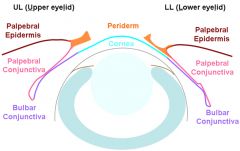
The sclera serves as a protective coat and a stable support for the intraocular tissues . Its thickness is not uniform, being the thickest at the posterior pole (1-1.35 mm), gradually decreasing to be the thinnest immediately posterior to the rectus muscle insertion (0.3 mm), and increasing again towards the limbus (0.8 mm). The scleral matrix is compact and made of collagen fibers and interfibrillar proteoglycans. In a normal healthy eye, the scleral stroma is avascular, receiving its nutrition from choroidal blood vessels and the vascular plexus in the Tenon’s capsule and on the episcleral surface. Scleral melt is a serious and challenging clinical problem as it threatens the integrity of the eye. Clinically, scleral melt is almost always the result of ischemia which interrupts the blood flow of episcleral blood vessels. Therefore, scleral ischemia and melt can be caused by a number of diseases that interrupt the blood circulation. Acutely, scleral ischemia can occur in chemical or thermal burns. When such ischemia extends near the limbus, it further compromises the limbal epithelial stem cells. Chronically, scleral ischemia can happen when excessive‚ beta irradiation or Mitomycin C are used to treat pterygia or develop after systemic vasculitis and connective tissue disorders.
What is scleral melt?
Scleral ischemia, thinning and melt can occur in acute severe chemical or thermal burns and following ocular surgeries such as pterygium excision with a bare sclera technique, 1 especially if such adjuvant therapies as betairradiation and mitomycin C are used. 2 In addition, scleral melt has also been described after retinal detachment repair, glaucoma surgery, systemic vasculitis and connective tissue disorders. 3, 4 Reim et al first described the use of Tenonplasty as an excellent alternative to treat limbal and scleral ischemia in patients with severe chemical and thermal eye burns in 1989 to facilitate conjunctival healing and to halt progressive scleral melt. 5, 6 Since then, several reports have been published reassuring the effectiveness of this surgical approach.7-9 Lin et al 9 in 2002 reported the use of Tenonplasty and amniotic membrane transplantation in 6 patients with scleral perforation after pterygium surgery. There were no recurrences during a follow-up period of 12 to 24 months.
What is the purpose of Sharp Wescott scissors?
Sharp Wescott scissors are used to release the healthy conjunctiva along the border of the scleral melt (Fig. 3A) and to create relaxing incisions radially from the edge of the scleral melt toward the healthy fornix. This allows for subsequent isolation of the Tenon’s capsule located posterior to the melt and creates a pedicle graft (Fig. 3B).
What is the treatment for scleroderma?
Treating Scleroderma. Treatment typically focuses on inflammation, autoimmunity, vascular issues and tissue fibrosis (the thickening and scarring of the connective tissue that surrounds the internal organs). Your treatment may include some or all of the following:
What is the role of a scleroderma specialist?
Scleroderma can impact many important aspects of life, which makes it critical for you to have a reliable team of people to help manage challenges. At different points in time, patients might want support from members of the family and friends or from specialists such as a physical therapist or a personal assistant.
How to reduce itchiness in the body?
Easing skin itchiness with skin lotions and moisturizers. Slowing skin thickening and minimizing damage to the internal organs with medication that suppresses the immune system. Maintaining muscle strength through physical therapy and exercise. Managing digestive tract function to optimize nutritional intake.
Is scleroderma a chronic disease?
Scleroderma is a chronic disease that can affect both the patient’s physical and mental health. The key to feeling better is to tailor the scleroderma treatment to meet the specific needs, taking into account symptoms, type of scleroderma, age and overall health of the patient.
Is scleroderma long term?
The Long-Term Prognosis for Scleroderma. Many scleroderma patients, even those with more invasive systemic scleroderma, can expect to have a normal life expectancy. But to remain as healthy as possible, you need to be open with the doctor about how you feel.
What is scleral buckle surgery?
Scleral buckle surgery, otherwise known as scleral buckling, is used to repair a retinal detachment. 4 The sclera is known as the white of the eye. It is the outer supporting layer of the eyeball. During this procedure, a surgeon fixes a piece of silicone (or a sponge) onto the white of the eye around the retinal tear.
How to prepare for scleral buckling surgery?
How to Prepare for Scleral Buckling. Before scleral buckling, you’ll need to arrange for someone to pick you up after the surgery. Your doctor will inform you of any medications/foods you need to stop taking beforehand. You’ll also have the option to use general anesthesia, which helps you sleep during the surgery.
What is retinal detachment?
Retinal detachment is a medical emergency that requires immediate medical attention and care. Scleral buckling surgery is one of the treatment options. Symptoms and side effects of detachment include a rise in the number of eye floaters. These are small, tiny specks that appear in your vision field.
How does a buckle help with retinal detachment?
The buckle helps repair retinal detachment by pushing the sclera toward the retinal break or tear. The retina is a layer of tissue located on the inside of the eye. It sends visual information from the optic nerve to your brain. A detached retina moves from its usual position.
How long does it take for vision to change after scleral buckle surgery?
Vision may also change for several months following scleral buckle surgery. You should have a follow-up eye exam after about six months to check for vision changes. You may need glasses or contact lenses (or a new prescription) to fix the changes.
What to expect during cataract surgery?
Here are the procedure steps you can expect during surgery: 5. You may receive anesthesia before the surgery to fall asleep. If you are awake during surgery, your doctor or surgeon will apply eye drops or provide you with an injection to numb your eye. You will also receive eye drops to dilate your eyes.
What is the procedure to remove a tear from the retina?
One of these is laser photocoagulation. During this procedure, your surgeon uses a laser beam to burn the area surrounding the retinal detachment or tear. This produces scar tissue, which seals a break and prevents fluid leakage. Or, your doctor may perform cryopexy.
How long does it take for veins to fade after sclerotherapy?
After sclerotherapy, treated veins tend to fade within a few weeks, although occasionally it may take a month or more to see the full results. In some instances, several sclerotherapy treatments may be needed.
What is the treatment for varicose veins?
Overview. Sclerotherapy effectively treats varicose and spider veins. It's often considered the treatment of choice for small varicose veins. Sclerotherapy involves injecting a solution directly into the vein. The sclerotherapy solution causes the vein to scar, forcing blood to reroute through healthier veins.
What is the procedure to check veins in legs?
Ultrasound. Depending on which veins are involved, your doctor may request ultrasound imaging on the veins in your legs. Ultrasound is a painless procedure that uses sound waves to produce images of structures inside the body.
How does lidocaine help with veins?
The solution, usually in liquid form, works by irritating the lining of the vein, causing it to swell shut and block the flow of blood. Some solutions contain a local anesthetic called lidocaine. Eventually, the vein will become scar tissue and disappear.
What is a scleral lens?
Such hard contact lenses are referred to as scleral lenses. In general, these lenses are very comfortable to wear and provide excellent vision in select patients. PROSE Lenses. A special subtype of scleral lenses is called PROSE (prosthetic replacement of the ocular surface ecosystem).
How is a cornea transplant done?
In a corneal transplant, the cornea of the patient is carefully removed, and the cornea of a donor is sewn into place, using stitches about a third of the width of a human hair. Corneas are received from recently deceased persons, with the permission of their next of kin.
Does cross linking help with keratoconus?
The minimally-invasive, advanced therapy slows down or stops the progression of the corneal deformation of keratoconus, by making collagen bonds in the cornea stronger, allowing it to become stiffer and usually stop bulging out.