
What are the treatment options for the soleus muscle?
Here’s an outline of soleus muscle treatment options: Rest: Not bearing weight on the injured foot for a week or so can allow the muscle to begin to heal. Elevation: Using pillows to keep the soleus elevated above the heart for as long as possible for the first day can be effective in reducing any swelling.
What is the accessory soleus muscle?
The accessory soleus muscle is an anatomical variant characterized by an additional distinct muscle encountered along a normal soleus muscle. It is uncommon with a prevalence of ~3% (range 0.7-5.5%).
How do you get rid of stress in the soleus?
TREATMENT: Chronic cases of problems with the soleus are usually associated with medial tibial stress syndrome (shin splints). The treatment protocol includes activity modification and changes in the running surfaces. Shoes with more support and a lift to lessen the stress to the soleus and prevent the foot from pronating.
What is Active Release Therapy for soleus pain?
Active Release Technique (ART): This is a hands-on technique to treat muscle, ligament, fascia, tendon, nerve, or capsule pain. In the case of soleus pain, the muscle is held with tension applied to the tissue. It is different from massage in that there is no skin tension or sliding on the skin with the hand.
How do you heal the soleus muscle?
TreatmentRest: Avoid moving the strained muscle as much as possible. ... Ice: Apply ice to the affected area to reduce inflammation and pain. ... Compression: Wrap the affected area with a medical bandage to prevent swelling. ... Elevation: Keep the leg elevated above the heart when possible to reduce bruising and pain.
How long does it take for the soleus muscle to heal?
In the less severe cases it usually takes up to three days for a pulled calf muscle to start feeling better. In the most severe cases that don't require surgery a full recovery may take up to six weeks.
What is accessory soleus?
Accessory soleus muscle is a rare anatomical variation in the posteromedial aspect of the ankle. It is an anomalous muscle that mimics soft tissue tumour. It may be a cause of exertional pain and swelling secondary to increased physical activity, especially in athletes.
What does soleus pain feel like?
Soleus strains also tend to be less dramatic in clinical presentation and more subacute when compared to injuries of the gastrocnemius. The classic presentation is of calf tightness, stiffness, and pain that worsen over days to weeks. Walking or jogging tends to provoke symptoms [3].
Why does my soleus hurt when walking?
A muscle strain occurs when muscle fibers are damaged by the loads placed on them by activity. A gradual onset of pain is commonly reported during soleus strain and often with no specific mechanism of injury (MOI). This may be due to the limited sensory innervation to the intramuscular aponeurosis.
How do you release a tight soleus?
0:371:27Self-Myofascial Release: Soleus - YouTubeYouTubeStart of suggested clipEnd of suggested clipThe ankle position in exactly the same way so dual finger back up towards the knee tension. AndMoreThe ankle position in exactly the same way so dual finger back up towards the knee tension. And bring it through into dorsiflexion.
Where are the accessory muscles?
The accessory muscles used when breathing in -- called accessory muscles of inspiration -- include the scalene, sternocleidomastoid, trapezius and pectoralis major muscles. These muscles are found around the shoulders, neck and upper chest.
What is a low lying soleus?
A low-lying soleus, with its normal muscle fiber extending to within an inch of the insertion into the calcaneal tuberosity (3), closely resembles the accessory soleus. On MR imaging, however, the accessory soleus, invested by its own fascia, is seen as a distinct muscle mass that is separate from the normal soleus.
What are the attachments of the affected muscle that allow for dorsiflexion of the foot?
DorsiflexionMUSCLEACTIONDISTAL ATTACHMENTExtensor Hallucis LongusDorsiflexion Extends Big ToeDorsal Aspect of Base Distal Phalanx of Big ToePeroneus TertiusDorsiflexion Aids EversionDorsum Base 5th Metatarsal3 more rows
How do you massage a soleus?
1:308:35Massage for Soleus Muscle and Achilles Tendon - YouTubeYouTubeStart of suggested clipEnd of suggested clipGoes is to really get on to the edge of the tibia as well because the muscles plays such a big roleMoreGoes is to really get on to the edge of the tibia as well because the muscles plays such a big role in the transverse plane we want to make sure we get on to this medial aspect as well.
What is soleus syndrome?
The superficial posterior compartment contains the distal portion of the sural nerve with the gastrocnemius and soleus muscles. Increased pressure in this compartment, or soleus syndrome, manifests itself as plantar flexion weakness and paresthesias of the lateral foot and distal calf.
How do you tape soleus?
0:103:47Ankle Taping - Soleus Muscle - YouTubeYouTubeStart of suggested clipEnd of suggested clipRun. The piece of tape along the inside of the leg. And down the inside of the ankle. So it's almostMoreRun. The piece of tape along the inside of the leg. And down the inside of the ankle. So it's almost coursing. Along the line of pull of the tibialis posterior tendon.
What is accessory soleus muscle?
The accessory soleus muscle is an anatomical variant characterized by an additional distinct muscle encountered along a normal soleus muscle. It is uncommon with a prevalence of ~3% (range 0.7-5.5%).
Where is tendinous insertion?
tendinous insertion into the upper calcaneal surface. tendinous insertion into the medial surface of the calcaneus. arterial supply : posterior tibial artery. innervation : tibial nerve.
Where is the calcaneus inserted?
insertion: calcaneus, at either the upper surface or the medial cortex. into the Achilles tendon distally. fleshy insertion into the upper surface of the calcaneus. fleshy insertion into the medial cortex of the calcaneus. tendinous insertion into the upper calcaneal surface.
Can accessory soleus muscle be asymptomatic?
Patients with an accessory soleus muscle can be asymptomatic and thus it will be detected incidentally during imaging performed for an unrelated reason 4. If symptomatic, the usual presentation is a soft mass in the posteromedial distal third of leg 5.
How to diagnose accessory soleus?
Before the advent of MRI, plain radiographs, ultrasound, and CT scans were used to diagnose an accessory soleus. 2,9,12,17,29 Plain radiography generally shows soft tissue swelling between the deep compartment musculature and the Achilles tendon, which obscures or obliterates Kager's triangle on the lateral radiograph of the ankle. 5 Ultrasound shows signal consistent with normal appearing muscle. 12 Computed tomography demonstrates a well circumscribed soft tissue mass, with attenuation signals equivalent to the surrounding normal musculature. 19 Magnetic resonance imaging is the most sensitive test of the available noninvasive diagnostic armamentarium. This test will demonstrate a well circumscribed mass that is isointense with normal muscle on the T1 and T2 weighted images. 29 There was no reported difference on MRI between patients with and those without symptoms. 17
What is accessory soleus?
The accessory soleus can vary in its anatomic configuration, particularly its insertion. This can be via a muscle or tendon into the tendoachilles or into the medial or superior calcaneus. 16,29 This anomalous muscle is contained in its own fascial sleeve and generally has its own blood supply from the posterior tibial artery. 17,28 The usual presentation in late adolescence may be attributable to an increase in muscle mass and muscle activity at this age. 24 Diminished blood supply because of growth that results in an ischemic type of pain has been suggested by several authors. 3,19,22,24 Trosko 27 states that an increase in the size of the accessory muscle can lead to a compartment syndrome or cause a compressive neuropathy of the posterior tibial nerve. Sekiya et al 25 suggest that the pain may be caused by a traction phenomenon on the nerve supplying the accessory soleus around the time that patients are in their adolescent growth spurt.
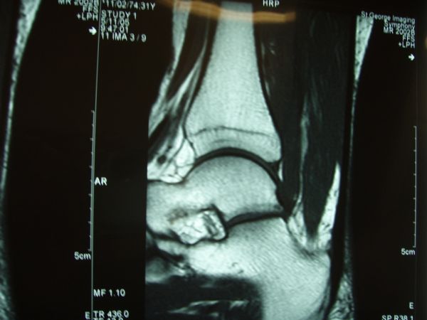
What muscle is anterior to the Achilles tendon?
Axial MRI (T1 weighted image) of Case 4 shows large accessory soleus muscle (arrow) anterior to the Achilles tendon on the left side.
What is accessory soleus muscle?
CONCLUSIONS. Accessory soleus muscle is a rare anatomical variation in the posteromedial aspect of the ankle. It is an anomalous muscle that mimics soft tissue tumour. It may be a cause of exertional pain and swelling secondary to increased physical activity, especially in athletes.
How to diagnose accessory soleus?
3 Before the advent of MRI, plain radiographs, ultrasound, and computed tomography (CT) scans were used to diagnose an accessory soleus. 11,16–18 Plain radiography generally shows soft tissue swelling between the deep compartment musculature and the tendon Achilles, which obscures or obliterates Kager’s triangle on the lateral radiograph of the ankle. Ultrasound shows a mass identical with the adjacent muscles. The characteristic appearances on CT/MRI allow a confident diagnosis without the need to resort to biopsy. 5 MRI allows accurate diagnosis of an accessory soleus muscle because normal muscle has a distinctly different signal intensity from that of both abnormal muscle and soft tissue tumours. 5,19 If the patient is asymptomatic, no treatment is required. If pain or other discomfort is provoked such as swelling and/or functional disorder (varus or equinovarus) especially during or after exercise, surgical treatment is recommended by means of fasciotomy or excision of the accessory muscle. 2 A fasciotomy may be required to relieve an exercise induced compartment syndrome. If nerve compression or claudication is the patient’s main complaint, total excision of the accessory muscle may be needed. 13,20,21 Dokter and Linclau 22 have described treatment of this condition by ligating the feeding artery, which led to atrophy of the muscle, but this procedure requires further intervention such as angiography. We believe that this method is not ideally suited, especially when other options such as surgical excision are more readily available, especially in young professional athletes who need early and definite pain-free return to athletic activity.
What muscle causes resistance to correction in congenital club foot?
Chotigavanichaya C , Scaduto AA, Jadhav A, et al. Accessory soleus muscle as a cause of resistance to correction in congenital club foot: a case report. Foot Ankle Int 2000;21:948–50.
What is the treatment for compartment syndrome?
If pain or other discomfort is provoked such as swelling and/or functional disorder (varus or equinovarus) especially during or after exercise, surgical treatment is recommended by means of fasciotomy or excision of the accessory muscle. 2 A fasciotomy may be required to relieve an exercise induced compartment syndrome.
Where is the soleus muscle inserted?
In one case, the muscle originated from the soleus muscle and was inserted in the superior aspect of the calcaneus in front of the Achilles tendon. The other case originated from the anterior fascia of the soleus muscle and was inserted in the medial aspect of the calcaneus.
Why was the musculotendinous insertion not explored proximally?
The musculotendinous insertion was not explored proximally because of operative restrictions. Blood supply was derived from the posterior tibial artery by two arteries. One accessory branch of the tibial nerve was responsible for the innervation (fig 2). Histopathology confirmed that it was normal muscle tissue.
Where does the accessory muscle come from?
This accessory muscle (typical location) arises from the anterior surface of the soleus, which corresponds to our case, from the soleal line of the tibia or from the fibula and is attached with a separate tendon to the calcaneus anteromedial to the tendon Achilles. The innervation of this muscle by two nerves from the tibial nerve 12 has been described, but in our case only one branch of the tibial nerve was identified as its nerve supply (fig 2).
What is the best treatment for soleus pain?
In cases that are unresponsive to the conservative care of if pain precludes normal weight bearing, a boot (Cam Walker) would be very helpful to take away stress from the soleus.
How to treat a sore soleus?
Treatment for acute injuries of the soleus includes rest by placing the foot in a plantarflexed or pointing down posture. Ice can be applied over a sock or a towel on the back part of the leg to lessen the pain and swelling.
What is the soleus muscle?
The function of the soleus muscle is to assist the gastrocnemius and other muscles in the posterior leg to lift the heel off the ground during propulsion. It also stabilizes the ankle by preventing it from flexing as it decelerates forward momentum of the tibia. The muscle also has a very strong force in turning the foot in (supination), which stabilizes the outside of the foot to the ground. Its action also helps the knee. When the tibia is moving forward it stops excessive movement so that the knee joint can be extended.
Why does the soleus help the foot?
As the foot rolls in during shock absorption, the soleus helps to prevent the foot from excessively pronating. The muscle may pull excessively on the bone causing inflammation at the muscle periosteal bone junction. Accessory soleal muscles have also been implicated in tarsal tunnel syndrome.
What muscle is involved in tarsal tunnel syndrome?
Accessory soleal muscles have also been implicated in tarsal tunnel syndrome. The muscle can extend more distally into the inner portion of the ankle. It would take up space into the tarsal tunnel and during activity and fill with blood, causing compression of the posterior tibial nerve.
Why is the soleus important in shin splints?
Injuries to the soleus are relatively rare. Because of its attachment to the tibia, the soleus is most often implicated in shin splints (medial tibial stress syndrome).
Where does soleus syndrome originate?
Soleus Syndrome. SOLEUS SYNDROME. By: Robert H. Sheinberg, D.P.M.,D.A.B.F.A.S., F.A.C.F.A.S. The soleus muscle originates on the backside of the tibia and fibula and runs deep to the inner and outer heads of the calf muscle (gastrocnemius). The soleus extends down beyond the gastric muscle and joins the covering of the gastrocnemius called ...
