What is intravenous urography for calyceal rupture?
Intravenous urography is a very sensitive and specific method to confirm the diagnosis of calyceal rupture.
How are obstructive stones with calyceal rupture treated?
According to the majority of the related literature, the management of obstructive stones with calyceal rupture is conservative and antibiotics are used to prevent infection. Recovery is most often uneventful. As the first therapeutic step, a low pressure system should be established [9].
What is the mechanism of calyceal rupture?
The mechanism of calyceal rupture is assumed to be related to the increased pressure 2. Although the management of small urinoma is generally conservative, obstructive calculi may require ureteral stent and lithotripsy 1.
When should spontaneous calyceal rupture be suspected in urolothiasis patients?
This case demonstrates that spontaneous calyceal rupture should be suspected in urolothiasis patients presenting for a severe pain even if the calculus is small (less than 5 mm) and the laboratory markers are normal. An immediate management is required to relief symptoms and prevent further complica …

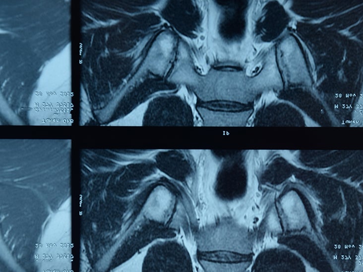
What is a Calyceal rupture?
Renal forniceal or calyceal rupture is the radiographic finding of a perirenal urine leak as a result of ureteric obstruction.
What happens when a kidney stone ruptures?
In conclusion, rupture of the ureter can cause serious complications, including urinoma, sepsis and sometimes kidney loss, thus prompt recognition, treatment and follow-up of the condition is necessary.
How do you treat Forniceal rupture?
A study by Doehn et al published in 2010 looked at 162 cases of forniceal rupture; 97% of patients were treated with primary endoscopic therapy and 92% were started on antibiotic therapy. Preantibiotic urine cultures were positive only in 27% of cases.
What causes a ruptured ureter?
Ureteric rupture is rare and can be traumatic or, less commonly, nontraumatic in nature [1]. The most common cause of traumatic ureteric rupture is iatrogenic trauma, followed by penetrating trauma, and occasionally, blunt abdominal trauma.
How long does a ruptured kidney take to heal?
The average time to heal is about six weeks; with minor lacerations, healing may occur even faster. Severe lacerations that may require surgery will require more time to heal.
Can a ruptured kidney be repaired?
Even with significant renal injuries, patients can fully recover with conservative management alone. Kidney rupture is similar to splenic rupture in that it is usually caused by a direct blow to the abdomen, side, or mid-to-low back which causes damage or a tear to the organ.
Can a kidney stone cause a rupture?
Spontaneous rupture of the urinary collecting system with extravasation of the urine is a very rare condition. This situation is commonly associated with an obstructing urinary stone.
What is a fornix rupture?
Renal forniceal rupture is a common finding in patients with ureteral obstruction. It is thought to be due to increased renal pelvis pressure from backup of urine, causing one or more renal fornices to leak urine. This phenomenon has not been systematically studied.
Where is a minor calyx located?
renal pyramidsThe minor calyces surround the apex of the renal pyramids. Urine formed in the kidney passes through a renal papilla at the apex into the minor calyx; two or three minor calyces converge to form a major calyx, through which urine passes before continuing through the renal pelvis into the ureter.
Can a damaged ureter be fixed?
Ureteral Trauma The ureters are tubes made of muscle to move urine from the kidneys to the bladder. If one or both are damaged, you will need surgery to repair them. If there is a limited tear to the ureters, stents or surgery can fix the tear.
What happens if a ureter bursts?
As rupture of the ureter can cause serious complications, including urinoma, retroperitoneal abscess formation and sepsis, prompt treatment of the condition is therefore necessary.
How do you fix a torn ureter?
A uretero-ureterostomy (UU) is an end to end reconnection of the ureter tube. This surgery entails removal of the diseased portion of the tube, opening up the cut ends along with freeing up the length of the tube to gain mobility, and sewing them together.
What is a rupture of the urinary collecting system?
Rupture of the urinary collecting system with perirenal and retroperitoneal extravasation of the urine is an unusual condition that is typically caused by ureteral-obstructing calculi. We report a case of calyceal rupture with urinoma formation, due to a stone in the distal ureter. The diagnosis was confirmed by computed tomography. Diagnosis, follow-up, and therapeutic approach are discussed.
Is spontaneous calyceal rupture a differential diagnosis?
Spontaneous calyceal rupture should always be considered in the differential diagnosis of a patient presenting with complex symptoms after renal colic. Symptoms will regress with conservative management. Diagnosis is suspected on ultrasonography, and confirmed by computed tomography.
How to treat a knee injury?
Prompt first-aid care can reduce pain and swelling immediately after an injury to your knee. Follow the R.I.C.E. model of self-care at home: 1 Rest. General rest is necessary for healing and limits weight bearing on your knee. 2 Ice. When you're awake, try to ice your knee at least every two hours for 20 minutes at a time. 3 Compression. Wrap an elastic bandage or compression wrap around your knee. 4 Elevation. Lie down with your knee propped up on pillows.
What causes a knee to buckle?
More than one ligament or the fibrous cartilage in your knee also is injured. The injury is causing your knee to buckle during everyday activities. During ACL reconstruction, the surgeon removes the damaged ligament and replaces it with a segment of tendon — tissue similar to a ligament that connects muscle to bone.
What happens after ACL surgery?
Your surgeon will use a piece of tendon from another part of your knee or a tendon from a deceased donor. After surgery you'll resume another course of rehabilitative therapy. Successful ACL reconstruction paired with rigorous rehabilitation can usually restore stability and function to your knee.
What is the best way to check for ACL damage?
An MRI can show the extent of an ACL injury and signs of damage to other tissues in the knee, including the cartilage. Ultrasound. Using sound waves to visualize internal structures, ultrasound may be used to check for injuries in the ligaments, tendons and muscles of the knee.
What is the physical exam for knee injury?
Diagnosis. During the physical exam, your doctor will check your knee for swelling and tenderness — comparing your injured knee to your uninjured knee. He or she may also move your knee into a variety of positions to assess range of motion and overall function of the joint.
How to reduce swelling and pain in knee?
Prompt first-aid care can reduce pain and swelling immediately after an injury to your knee. Follow the R.I.C.E. model of self-care at home: Rest. General rest is necessary for healing and limits weight bearing on your knee. Ice.
Can X-rays show ligaments?
X-rays. X-rays may be needed to rule out a bone fracture. However, X-rays don't show soft tissues, such as ligaments and tendons. Magnetic resonance imaging (MRI). An MRI uses radio waves and a strong magnetic field to create images of both hard and soft tissues in your body. An MRI can show the extent of an ACL injury and signs ...
Calyceal Stone Symptoms
The patients who suffer from the calyceal stone will not show symptoms until the stone migrates to the ureter. The symptoms can be acute, moderate, or chronic. When the symptoms start to appear it is advised to consult your healthcare professional as soon as possible. These symptoms are common in patients with ureteral coli:
Calyceal Stone Causes
The direct cause of calyceal stone is still not discovered. However, some factors need to be overlooked when diagnosing the patient with calyceal stones. If the body produces less than 1 liter of urine in a day, it might lead to calyceal stones.
