Gram stain procedural steps.
| Step | Procedure | Outcome |
| Primary stain (crystal violet) | Add several drops of crystal violet to t ... | Both Gram-positive and Gram-negative cel ... |
| Mordant (iodine) | Add several drops of iodine to the smear ... | Iodine “sets” the crystal violet, so bot ... |
| Decolorization (ethanol) | Add drops of ethanol oneatatime until th ... | Gram-positive cells resist decolorizatio ... |
| Counterstain (safranin) | Add several drops of safranin to the sme ... | Gram-negative cells will be stained pink ... |
Why is iodine added to gram stain?
Iodine is subsequently added as a mordant to form the crystal violet-iodine complex so that the dye cannot be removed easily. Gram staining is based on the ability of bacteria cell wall to retaining the crystal violet dye during solvent treatment. ... Bacteria cell walls are stained by the crystal violet.
What is the role of iodine in gram staining?
In Gram staining, the CV and iodine form an insouble complex ...
What is iodine used for?
Iodine is used as a mordant to bind the dye (primary stain) to the organism making it difficult to lose the stain, especially in gram positive bacteria with thick peptidoglycan layers. 4.7K views. Related Answer. Akshika Sharma. , Mba from Chitkara University. Answered 2 years ago.
What is Gram staining?
Gram staining is based on the ability of bacteria cell wall to retaining the crystal violet dye during solvent treatment. ... Bacteria cell walls are stained by the crystal violet. Iodine is subsequently added as a mordant to form the crystal violet-iodine complex so that the dye cannot be removed easily. source: Google.
Why are Gram positive and Gram negative cells pink?
The same result would occur if you destain for to. Continue Reading. The purpose of Gram’s iodine in the Gram stain is to act as a mordant.
What is the stain history of Gram positive bacteria?
Gram stain history and mechanisms. The cell walls for Gram-positive microorganisms have a higher peptidoglycan and lower lipid content than gram-negative bacteria. Bacteria cell walls are stained by the crystal violet.
Why is iodine used in Gram positive cells?
It is used to form Crystal Violet Iodine complex in cell, mostly in Gram positive bacterial cell because of thick peptidoglycan layer. It increases the affinity for crystal violet in cell.Such that on decolorizing with alcohol the Gram’s positive bacteria retain stain but Gram’s negative bacteria doesn't retain the stain and become colourless.
Why do Gram positive bacteria stain violet?
Gram positive bacteria stain violet due to the presence of a thick layer of peptidoglycan in their cell walls, which retains the crystal violet these cells are stained with. Alternatively, Gram negative bacteria stain red, which is attributed to a thinner peptidoglycan wall, which does not retain the crystal violet during the decoloring process.
How to stain a cell?
How To- Staining Protocol and Concerns: 1 Make a slide of cell sample to be stained. Heat fix the sample to the slide by carefully passing the slide with a drop or small piece of sample on it through a Bunsen burner three times. 2 Add the primary stain (crystal violet) to the sample/slide and incubate for 1 minute. Rinse slide with a gentle stream of water for a maximum of 5 seconds to remove unbound crystal violet. 3 Add Gram's iodine for 1 minute- this is a mordant, or an agent that fixes the crystal violet to the bacterial cell wall. 4 Rinse sample/slide with acetone or alcohol for ~3 seconds and rinse with a gentle stream of water. The alcohol will decolorize the sample if it is Gram negative, removing the crystal violet. However, if the alcohol remains on the sample for too long, it may also decolorize Gram positive cells. 5 Add the secondary stain, safranin, to the slide and incubate for 1 minute. Wash with a gentle stream of water for a maximum of 5 seconds. If the bacteria is Gram positive, it will retain the primary stain (crystal violet) and not take the secondary stain (safranin), causing it to look violet/purple under a microscope. If the bacteria is Gram negative, it will lose the primary stain and take the secondary stain, causing it to appear red when viewed under a microscope.
What is the process of staining cells with crystal violet?
The process involves three steps: Cells are stained with crystal violet dye. Next, a Gram's iodine solution (iodine and potassium iodide) is added to form a complex between the crystal violet and iodine. This complex is a larger molecule than the original crystal violet stain and iodine and is insoluble in water.
How to remove crystal violet from slide?
Rinse slide with a gentle stream of water for a maximum of 5 seconds to remove unbound crystal violet. Add Gram's iodine for 1 minute- this is a mordant, or an agent that fixes the crystal violet to the bacterial cell wall. Rinse sample/slide with acetone or alcohol for ~3 seconds and rinse with a gentle stream of water.
Does alcohol decolorize a cell?
However, if the alcohol remains on the sample for too long, it may also decolorize Gram positive cells. Add the secondary stain, safranin, to the slide and incubate for 1 minute.
What is Gram staining?
Gram staining is initially established by the physician Hans Christian Gram, which was from Denmark. He help to distinguish Klebsiella pneumonia to pneumococci. In short, the process of gram staining comprises the use of a solution of Gram iodine or Potassium iodide to the cells which are use to stain before with Crystal violet or Gentian violet.
How to get purple color out of a gram positive cell?
Rinse the slide with water. Gram-positive cells resist decolorization and remain purple. The dye is released from Gram-negative cells. Counterstain (safranin) Add several drops of safranin to the smear and allow it to sit for one minute. Rinse the slide with water and blot dry.
How to make Gram positive and Gram negative cells purple?
Both Gram-positive and Gram-negative cells will be stained purple by the crystal violet dye. Add several drops of iodine to the smear and allow it to sit for 1 minute. Rinse the slide with water. Iodine “sets” the crystal violet, so both types of bacteria will remain purple.
What are the structural changes between the cell walls of Gram positive bacteria and Gram negative bacteria?
Some the structural changes between the cell walls of gram positive bacteria and gram negative answerable in a way for the Gram stain response ? In the Gram staining , an impenetrable crystal violet with gram iodine complexes is made inside the cell of bacteria, and this complexes is extracted by alcohol from gram negative bacteria but not from gram positive. Gram positive organisms are dehydrated by alcohol, they have very dense cell wall containing of many coatings of peptido-glycan. This creates the pores/holes in the cell wall to near, avoiding the insoluble crystal violet + gram iodine complexes from evasion. In gram negative alcohol eagerly infiltrates the lipid rich external layer, and the thin peptido-glycan coating does not stop solvent channel, therefore, the crystal violet + gram iodine complexes is simply detached or removed.
What is Gram positive bacteria?
Bacteria retain the crystal violet and gram iodine mixtures complexes after the wash in 95% ethanol solution in purple color and are named as Gram positive Bacteria, those which miss these complexes of solution are red in color by counter stain the safranin or 10% fuchsine are named as Gram negative Bacteria. Gram Staining Results.
How long to decolorize a slide?
Decolorizer is use only for 15 Seconds. Pour the slide with Counter stain safranin or 10% Fuchsine for 1 Min or 60 Seconds. Rinse in tap water. Air dry the glass slide, or blot by Absorbent paper.
How many bacteria are in a glass slide?
To be observable on a glass slide, bacteria which are stained by the Gram staining procedure, it is essentially that concentrations of at least of 104 – 10 bacteria/micro liter of concentrated staining reagent. At lesser concentrations, the Gram stain solution of a clinical samples infrequently tells bacteria/organisms even, ...
How does Gram stain work?
How the Gram Stain Works 1 The primary stain ( crystal violet) binds to peptidoglycan, coloring cells purple. Both gram-positive and gram-negative cells have peptidoglycan in their cell walls, so initially, all bacteria stain violet. 2 Gram's iodine ( iodine and potassium iodide) is applied as a mordant or fixative. Gram-positive cells form a crystal violet-iodine complex. 3 Alcohol or acetone is used to decolorize the cells. Gram-negative bacteria have much less peptidoglycan in their cell walls, so this step essentially renders them colorless, while only some of the color is removed from gram-positive cells, which have more peptidoglycan (60-90% of the cell wall). The thick cell wall of gram-positive cells is dehydrated by the decolorizing step, causing them to shrink and trapping the stain-iodine complex inside. 4 After the decolorizing step, a counterstain is applied (usually safranin, but sometimes fuchsine) to color the bacteria pink. Both gram-positive and gram-negative bacteria pick up the pink stain, but it is not visible over the darker purple of the gram-positive bacteria. If the staining procedure is performed correctly, gram-positive bacteria will be purple, while gram-negative bacteria will be pink.
What is Gram staining?
The Gram stain is a differential method of staining used to assign bacteria to one of two groups (gram-positive and gram-negative) based on the properties of their cell walls. It is also known as Gram staining or Gram's method.
What is the primary stain for Gram positive bacteria?
The primary stain ( crystal violet) binds to peptidoglycan, coloring cells purple. Both gram-positive and gram-negative cells have peptidoglycan in their cell walls, so initially, all bacteria stain violet. Gram's iodine ( iodine and potassium iodide) is applied as a mordant or fixative. Gram-positive cells form a crystal violet-iodine complex.
How to get a crystal violet stain off a slide?
If too little heat is applied, the bacteria will wash off the slide during staining. Use a dropper to apply the primary stain (crystal violet) to the slide and allow it to sit for 1 minute. Gently rinse the slide with water no longer than 5 seconds to remove excess stain.
What is the primary stain for Gram staining?
The Gram stain involves staining bacteria, fixing the color with a mordant, decolorizing the cells, and applying a counterstain. The primary stain ( crystal violet) binds to peptidoglycan, coloring cells purple. Both gram-positive and gram-negative ...
Why is Gram stain important?
Because the bacteria are colored, not only is their Gram stain group identified, but their shape, size, and clumping pattern may be observed . This makes the Gram stain a valuable diagnostic tool for a medical clinic or lab.
What is the process of dehydrating gram positive cells?
The thick cell wall of gram-positive cells is dehydrated by the decolorizing step, causing them to shrink and trapping the stain-iodine complex inside. After the decolorizing step, a counterstain is applied (usually safranin, but sometimes fuchsine) to color the bacteria pink.
Why do Gram stainings help?
It stains the bacterial cell wall differently, by giving violet colour to the gram-positive bacteria and pink colour to the negative bacteria. Therefore, it adds colour to the cell wall of bacteria, which increases the visibility under the light microscope. Besides, gram staining also helps in the study of the morphology and arrangement ...
What is Gram stain?
The Gram stain is the differential stain that stains the bacterial cells differently according to the type of cell wall. Gram stain is used to differentiate the bacterial cells by staining the cell wall and distinguish two major groups of bacteria that are gram-positive and gram-negative. Gram-positive bacteria appear violet in colour, ...
What is the principle of Gram staining?
Principle of Gram Staining. The principle of gram staining relies on the reaction of a bacterial cell with the Gram stain, which finally differentiates the bacteria into gram-positive and gram-negative. First, the Crystal violet dissociates into CV + and Cl – that further penetrates the bacterial cell and stains the cell violet.
What is the primary stain of Gram?
Gram stain is the differential stain, which makes the use of four reagents that are given below: Crystal violet: It is the basic dye that acts as the “ Primary stain ”, and it stains all the microorganisms to appear violet in colour. Gram’s iodine: It acts as a “ Mordant dye ” which increases the affinity between the cell ...
What is the most common method of staining?
Gram staining is used widely and the most popular method in laboratories. It is the type of differential staining , which makes the use of more than one stains to differentiate the bacteria. The gram staining method was first given in 1884 by the Danish scientist and Physician Han’s Christian Gram. The Gram stain is the differential stain ...
What color does a Gram positive stain?
Gram-Positive: If the bacteria cell gives a positive result for the gram reaction, it will stain Violet. Gram-negative: If the bacterial cell gives a negative result for the gram reaction, it will stain Pink.
Why is gram staining important?
Besides, gram staining also helps in the study of the morphology and arrangement of the bacteria. It also helps us to know about the chemical and physical properties of the gram-positive and gram-negative bacteria, based on the cell wall difference.
What is Gram staining?
Gram staining is a bacteriological laboratory technique used to differentiate bacterial species into two large groups ( gram-positive and gram-negative) based on the physical properties of their cell walls . Gram staining is not used to classify archaea, formerly archaeabacteria, since these microorganisms yield widely varying responses ...
Why is Lugol's iodine added to the gram stain?
Lugol's iodine solution is always added after addition of crystal violet to strengthen the bonds of the stain with the cell membrane. Gram staining is almost always the first step in the preliminary identification of a bacterial organism.
What is the name of the stain that differentiates bacteria?
The name comes from the Danish bacteriologist Hans Christian Gram, who developed the technique. Gram staining differentiates bacteria by the chemical and physical properties of their cell walls. Gram-positive cells have a thick layer of peptidoglycan in the cell wall that retains the primary stain, crystal violet.
What is the most common gram stain?
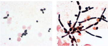
A Gram stain of mixed Staphylococcus aureus ( S. aureus ATCC 25923, gram-positive cocci, in purple) and Escherichia coli ( E. coli ATCC 11775, gram-negative bacilli, in red), the most common Gram stain reference bacteria. Gram stain or Gram staining, also called Gram's method, is a method of staining used to distinguish ...
What did Gram do to make bacteria more visible?
Gram devised his technique not for the purpose of distinguishing one type of bacterium from another but to make bacteria more visible in stained sections of lung tissue. He published his method in 1884, and included in his short report the observation that the typhus bacillus did not retain the stain.
What is the difference between a purple and a pink gram positive?
Purple-stained gram-positive (left) and pink-stained gram-negative (right) Gram-positive bacteria have a thick mesh-like cell wall made of peptidoglycan (50–90% of cell envelope), and as a result are stained purple by crystal violet, whereas gram-negative bacteria have a thinner layer (10% of cell envelope), so do not retain ...
Which phyla have monoderms?
Gram-positive bacteria generally have a single membrane ( monoderm) surrounded by a thick peptidoglycan. This rule is followed by two phyla: Firmicutes (except for the classes Mollicutes and Negativicutes) and the Actinobacteria. In contrast, members of the Chloroflexi (green non-sulfur bacteria) are monoderms but possess a thin or absent (class Dehalococcoidetes) peptidoglycan and can stain negative, positive or indeterminate; members of the Deinococcus–Thermus group stain positive but are diderms with a thick peptidoglycan.