What are the opacities behind the lungs on a chest X-ray?
In a chest X-ray, viewing the lungs from the prone side, the heart and ribs form defined opacities behind or around the lungs, and the lungs are translucent, explains Health Hype.
What does diffuse opacities in the lungs mean?
Diffuse: Diffuse opacities show up in multiple lobes of one or both lungs. This pattern occurs when the air in the lungs is replaced with fluid, inflammation, or damaged tissue. Nodular: This type can indicate both benign and malignant conditions.
What are ground glass opacities in the lungs?
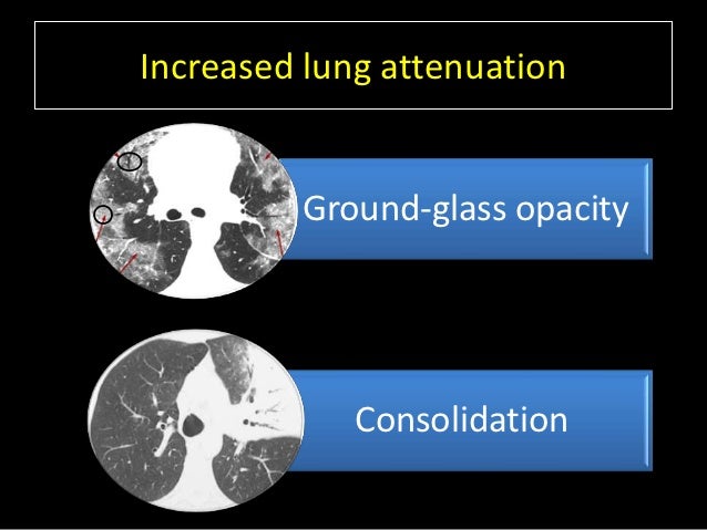
Ground-glass opacities (GGO) are gray areas that computed tomography scans or X-rays of the lungs pick up. The normal lungs appear black in such scans. GGOs can be seen mostly in patients with moderate to severe respiratory conditions, such as infections, cancers, and inflammation.
What is the prevalence of opacities in CTCT thorax?
CT thorax showed reticular opacities in 19 (52.8%), nodular opacities in 12 (33.3%), cystic opacities in 8 (22.2%) [Fig. 1], calcific opacities in 7 (19.4%) and acinar opacities in 6 (16.7%) cases.
What is diffuse opacity?
Diffuse: Diffuse opacities show up in multiple lobes of one or both lungs. This pattern occurs when the air in the lungs is replaced with fluid, inflammation, or damaged tissue. Nodular: This type can indicate both benign and malignant conditions.
What is the hexagonal division of the lung?
Lobules are the hexagonal divisions of the lung. The connective tissue between the lobules is unaffected. Mosaic: This pattern develops when small arteries or airways within the lung are blocked. The opaque areas vary in intensity. Crazy paving: Crazy paving shows up as a linear pattern.
What does GGO mean in a CT scan?
Summary. Ground glass opacity (GGO) refers to the hazy gray areas that can show up in CT scans or X-rays of the lungs. These gray areas indicate increased density inside the lungs. The term comes from a technique in glassmaking during which the surface of the glass is blasted by sand. This technique gives the glass a hazy white or frosted ...
What are the two types of opacities in the lung?
There are two types of opacities in the lung. These two opacities have some differences,... There are two types of opacities in the lung. One is an alveolar opacity, which lies in the alveoli of the lung and another is an interstitial opacity, which lies in the wall of the alveoli.
Which lung is a coalescing opacity?
Note: the unilateral coalescing opacities in the right lung . These are alveolar opacities.
Which opacities coalesce to form the acinus?
These opacities usually tend to coalesce, like the alveoli coalesce to form the acinus. These opacities have a predilection for a particular region of the lung, like a lobe or segment of the lung, since they coalesce and spread in the pattern.
Do opacities show rapid response?
These opacities don’t have any particular predilection to a lobe of the lung and tend to be diffusely located, and bilateral in distribution. With treatment, they usually show a rapid response. They do not show rapid response. Examples include consolidation, ground-glass opacities.
What is the treatment for haziness in the lung?
In case of bacterial or viral infections, treating with intravenous antibiotics and other supportive medicine relieves the symptoms, and the haziness resolves spontaneously without any further management. In severe cases of ground-glass opacities, lobectomy surgery is used to remove the affected part of the lung.
What is ground glass opacity?
Ground-glass opacities (GGO) are gray areas that computed tomography scans or X-rays of the lungs pick up. The normal lungs appear black in such scans. GGOs can be seen mostly in patients with moderate to severe respiratory conditions, such as infections, cancers, and inflammation.
What is COPD pulmonary disorder?
COPD is a pulmonary disorder caused by obstructions in the airways of the lungs leading to breathing problems. Learn about COPD symptoms, diagnosis, and treatment options.
Is ground glass opacity benign?
Ground-glass opacities are usually benign and resolve spontane ously without any complications in patients with short-term illnesses. Most of these patients may not even know that it is present. Others may complain of cough, tiredness, and shortness of breath .
When was ground glass opacity first used?
The first usage of "ground-glass opacity" by a major radiological society occurred in a 1984 publication of the American Journal of Roentgenology. It was published as part of a glossary of recommended nomenclature from the Fleischner Society, a group of thoracic imaging radiologists.
What is ground glass opacity?
Ground-glass opacity ( GGO) is a finding seen on chest x-ray ( radiograph) or computed tomography (CT) imaging of the lungs. It is typically defined as an area of hazy opacification (x-ray) or increased attenuation (CT) due to air displacement by fluid, airway collapse, fibrosis, or a neoplastic process. When a substance other than air fills an area of the lung it increases that area's density. On both x-ray and CT, this appears more grey or hazy as opposed to the normally dark-appearing lungs. Although it can sometimes be seen in normal lungs, common pathologic causes include infections, interstitial lung disease, and pulmonary edema.
What is mosaic pattern on CT?
A mosaic pattern of GGO refers to multiple irregular areas of both increased attenuation and decreased attenuation on CT. It is often the result of occlusion of small pulmonary arteries or obstruction of small airways leading to air trapping. Sarcoidosis is an additional cause of a mosaic GGOs due to the formation of granulomas in interstitial areas. This may coexist with granulomatosis with polyangiitis, leading to diffuse areas of increased attenuation with ground-glass appearance.
How many patterns of ground glass opacities are there?
There are seven general patterns of ground-glass opacities. When combined with a patient's clinical signs and symptoms, the GGO pattern seen on imaging is useful in narrowing the differential diagnosis. It is important to note that while some disease processes present as only one pattern, many can present with a mixture of GGO patterns.
What causes mosaic GGOs?
Sarcoidosis is an additional cause of a mosaic GGOs due to the formation of granulomas in interstitial areas. This may coexist with granulomatosis with polyangiitis, leading to diffuse areas of increased attenuation with ground-glass appearance.
Why do lungs look dark?
Definition. In both CT and chest radiographs, normal lungs appear dark due to the relative lower density of air compared to the surrounding tissues. When air is replaced by another substance (e.g fluid or fibrosis), the density of the area increases, causing the tissue to appear lighter or more grey. Ground-glass opacity is most often used ...
Can atypical pneumonia cause GGOs?
It is important to note that while many of the pulmonary infections listed below may lead to GGOs, this does not occur in every case.
What is the opacity of the lungs?
An ill-defined opacity is a shaded area that does not have the qualities of these normal internal structures, but instead is often an indication of fluid, nodules, infection, swelling or other abnormalities in the lung tissue itself.
What does ill defined opacity mean?
What Is the Meaning of an "ill-Defined Opacity of the Lung"? In a chest X-ray, an ill-defined opacity of the lung refers to an area of the lung that is shaded rather than translucent. It does not have the clearly defined edges of an anatomical structure such as the heart, according to Health Hype. In a chest X-ray, viewing the lungs from ...
What is lung consolidation?
What is a lung consolidation? Lung consolidation occurs when the air that usually fills the small airways in your lungs is replaced with something else. Depending on the cause, the air may be replaced with: The appearance of your lungs on a chest X-ray, and your symptoms, are similar for all these substances.
When to see a doctor for lung consolidation?
Starting treatment early in your illness usually gives you a better outcome. Last medically reviewed on January 22, 2018.
How to treat pulmonary edema?
Treatment may include medication to remove the extra fluid, lower the pressure in your blood vessels, or make your heart pump better.
What tests are needed for pneumonia?
These include: Blood tests. These tests can help determine if: you have pneumonia and what’s causing it. your red blood cell level is low. you’re bleeding into your lung.
Do chest X-rays show lungs?
The appearance of your lungs on a chest X-ray, and your symptoms, are similar for all these substances. So, you’ll typically need more tests to find out why your lungs are consolida ted. With appropriate treatment, the consolidation usually goes away and air returns.
Can lung cancer be removed?
Cancer. Lung cancer is hard to treat. Removing the tumor with surgery may give you the best chance to be cured, but not all lung cancers can be removed. Once the cancer starts to spread, it can’t be cured, and treatment is given only to help your symptoms. Early detection is key.
How much do you know about GGO?
Pulmonary nodules with ground-glass opacity (GGO nodule) are a kind of nodules observed in lungs and a lung lesion often found in physical examinations in recent years.
What kind of Symptoms will GGO present?
Most pulmonary nodules with GGO are asymptomatic. Most patients pathologically diagnosed with early lung adenocarcinoma are asymptomatic. Most lung cancer patients have no symptoms until the disease has progressed into clinical stages 3 to 4.
How to deal with GGO?
Compared with chest X-ray, CT can find pulmonary nodules blocked by organs and has better resolution and contrast, so it is more helpful to find small lesions suspected of early lung cancer.
How GGO be found on imaging?
The pulmonary nodules with GGO on CT show higher density than normal lung tissue, but the broncho-vascular bundle inside the lesion is distinguishable.
How to treat GGO?
There are no definite guidelines for the treatment of pulmonary nodules with GGO at present. Generally, whether a surgery is performed depends on a regular follow-up of chest CT scan. However, there are no definite guidelines on the timing and method of treatment, so they often depend on the patient’s condition and the physician’s judgment.
