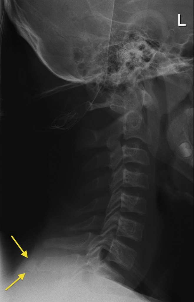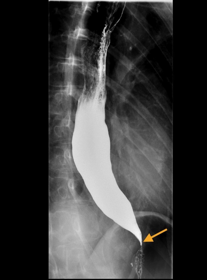
What is a fluoroscopy used for?
Fluoroscopy is used to examine a wide range of internal structures, including bones, the lungs, heart, kidneys, bladder, digestive system, muscles, reproductive system, and joints. The device used in this procedure is called a fluoroscope or, sometimes, a C-arm.
What is fluoroscopy and how to prepare?
You can generally prepare for fluoroscopy by:
- Answering all questions about your medical history, allergies, and medications. ...
- Following instructions to clean stool out of your colon before a barium enema. ...
- Leaving all jewelry and metal objects at home
- Telling your doctor and your radiologic technologist if you feel nervous or anxious about lying still or having the fluoroscopy procedure
Does fluoroscopy use radiation?
Fluoroscopy and X-rays are both imaging tests that use radiation to take images of your internal tissues. The difference is that X-rays take snapshots of internal tissues in a single moment, whereas fluoroscopy can provide continuous, real-time images of your internal tissues using several pulses (brief bursts) of radiation.
How does a fluoroscopy work?
- mode
- pulse rate (if pulsed fluoroscopy was used)
- field of view
- dose rate

What is fluoroscopy and why is it used?
Fluoroscopy is a type of imaging procedure that uses several pulses of an X-ray beam to take real-time footage of tissues inside your body. Healthcare providers use fluoroscopy to help monitor and diagnose certain conditions and as imaging guidance for certain procedures.
What is a fluoroscopy procedure?
During a fluoroscopy procedure, an X-ray beam is passed through the body. The image is transmitted to a monitor so the movement of a body part or of an instrument or contrast agent (“X-ray dye”) through the body can be seen in detail.
Does a fluoroscopy hurt?
Though the fluoroscopy itself is completely painless, the main procedure that the fluoroscopy technology is assisting with may be painful, in which case your doctor will recommend either sedation, local or general anesthesia.
What can a fluoroscopy detect?
Fluoroscopy allows your doctor to see your organs and tissues working on a video screen, similar to watching a movie. Fluoroscopy helps diagnose and treat many conditions of the blood vessels, bones, joints, and digestive, urinary, respiratory and reproductive systems.
How long does a fluoroscopy take?
The exam is painless and typically takes 15 to 20 minutes to complete. You will be given a liquid contrast medium to drink during the exam. The contrast medium is a flavored mixture of barium sulfate and water.
What is required to be recorded during a fluoroscopy procedure?
Each facility that uses fluoroscopic x-ray systems shall maintain a record of the cumulative fluoroscopic exposure time used and the number of spot films for each examination. This record shall indicate patient identification, type of examination, date of examination, and operator's name.
What is the cost of fluoroscopy?
How Much Does Fluoroscopy Cost? On MDsave, the cost of Fluoroscopy ranges from $145 to $1214. Those on high deductible health plans or without insurance can shop, compare prices and save.
Where do you stand during fluoroscopy?
Measurements have shown that scattered radiation from a patient's body is more intense at the entrance side of X-ray beam, i.e. on the side where the X-ray tube is located. Therefore it is better to stand on the side of the detector, that is the exit side, and not on the X-ray tube side during a fluoroscopic procedure.
Can you eat before a fluoroscopy?
Don't drink or eat anything for four hours before this procedure. Let the doctor know if you have allergies in general and an allergy to contrast dye or iodine. This injection does require contrast material containing iodine.
Why is it called fluoroscopy?
Both live moving images and recorded still images were available from the beginning with simple equipment; thus, both "looking with a fluorescent screen" (fluoro- + -scopy) and "recording/engraving with radiation" (radio- + -graphy) were immediately named with New Latin words—both words are attested since 1896.
What are the types of fluoroscopy?
Types of FluoroscopyMusculoskeletal Fluoroscopy. ... Barium Swallow. ... Fluoroscopic Enteroclysis. ... Fluoroscopic Defecography. ... Fluoroscopic Small Bowel Follow Through. ... Fluoroscopic IVP (Intravenous Pyelogram) ... A Fluoroscopic VCUG (voiding cystourethrogram) ... Fluoroscopic HSG (hysterosalpingogram)
Overview
Fluoroscopy is a medical imaging procedure that uses several pulses (brief bursts) of an X-ray beam to show internal organs and tissues moving in real time on a computer screen. Standard X-rays are like photographs, whereas fluoroscopy is like a video.
Test Details
Your preparation will depend on the type of fluoroscopy procedure and why you’re getting it. Some procedures don’t require any special preparations. For others, your provider may have you avoid certain medications and/or fast (not eat or drink anything except water) for several hours before the imaging procedure.
Results and Follow-Up
The type and interpretation of your fluoroscopy results will depend on which part of your body was examined or treated and why your healthcare provider had you undergo it. Fluoroscopy can help diagnose several different health conditions.
What is fluoroscopy used for?
Fluoroscopy is used in a wide variety of examinations and procedures to diagnose or treat patients. Some examples are: Barium X-rays and enemas (to view the gastrointestinal tract) Catheter insertion and manipulation (to direct the movement of a catheter through blood vessels, bile ducts or the urinary system)
What is fluoroscopy in medical terms?
Description. Fluoroscopy is a type of medical imaging that shows a continuous X-ray image on a monitor, much like an X-ray movie. During a fluoroscopy procedure, an X-ray beam is passed through the body. The image is transmitted to a monitor so the movement of a body part or of an instrument or contrast agent ...
What is the procedure called when you open a narrowed blood vessel?
Placement of devices within the body, such as stents (to open narrowed or blocked blood vessels) Angiograms (to visualize blood vessels and organs) Orthopedic surgery (to guide joint replacements and treatment of fractures) Fluoroscopy carries some risks, as do other X-ray procedures.
What is the procedure performed in the same day hospital?
Other procedures are performed as same-day hospital procedures or sometimes as inpatient procedures, typically while the patient is sedated – for example, cardiac catheterization to examine the heart and the coronary arteries that supply blood to the heart muscle.
Can fluoroscopy be used for long periods of time?
The radiation dose the patient receives varies depending on the individual procedure. Fluoroscopy can result in relatively high radiation doses, especially for complex interventional procedures (such as placing stents or other devices inside the body) which require fluoroscopy be administered for a long period of time.
Who regulates fluoroscopic X-rays?
The FDA regulates the manufacturers of all X-ray imaging devices, including fluoroscopic X-ray systems to assure that these medical devices are safe and effective when used as indicated (see the section "Information for Industry"). Individual states and other federal agencies regulate the use of fluoroscopy systems through recommendations and requirements for personnel qualifications, quality assurance and quality control programs, and facility accreditation.
Is fluoroscopy radiation risk?
In fact, the radiation risk is usually far less than other risks not associated with radiation, such as anesthesia or sedation, or risks from the treatment itself. To minimize the radiation risk, fluoroscopy should always be performed with the lowest acceptable exposure for the shortest time necessary.
What is fluoroscopy in medicine?
Radiation in Medicine - Fluoroscopy. Fluoroscopy is a medical imaging test that uses an x-ray beam that passes continuously through the body to create an image. The image is projected on a monitor which allows doctors to see the movement of internal organs in real-time. Medical imaging procedures such as fluoroscopy play a valuable role in ...
Why do you need fluoroscopy?
Medical imaging procedures such as fluoroscopy play a valuable role in preventing health problems and diagnosing diseases. During a hospital stay or outpatient procedures your doctor may request that you undergo fluoroscopy to determine treatment procedures for a particular health concern. Fluoroscopy procedures involve exposure to ionizing ...
What is ultrasound imaging?
Ultrasound imaging uses high-frequency sound waves to see inside the body. There is no ionizing radiation used and in most ultrasound examinations, no contrast is given. Page last reviewed: November 8, 2016. Content source: Centers for Disease Control and Prevention.
Why do doctors use ultrasounds?
Many doctors use ultrasound to examine the abdomen, pelvic area, or heart. Ultrasound does not use ionizing radiation, so it does not expose women of childbearing age to radiation in the pelvic area. This is particularly important in pregnancy. For more information, please see the Image Wisely external icon website.
What are some examples of imaging tests?
Some other common examples of imaging tests include: X-rays (including dental x-rays, chest x-rays, spine x-rays) CT or CAT (computed tomography) scan . If your doctor suggests x-rays or other medical imaging procedures you should consider the following: Medical imaging tests should be performed only when necessary.
Why is it important to use the lowest exposure setting for x-rays?
It is important that x-rays and other medical imaging procedures performed on children use the lowest exposure setting needed to obtain a good quality image. The Image Gently Alliance external icon, part of the Alliance for Radiation in Pediatric Imaging, suggests the following for imaging of children:
What do you do on an x-ray table?
You will be positioned on the x-ray table, and depending on the type of procedure, you may be asked to do the following: assume different positions, move a specific body part, or hold your breath at intervals while the fluoroscopy is being performed.
Why do doctors do fluoroscopy?
Your doctor may recommend a fluoroscopy to diagnose a disease and to guide invasive treatments. Doctors use fluoroscopy by itself or combine it with other procedures. The following common procedures use fluoroscopy: Arthrogram shows joint structures including tendons, ligaments and cartilage.
Where is fluoroscopy performed?
Your fluoroscopy will be performed in a hospital or outpatient setting. Fluoroscopy techniques vary depending on the particular procedure but generally include these steps: You will undress, remove any jewelry, and put on a patient gown. You will be positioned on a table to make the best images.
What type of doctor performs fluoroscopy?
Doctors who commonly use fluoroscopy include: Cardiologists specialize in diagnosing and treating heart diseases.
What is a hysterosalpingogram?
It diagnoses and treats coronary artery disease and other heart conditions. Hysterosalpingogram shows the shape of the uterus and fallopian tubes, and fallopian tube blockages. It diagnoses the cause of infertility. Intravenous pyleogram (IVP) shows the structure and function of the kidneys, ureters and bladder.
What is the specialty of a gastroenterologist?
Gastroenterologists are internists or pediatricians who specialize in diagnosing and treating diseases, disorders and conditions of the gastrointestinal (GI) tract or digestive system. Obstetricians-gynecologists (Ob/Gyns) specialize in the medical and surgical care of the female reproductive system.
How to prepare for fluoroscopy?
You can generally prepare for fluoroscopy by: Answering all questions about your medical history, allergies, and medications. This includes prescriptions, over-the-counter drugs, herbal treatments, and vitamins.
What is needed for fluoroscopy?
A contrast agent or dye is often necessary to create the fluoroscopy images. A radiologist will review your fluoroscopy images and discuss them with your doctor. Your doctor will then discuss the results with you. Together, you will decide what next steps, if any, you need to take based on the fluoroscopy results.
What is fluoroscopy used for?
Fluoroscopy can be used to examine the digestive system using a substance which is opaque to X-rays (usually barium sulfate or gastrografin ), which is introduced into the digestive system either by swallowing or as an enema. This is normally as part of a double contrast technique, using positive and negative contrast.
What revolutionized fluoroscopy?
Analog electronics revolutionized fluoroscopy. The development of the X-ray image intensifier by Westinghouse in the late 1940s in combination with closed circuit TV cameras of the 1950s allowed for brighter pictures and better radiation protection. The red adaptation goggles became obsolete as image intensifiers allowed the light produced by the fluorescent screen to be amplified and made visible in a lighted room. The addition of the camera enabled viewing of the image on a monitor, allowing a radiologist to view the images in a separate room away from the risk of radiation exposure. The commercialization of video tape recorders beginning in 1956 allowed the TV images to be recorded and played back at will.
What was the surgical procedure used during World War I?
Surgical operation during World War I using a fluoroscope to find embedded bullets, 1917. Thoracic fluoroscopy in 1940. Adrian shoe-fitting fluoroscope used prior to 1950 in shoe stores for testing the fit of shoes. A high-tech sales gimmick, these were phased out due to concerns about unnecessary radiation exposure.
What is fluoroscope in medical terms?
Fluoroscopy ( / flʊəˈrɒskəpi /) is an imaging technique that uses X-rays to obtain real-time moving images of the interior of an object. In its primary application of medical imaging, a fluoroscope ( / ˈflʊərəskoʊp /) allows a physician to see the internal structure and function of a patient, so that the pumping action of the heart or the motion ...
Is fluoroscopy the same as radiography?
For many decades, fluoroscopy tended to produce live pictures that were not recorded, but since the 1960s, as technology improved, recording and playback became the norm. Fluoroscopy is similar to radiography and X-ray computed tomography (X-ray CT) in that it generates images using X-rays.
When did X-rays start being used?
X-ray shoe fitting. More trivial uses of the technology emerged in the early 1920s, including a shoe-fitting fluoroscope that was used at shoe stores and department stores. Concerns regarding the impact of frequent or poorly-controlled use were expressed in the late 1940s and 1950s.
Who invented the fluoroscope?
In the late 1890s, Thomas Edison began investigating materials for ability to fluoresce when X-rayed, and by the turn of the century he had invented a fluoroscope with sufficient image intensity to be commercialized. Edison had quickly discovered that calcium tungstate screens produced brighter images.
Why do you need a fluoroscopy?
You may need chest fluoroscopy if your healthcare provider needs to see how well your lungs, diaphragm, or other parts of your chest are working. Your provider may order this test if he or she thinks you may have: Less movement or no movement in your diaphragm because of lung disease or injury.
What is fluoroscopy X-ray?
Fluoroscopy is a kind of X-ray "movie.". This test uses more radiation than a standard chest X-ray. So your healthcare provider will make sure that this test is important for a diagnosis.
Can you hold your breath during a fluoroscopy?
You may be asked to move into different positions, cough, or hold your breath while the fluoroscopy is being done. The radiologist will use a special X-ray scanner to make images of your chest. The fluoroscopy images may be seen on a monitor. This lets the radiologist see how parts of your chest move during the test.
What are the different types of fluoroscopy?
Below are common types of fluoroscopy: 1 Upper gastrointestinal tract (GI) radiography: It produces images of the lining of the esophagus, stomach, and proximal small intestine with a barium-based contrast material. It is used to diagnose ulcers, foreign bodies in the gut, etc. When the upper GI tract is coated with the barium-based contrast (which is given to the patient as a drink), the radiologist is able to view and assess the anatomy and function of the esophagus, stomach, and duodenum (proximal small intestine). 2 Lower GI radiography or barium enema: It produces images of the inner lining of the large intestine to detect ulcerations, cancerous growths, and foreign bodies. 3 Hysterosalpingography (HSG): It uses fluoroscopy to examine the uterus and fallopian tubes of a woman who is having difficulty becoming pregnant. It’s also used to investigate miscarriages resulting from abnormalities within the uterus and to determine the presence and severity of adhesions or uterine fibroids tumor masses, adhesions, and uterine fibroids. A contrast material is administered through a catheter.
What are the systems that can be seen by fluoroscopy?
Doctors and radiologists can view many systems of the body using fluoroscopy: Skeletal system. Digestive system. Cardiac system.
What is the name of the procedure that uses fluoroscopy to examine the uterus and fallopian tubes
Lower GI radiography or barium enema: It produces images of the inner lining of the large intestine to detect ulcerations, cancerous growths, and foreign bodies. Hysterosalpingography (HSG): It uses fluoroscopy to examine the uterus and fallopian tubes of a woman who is having difficulty becoming pregnant.
How long does it take to get fluoroscopy?
These procedures generally take 45-60 minutes.
Can fluoroscopy be used alone?
Fluoroscopy can be used alone or in conjunction with other imaging modalities such as computed tomography (CT) and magnetic resonance imaging ( MRI) to perform a wide variety of diagnostic examinations and procedures. Below are its few common uses:
Is fluoroscopy safe?
Fluoroscopy exams are safe, easy, and generally painless. The main risk of fluoroscopy is its use of ionization radiation to create images; the amount of radiation required varies depending upon the exam.
Understanding Cancer: Metastasis, Stages of Cancer, and More
Learn the basics about cancer including types, causes, how it spreads, symptoms and signs, stages and treatment options. Read...
Signs of Cancer in Women: Symptoms You Can't Ignore
Colon and stomach cancer symptoms can surprise women but can be treated if detected early. Learn about breast cancer signs and...
Cancer: Symptoms of Common Cancers in Men
Can men get breast cancer? Cancer symptoms men need to watch out for include skin changes, difficulty swallowing, rapid weight...
Top 10 Cancers Quiz
Take this quiz to learn the causes of cancer. Get the facts about the causes, symptoms, and treatments for the world's most...

Description
Benefits/Risks
- Fluoroscopy is used in a wide variety of examinations and procedures to diagnose or treat patients. Some examples are: 1. Barium X-rays and enemas (to view the gastrointestinal tract) 2. Catheter insertion and manipulation (to direct the movement of a catheter through blood vessels, bile ducts or the urinary system) 3. Placement of devices within the body, such as stents (to ope…
Information For Patients
- Fluoroscopy procedures are performed to help diagnose disease, or to guide physicians during certain treatment procedures. Some fluoroscopy procedures may be performed as outpatient procedures while the patient is awake – for example, upper gastrointestinal series to examine the esophagus, stomach and small intestine, or a barium enema to examine the colon. Other proced…
Information For Health Care Providers
- Concerns about radiation-related injuries to patients have increased since the mid-1990s due to the increasing complexity and radiation dose of some fluoroscopically-guided interventions. In 2005, the FDA revised the radiation safety performance standard for diagnostic X-ray systems, including fluoroscopy to improve the display of dose information to the physicians (21 CFR 1020…
Information For Industry
- The FDA regulates manufacturers of fluoroscopic X-ray systems through the Electronic Product Radiation Control (EPRC) and the medical device provisions of the Federal Food, Drug, and Cosmetic Act. The FDA specifies mandatory requirements as well as related recommendations through the issuance of “guidance.” Recent guidance documents related to EPRC and fluorosco…
Reporting Problems to The FDA
- Prompt reporting of adverse events can help the FDA identify and better understand the risks associated with the product. We encourage health care providers and patients who suspect a problem with a medical imaging device to file a voluntary report through MedWatch, the FDA Safety Information and Adverse Event Reporting Program. Health care personnel employed by fa…