
Can you fully recover from a subdural hematoma?
The speed of recovery often depends on the extent of damage the subdural hematoma has caused to the brain. Only between 20 and 30 percent of people can expect to see a full or nearly full recovery of brain functioning. Often, people treated quickly have the best chances of full recovery.
What is the prognosis for a subdural hematoma?
- Acute subdural hematoma – the manifestations appear during the first 3 days
- Subacute subdural hematoma – clinically manifests between 4 and 21 days
- Chronic subdural hematoma – the clinical manifestations appear after 21 days
- 2 minimal craniotomies technique – with a drainage system (a variant of the previous technique)
What to expect after a subdural hematoma?
Signs and symptoms of a subdural hematoma include:
- Headache that doesn’t go away. ...
- Confusion and drowsiness.
- Nausea and vomiting.
- Slurred speech and changes in vision.
- Dizziness, loss of balance, difficulty walking.
- Weakness on one side of the body.
- Memory loss, disorientation, and personality changes, especially in older adults with chronic subdural hematoma.
What is the mortality rate for subdural hematoma (SDH)?
The mortality rate for patients with an acute SDH ranges from 50 percent to 90 percent. A significant percentage of these deaths result from the underlying brain injury and pressure on the brain that develops in the days after injury. Approximately 20 percent to 30 percent of patients will recover full or partial brain function.
What is subdural hematoma?
What are the effects of chronic subdural hematoma?
How does a subdural hematoma affect the prognosis?
How long does it take for a subdural hematoma to appear?
How many people survive a subdural hematoma?
Why do older people have a higher risk of developing a hematoma?
How to treat a hematoma?
See more
About this website
Is it possible to cure subdural hematoma?
People with an acute subdural hematoma typically do not need treatment because the hematoma will break down in the body over time. However, in some cases, following a head injury, an acute subdural hematoma will need to be treated immediately with surgery to relieve pressure on the brain.
How long does it take for a subdural hematoma to dissolve?
In some cases, a subdural haematoma can cause damage to the brain that requires further care and recovery time. How long it takes to recover varies from person to person. Some people may feel better within a few weeks or months, while others may never make a full recovery even after many years.
How do you treat a subdural hematoma without surgery?
Chronic subdural hematoma can be treated with tranexamic acid without concomitant surgery. Tranexamic acid might simultaneously inhibit the fibrinolytic and inflammatory (kinin-kallikrein) systems, which might consequently resolve CSDH.
What happens after a subdural hematoma?
Some subdural hematomas can bring on serious complications, including coma or even death. This can happen if the hematoma is not treated, or even sometimes after treatment. Possible complications include: Brain herniation.
Can a brain bleed heal itself?
Diagnosis & treatment Many hemorrhages do not need treatment and go away on their own. If a patient is exhibiting symptoms or has just had a brain injury, a medical professional may order a computerized tomography (CT) scan or a magnetic resonance imaging (MRI) scan to check for brain hemorrhages.
What helps a hematoma heal faster?
Ice (Apply the ice or cold pack for 20 minutes at a time, 4 to 8 times a day.) Compress (Compression can be achieved by using elastic bandages.) Elevate (Elevation of the injured area above the level of the heart is recommended.)
How is a hematoma drained surgically?
The skin is punctured using a sharp needle, a lancet, or a scalpel. The depth of the incision is dependent on the presence of blood vessels and nerves in the surgical site. A pair of sinus forceps is then inserted into the incision and the opening is gradually widened. The accumulated fluid is then drained.
How long does a brain bleed take to heal?
Adults will have the majority of their recovery during the first six months. Then you might have smaller, more-gradual improvements for up to two years after the hematoma. To aid your recovery: Get enough sleep at night, and rest in the daytime when you feel tired.
How do you drain blood from the brain?
Decompression may be done through a burr hole procedure (drilling a hole in the skull to allow blood drainage), a craniectomy incision (partial removal of the skull to allow the swelling brain to expand), or a craniotomy (opening of the skull cavity).
How is a small brain bleed treated?
Surgery may be needed to alleviate swelling and prevent bleeding. Certain medications may also be prescribed. These include painkillers, corticosteroids, or osmotics to reduce swelling, and anticonvulsants to control seizures.
Can a hematoma cause permanent damage?
If you have a subdural hematoma, your prognosis depends on your age, the severity of your head injury and how quickly you received treatment. About 50% of people with large acute hematomas survive, though permanent brain damage often occurs as a result of the injury.
Does a hematoma go away on its own?
Hematomas usually clear on their own, slowly getting smaller over time as the accumulated blood is absorbed. It might take months for a large hematoma to be fully absorbed.
Subdural Hematoma: Symptoms, Causes, and Treatments
Subdural Hematoma Complications. Some subdural hematomas can bring on serious complications, including coma or even death. This can happen if the hematoma is not treated, or even sometimes after ...
NCBI Bookshelf
NCBI Bookshelf
Management of Subdural Hematomas: Part I. Medical Management of ...
Initial management of patients with concern for altered mental status with or without trauma starts with Emergency Neurological Life Support (ENLS) guidelines, with a focus on maintaining ICP < 22 mmHg, CPP > 60 mmHg, MAP 80-110 mmHg, and PaO 2 > 60 mmHg, followed by rapid sequenc …
Dr.Jane Gillett-Whats the Difference Between a Subdural and Epidural ...
Page: 2 of 2 because they're stuck to the... adherent to the dura on the other side. So the veins stretch. Now if you've got too much space in your brain and they stretch, they'll stretch and tear, and
What is subdural hematoma?
What Is a Subdural Hematoma? A subdural hematoma is a common neurological condition that occurs after a head injury. It occurs when blood builds up between the outermost covering of the brain (the dura) and the brain itself.
Why do subdural hematomas occur?
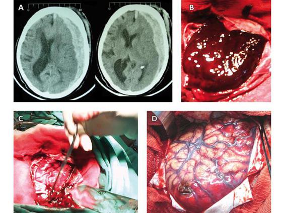
Acute subdural hematomas usually occur because of a head injury. In cases that need immediate treatment, patients will undergo traditional surgery in which a portion of the skull is removed, the outermost covering of the brain (the dura) is opened, and the acute subdural hematoma is evacuated.
How long does it take for a subdural hematoma to break down?
This allows the body to break down the chronic subdural hematoma on its own within the following days and weeks.
How to do a subdural in the skull?
He or she will drill a tiny hole into your skull and insert a device called a subdural evacuating port system to gently drain the blood out.
What is MMA in neurosurgeon?
Our neurosurgeons specialize in traditional surgery techniques and minimally invasive procedures such as middle meningeal artery ( MMA) embolization. We use MMA embolization as an alternative to brain surgery for some patients. At University of Utah Health, we provide our subdural hematoma patients with exceptional care and support every step of the way.
How long does subdural hematoma last?
This type of subdural hematoma typically goes away on its own over the span of a few weeks.
How long does it take to get MMA embolized?
It takes about 30 minutes and typically requires light sedation, not general anesthesia that puts you to sleep.
What is the best treatment for subdural hematoma?
Your doctor may prescribe anti-seizure medications to treat or prevent seizures that might be caused by the subdural hematoma. Medication may also be used to treat your brain injury. Corticosteroids are often prescribed to reduce inflammation in the brain.
What is the procedure to remove a subdural hematoma?
A surgical procedure called a craniotomy may be used to remove a large subdural hematoma. It’s normally used to treat acute subdural hematomas. In this procedure, your surgeon removes a part of your skull in order to access the clot or hematoma. They then use suction and irrigation to remove it.
How do burr holes work?
A burr hole can be used to drain chronic subdural hematomas as well as acute ones that are smaller than one centimeter at the thickest point. First, your surgeon creates small holes in your skull and then places rubber tubes in them. The blood from the hematoma drains out through these holes.
What is the most dangerous type of subdural hematoma?
If you sustain a major brain injury, this area can fill with blood and cause life-threatening symptoms. This is called an acute subdural hematoma. It’s the most dangerous type of subdural hematoma. Acute subdural hematomas are usually caused by: a car accident. a blow to the head.
What is subdural hematoma?
A subdural hematoma occurs when blood collects on your brain’s surface beneath the skull. Subdural hematomas can be life-threatening. They usually result from a head injury. Subdural hematomas are either acute or chronic. Acute subdural hematomas commonly form because of a severe head injury. Approximately 20 to 30 percent ...
Why do older people have subdural hematomas?
Chronic subdural hematomas are usually caused by mild or repeated head injuries. These are common in older adults who repeatedly fall and hit their heads. Some chronic subdural hematomas occur with no apparent cause. The higher rate of this condition in older adults may also be because the brain shrinks as people age.
How to diagnose subdural hematoma?
A subdural hematoma can be diagnosed using imaging tests, such as a CT or MRI scan. These scans provide your doctor with an in-depth look at your:
What is the procedure to treat subdural hematoma?
Surgeons can use various techniques to treat subdural hematomas: Burr hole trephination.
What Is a Subdural Hematoma?
A subdural hematoma is a collection of blood outside the brain. They’re usually caused by serious head injuries. Bleeding and added pressure on the brain from a subdural hematoma can be life-threatening. Some stop and go away suddenly; others need surgical drainage.
How long does it take for a subdural hematoma to show symptoms?
In very slow-growing subdural hematomas, there may be no noticeable symptoms for more than 2 weeks after the bleeding starts.
Why is the skull removed?
A larger section of the skull is removed, to allow better access to the subdural hematoma and reduce pressure. The removed skull is replaced shortly after the procedure. Craniectomy. A section of the skull is removed for an extended period of time, to allow the injured brain to expand and swell without permanent damage.
Why are elderly people at higher risk for subdural hematoma?
Elderly people are at higher risk for chronic subdural hematoma because brain shrinkage causes these tiny veins to be more stretched and more vulnerable to tearing.
What is a sudden blow to the head called?
The sudden blow to the head tears blood vessels that run along the surface of the brain. This is referred to as an acute subdural hematoma. People with a bleeding disorder and people who take blood thinners are more likely to develop a subdural hematoma.
What is the outermost layer of a subdural hematoma?
In a subdural hematoma, blood collects between the layers of tissue that surround the brain. The outermost layer is called the dura. In a subdural hematoma, bleeding occurs between the dura and the next layer, the arachnoid.
What are the key components of a treatment plan for acute SDH?
Clinical presentation, neurologic condition, and imaging findings are the key components in establishing a treatment plan for acute SDH. Location and size of the SDH and presence of midline shift can rapidly be determined by computed tomography of the head. Immediate laboratory work up must include …
Why do you need to reverse a bleeding diathesis?
Presence of a coagulopathy or bleeding diathesis requires immediate reversal and treatment with the appropriate agent(s), in order to lessen the risk of hematoma expansion. Reversal protocols used are similar to those for intracerebral hemorrhage, with institutional variations.
Where is subdural haematoma located?
Subdural haematoma represents an extracerebral blood collection, which can be met as a clot or in liquid form, located between the dura mater and the middle layer of the meninges (arachnoid) and, which does not expand in the subarachnoid area or in the basal cisterns (interpeduncular cisterns). Usually, this collection has a traumatic etiology (the acute and subacute ones are always traumatic) and a compressive effect on the brain, producing localization neurological signs, increased intracranial pressure signs and various alterations of the consciousness.
How thick is a subdural haematoma?
What is interesting to mention is the medium thickness of chronic subdural haematomas, which was of approximately 20,5mm +/- 5mm in unilateral haematomas and of approximately 29,6mm +/- 9mm in bilateral haematomas.
How long has subdural haematoma been diagnosed?
The diagnosis of chronic subdural haematoma has evolved in the last 20-30 years together with the introduction of the CT scan, the MRI and the access of the patients to this type of explorations.
Which hemisphere has the most chronic subdural haematomas?
As far as the localization of chronic subdural haematomas are concerned, the studies have demonstrated a higher frequency of chronic subdural haematomas of left brain hemisphere (52%) compared to the ones of right brain hemisphere (30%), in 18% of the cases being bilateral.
Can you diagnose chronic subdural haematoma on CT?
If the CT examination is currently very easily accessible, the diagnosis of chronic subdural haematoma has become quite simple, the main condition being that you only have to think about such a diagnosis. This is valid mostly in cases of elders, who progressively develop psychic disorders, representing the group of patients with chronic subdural haematoma.
Can a subdural haematoma be a latent period?
Moreover, in the context of a usual brain injury, which can pass unobserved and may frequently be ignored, the chronic subdural haematoma has a latent period until the appearance of the clinical symptoms, the diagnosis presenting many errors.
Can a haematoma mimic a stroke?
Regarding the differential diagnosis, it should not be forgotten that the clinical sign s of a haematoma mimic an intracranial neoformation or an ischemic stroke. In youngsters, chronic subdural haematomas often manifest with epilepsy crises and behavior disorders, which are due to alcohol consumption.
How to treat subdural hematoma?
In some cases, very small subdural hematomas that don’t produce signs or symptoms don’t have to be removed. Doctors may opt to simply observe the subdural hematoma with repeated head imaging tests.
What is the best treatment for subdural hematoma?
Doctors may opt to simply observe the subdural hematoma with repeated head imaging tests. Medications designed to reduce and control swelling in the brain are also used, such as diuretics and corticosteroids. If the subdural hematoma is severe and life-threatening, emergency surgery may be needed.
What is subdural hematoma recovery?
A subdural hematoma occurs when blood vessels between the brain and its outermost membrane rupture, causing leaking blood that results in compression of the brain tissue. Subdural hemotomas are classified as acute, characterized by immediate signs and symptoms; subacute, ...
What are the symptoms of subdural hematoma?
Confusion. Weakness in limbs on one side of the body. If the condition goes undiagnosed and more blood causes additional brain tissue compression, more severe signs and symptoms include: Lethargy. Seizures. Unconsciousness. Treatment for Subdural Hematoma.
What is a traumatic brain injury?
A traumatic brain injury (TBI) occurs when there is a “bump, blow, or jolt to the head” that causes issues with the functions of the ...
How long does it take to recover from a subdural hematoma?
As a general rule of thumb, adults experience most of their recovery within six months, while children recover more quickly and more completely.
How long does it take for hemotomas to appear?
Subdural hemotomas are classified as acute, characterized by immediate signs and symptoms; subacute, characterized by symptoms that appear within several hours; and chronic, characterized by signs and symptoms take days or even months to appear.
What is subdural hematoma?
Overview. A subdural hematoma is a type of brain bleed. Blood leaks out of a blood vessel into the space below the outermost membrane of the brain -- the dura mater.
What are the effects of chronic subdural hematoma?
Memory loss, disorientation, and personality changes, especially in older adults with chronic subdural hematoma.
How does a subdural hematoma affect the prognosis?
If you have a subdural hematoma, your prognosis depends on your age, the severity of your head injury and how quickly you received treatment. About 50% of people with large acute hematomas survive, though permanent brain damage often occurs as a result of the injury. Younger people have a higher chance of survival than older adults.
How long does it take for a subdural hematoma to appear?
Acute: This is the most dangerous type of subdural hematoma. Symptoms are severe and appear right after a head injury, often within minutes to hours. Pressure on the brain increases quickly as the blood pools. If not diagnosed and treated quickly, you could lose consciousness, become paralyzed or even die.
How many people survive a subdural hematoma?
If you have a subdural hematoma, your prognosis depends on your age, the severity of your head injury and how quickly you received treatment. About 50% of people with large acute hematomas survive, though permanent brain damage often occurs as a result of the injury.
Why do older people have a higher risk of developing a hematoma?
This is because older brains cannot re-expand and fill the space where the blood was, leaving them more vulnerable to future brain bleeds with even minor head injuries.
How to treat a hematoma?
Healthcare providers treat larger hematomas with decompression surgery. A surgeon drills one or more holes in the skull to drain the blood. Draining the blood relieves the pressure the blood buildup causes on the brain. Additional surgery may be needed to remove large or thick blood clots if present.
Clinical significance
- A subdural hematoma is a collection of blood outside the brain. Subdural hematomas are usually caused by severe head injuries. The bleeding and increased pressure on the brain from a subdural hematoma can be life-threatening. Some subdural hematomas stop and resolve spontaneously; others require surgical drainage.
Pathophysiology
- In a subdural hematoma, blood collects between the layers of tissue that surround the brain. The outermost layer is called the dura. In a subdural hematoma, bleeding occurs between the dura and the next layer, the arachnoid. The bleeding in a subdural hematoma is under the skull and outside the brain, not in the brain itself. As blood accumulates, however, pressure on the brain increases…
Causes
- Subdural hematoma is usually caused by a head injury, such as from a fall, motor vehicle collision, or an assault. The sudden blow to the head tears blood vessels that run along the surface of the brain. This is referred to as an acute subdural hematoma.
Prognosis
- People with a bleeding disorder and people who take blood thinners are more likely to develop a subdural hematoma. A relatively minor head injury can cause subdural hematoma in people with a bleeding tendency.
Symptoms
- In a chronic subdural hematoma, small veins on the outer surface of the brain may tear, causing bleeding in the subdural space. Symptoms may not be apparent for several days or weeks. Elderly people are at higher risk for chronic subdural hematoma because brain shrinkage causes these tiny veins to be more stretched and more vulnerable to tearing.