However, sclerotherapy is the primary nonsurgical intervention for venous malformations. Large-sized lesions are cured; with the help of 95% ethanol. Whereas smaller and cutaneous lesions are treated with 1% sodium tetradecyl sulfate, often performed by a radiologist under anesthesia.
What are the treatment options for a venous malformation?
These are usually caused by other vascular malformations that can be found with a venous malformation. Doctors typically treat seizures with medications. Some hemorrhages require surgery, but many hemorrhages can be treated with medical management and observation in a hospital.
Do developmental venous anomalies (DVAS) require treatment?
Generally, developmental venous anomalies (DVAs) do not require treatment. These veins do a necessary job of getting blood in and out of the brain, so they do not need to be surgically removed or closed. Because they are normal and not dangerous, long-term imaging is generally not necessary. Key points about a DVA
What is a developmental venous anomaly?
Key points about a DVA 1 A developmental venous anomaly (DVA) is an irregular arrangement of small veins that may look like the spokes of a wheel that drain into a larger central vein. 2 DVAs are congenital—a person is born with them. 3 DVAs are not dangerous, and most people do not know if they have them. More items...
How do you test for developmental venous anomalies?
Imaging tests may include MRI or MRA, conventional angiogram, or specific types of CT scans that show areas of blood flow. Most people may never know they have a DVA, and it will only be found after their death, if an autopsy is done. How are developmental venous anomalies treated?

What is the treatment for VM?
Other therapies, such as cryoablation (freezing therapy) and laser/radiofrequency ablation (heating therapy) are sometimes used to treat VM. VMs that involve the skin are sometimes treated with different types of lasers, such as a pulsed-dye laser, a long-pulsed Nd:YAG laser or others.
When does a VM need to be treated?
Once a VM starts causing problems, treatment will begin. If a VM is in a sensitive or dangerous area, it may need treatment even if it has not yet begun to cause problems. We recommend early evaluation by a specialist if you are concerned that you or your child may have a VM. Treatment is individualized.
How far apart should sclerotherapy treatments be?
This will cause the VM to shrink. Several sclerotherapy treatments are often needed. Treatments are usually at least six weeks apart.
What is laser therapy for VMs?
Sometimes laser therapy is used to treat VMs that affect the skin. On occasion surgery can help correct deformity or loss of function. For most vascular anomalies, a combination of treatment methods is best.
What doctor treats VMs?
Large VMs can lead to problems with blood clotting. A hematologist is a doctor who treats blood diseases and will make sure that blood is clotting properly before, during and after any procedures.
What is the function of veins?
Veins are part of the circulatory system that moves blood through the body. Veins carry blood from the body back to the heart. The heart pumps the blood through the lungs so that it can pick up oxygen. The body uses oxygen to make energy. Venous malformations (VMs) occur when veins do not form normally. VMs can be completely isolated ...
What is the best imaging study for a VM?
An MRI is the best imaging study to diagnose a VM. It provides a detailed scan or picture of the inside of the body to help see the size and location of the VM. An MRI will also show other important structures, such as nerves, muscles, arteries and veins that are near the VM and that may be affected by treatment.
What is the best treatment for large venous channels?
It is useful for treating large venous channels or spaces and is often combined with sclerotherapy . The combination of endovenous laser therapy and sclerosant injection appears to produce a quicker response and an easier recovery.
How to diagnose VM?
We can diagnose a VM in the skin and superficial tissue by physical examination. Magnetic resonance imaging (MRI) is the best imaging test to diagnose a VM, and to determine the extent of the condition. Ultrasonography is also useful when the VM is near the surface.
How does endovenous laser therapy work?
Endovenous laser therapy is similar to sclerotherapy, but involves placing a diode laser fiber through a needle or catheter.
What causes a VM to become hardened?
Several diseases and conditions involve various types of VMs. Glomovenous malformations contain nerve cells and cause the malformations to become hardened and tense.
What is a superficial VM?
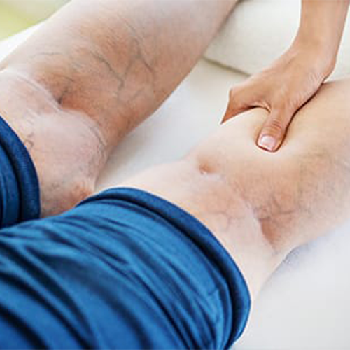
If the VM is superficial, it will be discolored blue and may appear in different areas of your body (called multifocal), especially around the mouth, lips, tongue, cheek, side of the face, scalp, and neck. Superficial VMs can range in size from tiny dots to large disfigurations. VMs are soft.
What does a VM look like?
VMs can be extremely painful and sensitive. A VM usually looks like a bluish discoloration. It can be a single lesion or it may be one of many. It can be confined to one specific area or spread out; and it can be superficial or deep. The walls of a vein that has a VM lack the smooth muscle cells of a normal vein.
Can a VM cause a pulmonary embolism?
Those involving the tongue or other structures around the airway may cause problems with breathing or speaking, while those in the arms and legs typically lead to painful swelling. Rarely, blood clots that form in a VM can travel to the lungs, creating a pulmonary embolism.
What is developmental venous anomaly?
What are developmental venous anomalies? A developmental venous anomaly (DVA) is a problem with the way small veins are arranged. They may look like the spokes of a wheel. The veins drain into a larger central vein.
Where are DVAs found?
This is a Latin term that means head of Medusa . These unusual vein formations can occur anywhere in the body. But they are found most often in the brain or spinal cord.
Do DVAs need to be removed?
DVAs often don't require treatment. These veins work correctly to get blood in and out of the brain. They don't need to be removed or closed. Because they are normal and not dangerous, you often don't need to get imaging tests over the long term.
Why do you inject contrast dye into your vein?
In some cases a contrast dye is injected into a vein to look at the brain tissue in a different way, and to evaluate your blood vessels (magnetic resonance angiography or magnetic resonance venography).
Can a vascular malformation be treated with medication?
These are usually caused by other vascular malformations that can be found with a venous malformation. Doctors typically treat seizures with medications. Some hemorrhages require surgery, but many hemorrhages can be treated with medical management and observation in a hospital.
Can a doctor treat intracranial venous malformations?
Doctors usually don't treat intracranial venous malformations because they rarely cause symptoms. If you have unrelated symptoms, such as headaches, your doctor might prescribe medications. Rarely, people who have intracranial venous malformations have seizures or bleeding in the brain (brain hemorrhage).
What is the etiology of developmental venous anomalies?
The etiology of developmental venous anomalies remains uncertain but may relate to arrested development of venous structures 2,3. Histologically they consist of a number of abnormally thickened veins with normal feeding arteries and capillaries 3.
What is a DVA?
Developmental venous anomaly (DVA), also known as cerebral venous angioma, is a congenital malformation of veins which drain normal brain. They were thought to be rare before cross-sectional imaging but are now recognized as being the most common cerebral vascular malformation, accounting for ~55% of all such lesions.
Can a venous anomaly be seen on MRI?
MRI. Developmental venous anomalies are often visible on most sequences but can be subtle and are most easily seen on postcontrast T1 sequences and susceptibility weighted imaging (SWI). If there is an associated cavernous hemangioma, then susceptibility weighted sequences will be most sensitive to this component.
Is imaging appearance atypical?
Generally, the appearances will be typical and no differential should be offered. In some instances, imaging appearances may be atypical or be confounded by concurrent pathology (e.g. hemorrhage). In such cases it is worth considering:
Can a DVA be treated?
If isolated developmental venous anomalies require no treatment. If part of a mixed vascular malformation then treatment will be predicated on the other component. Informing the surgeon of the presence of a DVA is, however, essential as cautery of the collecting vein can lead to venous infarction of the brain parenchyma it drains.
What is the name of the venous malformations on the skin?
Blue Rubber Bleb Nevus Syndrome: Also known as Bean Syndrome, this refers to the presence of multiple, isolated slow-flow venous malformations on the skin and in underlying tissue, as well as in the intestines and other internal organs.
Where are arteriovenous malformations most commonly found?
Arteriovenous malformations can occur anywhere in the body, but are most typically found in the brain, spinal cord and extremities.
What is vascular malformation?
A vascular malformation is an abnormal development of blood vessels. They might be found in the large arteries and veins, in smaller vessels called arterioles and venules, in microscopic capillaries, and/or in the lymphatic channels that carry lymphatic fluid and white blood cells outside of the arteries and veins.
What is fast flow arteriovenous malformation?
Fast-flow arteriovenous malformations develop as the result of an abnormal connection between arteries that supply the body’s organs, and the veins, which drain them. Picture these as being like short-circuits: Blood pumped from the heart to a given organ can’t get there and is instead sent back toward the heart.
What are the problems with lymphatic drainage?
These cysts, in turn, can develop problems such as infection, bleeding and erosion into adjacent organs.
What is the procedure to diagnose blood vessel disease?
Typically, doctors will order imaging studies to help with diagnosis. Imaging studies may include ultrasound, MRI, and/or angiography, an imaging procedure that involves the injection of dye that will outline the blood vessels on an X-ray.
Can a person have a single vascular malformation?
A person can have a single isolated vascular malformation or one that involves several vessels. In some cases, a vascular malformation turns out to be part of a more complex syndrome that features multiple disorders and affects multiple organs. Typically, doctors will order imaging studies to help with diagnosis.
