
Prostate MRI provides more clear and detailed images of the soft-tissue structures of the prostate gland than other imaging methods. The level of details and high resolution of images makes MRI an invaluable tool in early diagnosis and evaluation of prostate cancer (18).
What is the best MRI for prostate cancer?
· The accuracy of diagnosis for higher grade prostate cancer was similar for immediate biopsy vs. MRI first. Also, 37% of the men who had a completely normal MRI were able to avoid biopsy. These results support findings in prior studies showing that MRI may save some men with a high PSA from a biopsy and also helps identify the more aggressive types of …
How accurate is prostate MRI?
· A prostate MRI will help the healthcare team get a good biopsy so they can determine if you’re a candidate for active surveillance. There are many ongoing studies to see how else prostate MRIs can be used to help treat prostate cancer. In the future: Some people might be able to avoid unnecessary prostate biopsies if their prostate MRIs are normal.
Can a MRI detect prostate cancer?
· A Likert or PI-RADS score of 4-5 dictates the probability of prostate cancer that demands treatment, as well as the need for a prostate biopsy (which is known to traumatise prostate tissue). Where applicable, MRI scans can accurately identify and locate cancer cells ahead of the biopsy; this minimises prostate trauma from initial biopsy procedures and …
What to expect from a prostate MRI exam?
In prostate cancer patients, MRI may be used to examine the prostate and nearby lymph nodes to distinguish between benign (noncancerous) and malignant (cancerous) areas. Is MRI …
Is MRI effective in diagnosing prostate cancer?
Among the diagnostic strategies considered, the MRI pathway has the most favourable diagnostic accuracy in clinically significant prostate cancer detection. Compared to systematic biopsy, it increases the number of significant cancer detected while reducing the number of insignificant cancer diagnosed.
Why do you have an MRI scan for prostate cancer?
Doctors use Prostate MRI to evaluate the extent of prostate cancer and determine whether it has spread. They may also use it to help diagnose infection, conditions you were born with, or an enlarged prostate. Some exams may use an endorectal coil, a thin wire covered with a latex balloon.
Why would a urologist order an MRI?
MRI allows urologists to better stratify the patient with regards to biopsy grade and tumor extent, as well as treatment planning. The urologist should develop an understanding with his radiologist, as prostate mp-MRI has an increasing role in prostate cancer characterization, detection, and management.
Is MRI better than prostate biopsy?
Only a biopsy can determine for certain whether prostate cancer is present, but a new study suggests that using magnetic resonance imaging (MRI) can help to better identify patients who are more likely to need a biopsy versus those who aren't.
Can MRI show if prostate cancer has spread?
If prostate cancer has been found, MRI can be done to help determine the extent (stage) of the cancer. MRI scans can show if the cancer has spread outside the prostate into the seminal vesicles or other nearby structures. This can be very important in determining your treatment options.
What are the signs that prostate cancer has spread?
Prostate cancer can spread to the lymph nodes in the groin area, or to other parts of the body. The most common symptoms are swelling and pain around the area where the cancer has spread. Cancer cells can stop lymph fluid from draining away. This might lead to swelling in the legs due to fluid build up in that area.
How long does it take to get prostate MRI results?
You should get your results within 1 or 2 weeks. Waiting for results can make you anxious. Ask your doctor or nurse how long it will take to get them. Contact the doctor who arranged the test if you haven't heard anything after a couple of weeks.
What can I expect from a prostate MRI?
A series of images will be taken of the prostate and surrounding area. During the exam, the MRI machine can be very loud. Contrast, which is a special MRI dye, will be given by IV near the end of the exam but you will be given ear plugs or head phones to listen to the music of your choice.
What is the average cost of a prostate MRI?
Prostate MRI costs anywhere from $500 to $2,500 in the United States, depending upon a patient's insurance coverage. Approximately 1 million American men are currently sent for prostate biopsy every year. If all of those men received MRI instead, costs could reach $3 billion annually.
Should I get MRI before prostate biopsy?
“There is now sufficient data to support the use of prostate MRI in all men before their initial prostate biopsy when the MRI is of sufficient quality."
At what PSA level should a biopsy be done?
A lower percent-free PSA means that your chance of having prostate cancer is higher and you should probably have a biopsy. Many doctors recommend a prostate biopsy for men whose percent-free PSA is 10% or less, and advise that men consider a biopsy if it is between 10% and 25%.
Can you see a tumor on an MRI?
MRI creates pictures of soft tissue parts of the body that are sometimes hard to see using other imaging tests. MRI is very good at finding and pinpointing some cancers. An MRI with contrast dye is the best way to see brain and spinal cord tumors. Using MRI, doctors can sometimes tell if a tumor is or isn't cancer.
What are the risks and benefits of getting a prostate MRI?
MRI doesn’t use radiation, and it isn’t invasive or painful. In fact, it’s one of the safest medical procedures available. So there’s not a lot of risk that comes with getting an MRI, but there are a lot of benefits. This makes the prostate MRI a powerful tool.
How do I prepare for a prostate MRI, and how long does it last?
There’s not much you need to do to prepare for your MRI. If you’re getting sedation medication, you won’t be able to have anything to eat or drink for about 8 hours before your MRI. If your provider needs to use an endorectal coil during your MRI, they might ask you to take an enema a few hours before the procedure.
The bottom line
Prostate MRIs are becoming more common. They’re powerful tools that help your healthcare team get an accurate picture of your prostate. They also help guide prostate biopsies so that you can get accurate results and start treatment as fast as possible. And fortunately prostate MRIs are short and relatively painless.
What is MRI scan?
Magnetic Resonance Imaging (MRI) scans use magnets to create detailed digital images of your prostate and surrounding soft tissues. When looking for prostate cancer and the likelihood of it spreading, doctors typically use MRI scans to avoid invasive procedures such as biopsies, but when learning how diagnosed prostate cancer has spread, an initial biopsy may be required.
Can MRI scans detect prostate cancer?
Doctors can quickly identify prostate cancer and measure it’s growth rate using MRI scans (although non-problematic cancers are usually found via biopsy). Whenever biopsies do detect prostate cancer, MRI scans can also be used retrospectively to avoid unnecessary scanning, to facilitate quicker patient treatment or planning, and to clarify where biopsy samples should be taken.
Can you listen to music during an MRI?
Should they prefer, patients can usually be accompanied by a close friend or family member throughout their MRI scan; listening to music via headphones for the duration is also a popular option . It’s important to remember that the MRI scan procedure is physically painless, and that any mild discomfort that’s experienced during the scan should be short-lived and gone the same day.
Is MRI safe?
In-depth studies on MRI scanning and the radio waves and/or magnetic fields involved, have identified no risks to patients, making the procedure one of the safest stages of treatment available. Ongoing global research and development is already producing breakthrough techniques and technologies, with scanners that can accommodate a patient’s medical condition or limitation.
Can MRI scans be done with metal?
Due to their use of magnets, MRI scans require the removal of all metal factors including jewelry, zips and buttons; internal implants such as metal plating or pacemakers could make MRI scanning impossible. Initial discussions about a patient’s general health will account for such factors, as well as other conditions (such as claustrophobia) known to impact on the MRI procedure.
Is MRI loud?
The MRI procedure is both loud and noisy; mild side effects include feeling warm, queasiness, skin rashes, headaches and dizziness. Despite these factors, patients are usually allowed to return home on the same day and resume their usual activities; this is impacted if patients have undergone sedation during their scan.
What is MRI used for prostate cancer?
In prostate cancer patients, MRI may be used to examine the prostate and nearby lymph nodes to distinguish between benign (noncancerous) and malignant (cancerous) areas.
What is MRI prostate?
Medically Reviewed by Minesh Khatri, MD on October 15, 2019. An MRI is a test that produces very clear pictures of the human body without the use of X-rays. Instead, MRI uses a large magnet, radio waves, and a computer to produce these images. In prostate cancer patients, MRI may be used to examine ...
What is dye injection in MRI?
Certain MRI exams require an injection of a dye (contrast material). This helps identify certain anatomic structures on the scan images.
Do you have to wear a gown for an MRI?
You will be asked to wear a hospital gown during the MRI scan.
Can you bring a wallet into an MRI room?
Personal items such as your watch, wallet (including any credit cards with magnetic strips - should not be brought into the room with the MRI machine in it.The credit cards will be erased by the magnet), and jewelry should be left at home or removed prior to the MRI scan. Secured lockers should be available to store personal items.
Is an MRI good for back pain?
Some conditions may make an MRI examination not a good idea. Tell your doctor if you have any of the following conditions: Implanted insulin pump (for treatment of diabetes ), narcotics pump (for pain medication), or implanted nerve stimulators ( TENS) for back pain.
Can you take a prescription before an MRI?
People who get anxious when in tight spaces (claustrophobic) may benefit from talking to their doctor before the procedure. Some options include taking a prescription medication before the procedure to relieve anxiety or having the exam done in one of the newer and less confining MRI units, called an open MRI, when available.
Why is MRI important for prostate cancer?
Imaging studies play an important role in detection and management of prostate cancer and MRI especially with the use of endorectal coil because of high contrast resolution is recognized as the best imaging modality in evaluation of prostate cancer. Multiparametric MR study including T1 and T2 weighted images, diffusion weighted images, dynamic contrast study and MR spectroscopy is useful for detection and local staging of prostate cancer as well as posts treatment evaluation of patients either after surgery or radiation therapy for detection of local recurrence.
What are the properties of MRI for prostate?
In prostate imaging two property of MRI are important: 1. Strength of MRI magnet; 1.5 T or 3-T 2. Type of MRI coil use for imaging; endorectal coil or torso/pelvic coil or combination of these two coils.
What is a T1-weighted prostate image?
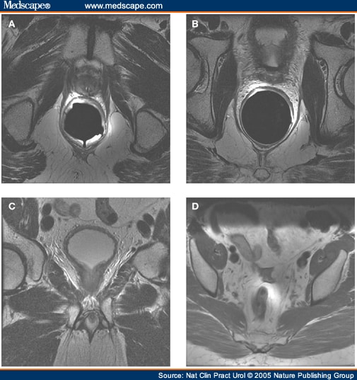
T1-weighted images illustrate prostate as homogeneous low signal intensity organ in which zonal anatomy and prostate cancer distinguishing is very difficult. T1-weighted is a useful image in detecting Post biopsy hemorrhage in prostate cancer (appears as high T1 signal intensity areas within the homogeneous prostate) and assess lymph nodes and osseous structures in pelvis (29-30). T2-weighted high resolution images are the mainstay in prostate cancer detection. The normal peripheral zone of prostate has high signal intensity in T2-weighted images. 70% of prostate cancers occur in peripheral zone and manifest as low signal intensity areas within the bright peripheral zone on T2-weighted MR images (Figure 1). Although T2-weighted MR images are useful in detecting prostate cancer but many pathologic features like post biopsy hemorrhage, hormone or radiation therapy effects, prostatitis, calcification and fibrosis appear as low signal intensity lesions in peripheral zone and mimic prostate cancer (29). In addition, heterogeneity in central glands signal intensity especially in patients with benign prostate hyperplasia overlap with tumoral tissue signal and makes detection of central glands cancer difficult. To overcome these limitations and to improve diagnostic accuracy of conventional MRI in prostate cancer detection, new techniques titled functional MRI have been developed. Functional MRI techniques include diffusion-weighted magnetic resonance (DW-MR) imaging, dynamic contrast-enhanced MR (DCE-MR) imaging and MR spectroscopy (30).
What is the gold standard for prostate cancer?
According to above statements it can be concluded that the gold standard approach for diagnosis, staging and management of prostate cancer is using 1.5 T MR machines with both endorectal and pelvic phased-array coils.
Can pelvic coils be used for prostate cancer?
Simultaneous using of endorectal and pelvic coils can best help in prostate cancer det ection and evaluation of tumor extension.
Can contrast enhanced US detect micro vascularization?
Contrast-enhanced US can detect micro-neovascularization and even is sensitive to low blood flow (Conventional color/power Doppler US cannot detect micro vascularization). Although contrast-enhanced US shows more sensitivity than conventional TURS in evaluating prostate cancer but performing it for detection of prostate cancer remains investigational (15, 16).
Does ultrasound increase accuracy?
Improvements of ultrasound technique in recent years lead to increase accuracy of TURS in detecting prostate cancers.
What is the purpose of MRI for prostate cancer?
Newer MRI techniques are being tested to improve the accuracy of prostate cancer diagnosis. Three presentations during the 2018 American Urological Association Annual Meeting discussed potential approaches to reduce the need for prostate biopsies for men with prostate cancer
What are some lab tests for prostate cancer?
Examples of such tests include the Prostate Health Index , 4Kscore test, PCA3 tests , and ConfirmMDx. These tests are discussed in Whats New in Prostate Cancer Research?
What are the two types of prostate biopsies?
There are two main types of prostate biopsies transrectal ultrasound guided biopsies and transperineal biopsies. As the former tends to be more common, well focus on this method.
Is prostate biopsy accurate?
Not only is the trans-rectal ultrasound part of the study blind and unable to identify high-grade prostate cancer but, despite the knowledge that prostate cancer often develops in more than one area of the prostate and or, at different times , this blind needle biopsy test samples randomly only some 0.1%0.3% of the prostate to leave one absolutely uninformed about the 99% rest of the prostate especially so for the anterior portion of the prostate. This highly inaccurate biopsy test is also responsible for all of the confusion related to so-called prostate cancer upgrading and progression.
Is a PSA test sensitive?
However, the PSA is neither specific and, is highly insensitive for detecting only significant prostate cancers .
How many biopsy results are false negatives?
Research shows that 35% of biopsy results are false negatives. This means despite the invasive sampling of tissues, cancer can go undetected and continue to grow while symptoms persist.;
Can you have a prostate biopsy if you have a previous negative biopsy?
If the patient had a prior negative biopsy, we then assess the markers, and if the levels are high or suspicious, the patient has a biopsy. If not, then the patient may avoid a prostate biopsy, Abreu said.
What tests can you get to see if you have prostate cancer?
Getting other lab tests (of blood, urine, or the prostate biopsy samples) to help get a better idea of whether or not you might have prostate cancer. Examples of such tests include the Prostate Health Index (PHI), 4Kscore test, PCA3 tests (such as Progensa), and ConfirmMDx.
What is the best way to diagnose prostate cancer?
A core needle biopsy is the main method used to diagnose prostate cancer. It is usually done by a urologist. During the biopsy, the doctor usually looks at the prostate with an imaging test such as transrectal ultrasound (TRUS) or MRI, or a ‘fusion’ of the two (all discussed below).
What happens if a biopsy is negative?
If the prostate biopsy results are negative (that is, if they don’t show cancer), and the chance that you have prostate cancer isn’t very high based on your PSA level and other tests, you might not need any more tests, other than repeat PSA tests (and possibly DREs) sometime later.
How long does it take to get a biopsy?
Your biopsy samples will be sent to a lab, where they will be looked at with a microscope to see if they contain cancer cells. Getting the results (in the form of a pathology report) usually takes at least 1 to 3 days, but it can sometimes take longer. The results might be reported as:
Why is a PSA test important?
PSA tests are often an important part of determining how well treatment is working, as well as in watching for a possible recurrence of the cancer after treatment (see Following PSA Levels During and After Treatment ).
What is the purpose of PSA?
The PSA level is used to help determine the stage of your cancer. This can affect your treatment options, since some treatments (such as surgery and radiation) are not likely to be helpful if the cancer has spread to other parts of the body.
Can you get a PSA test if you have prostate cancer?
The PSA test can also be useful if you have already been diagnosed with prostate cancer.
What is MRI for cancer?
MRI for Cancer. Other names for this test: magnetic resonance imaging, MRI, magnetic resonance, MR, and nuclear magnetic resonance (NMR) imaging. MRI helps doctors find cancer in the body and look for signs that it has spread. MRI also can help doctors plan cancer treatment, like surgery or radiation. MRI is painless and you don’t have ...
What is MRI used for?
MRI can also be used to look for signs that cancer may have metastasized (spread) from where it started to another part of the body. MRI images can also help doctors plan treatment such as surgery or radiation therapy. (A specific kind of MRI can be used to look inside the breast. Learn more about breast MRI .)
What does an MRI machine make?
The machine makes loud, thumping, clicking, and whirring noises, much like the sound of a washing machine, as the magnet switches on and off. You may be given earplugs or headphones with music to block noise out during the scan. Special, open MRI machines that are less restrictive may be easier for some people.
Can you scan for implants?
If you have any of these implants, you should not even enter the MRI scanning area unless told to do so by a radiologist or technologist who knows you have:
What is the contrast material used in MRI?
The contrast material used for an MRI exam is called gadolinium. (This is not the same as the contrast dye used in CT scans.) Let your doctor and the technologist know if you have any kind of allergies or have had problems with any contrast used in imaging tests in the past.
Can you relax in an MRI?
If being in a small, enclosed space is a problem for you (you have claustrophobia), you might need to take medicine to help you relax while in the scanner. Sometimes talking with the technologist or a patient counselor, or seeing the MRI machine before the test can help. In some cases, you can arrange to have an open MRI which allows more space around your body (see the next section).
Do you have to be in a hospital for an MRI?
MRI scans are most often done on an outpatient basis, so you don’t have to be in a hospital to get one. You don’t usually need to follow a special diet or do anything to get ready for an MRI, but follow any instructions you are given.
How does MRI help prostate cancer?
Magnetic resonance imaging or MRI uses strong magnetic fields instead of x-rays to produce images of parts of the body. MRI scans can be very helpful in looking at prostate cancer. By producing a very clear picture of the prostate gland, an MRI can show whether the cancer has spread outside the gland into the seminal vesicles or bladder. Unfortunately the cost of carrying out MRI examinations on every patient who might have prostate cancer extending beyond the prostate capsule would be enormous. Thus, from a diagnostic point of view, the important question is, “When is an MRI really appropriate?”
What is the most important imaging test for prostate cancer?
Although bone scans are the primary and most important imaging test used in the diagnosis of prostate cancer, several other imaging tests also have significant — if slightly less central — roles. If you are one of the people who needs one of these tests, it will help you to have some idea what is going on.
When did the Prostascint test become available?
The ProstaScint imaging technique first became available in the mid 1990s as a highly promising technique that appeared to be able to identify prostate cancer that had escaped from the prostate and into the pelvic lymph nodes and other surrounding tissues. As such, this test gave hope that it would be possible to better identify those patients in whom surgery alone as a treatment carried out with curative intent would not be feasible.
Can color Döppler be used for prostate biopsy?
A select group of subspecialists have been strong advocates for the use of color Döppler ultrasound imaging in the diagnosis of prostate cancer. They have claimed that the use of this technique permits much greater accuracy in the identification of prostate cancer, and also in carrying out appropriate prostate biopsies.
Do CT scans show bone metastases?
The one place where CT scans appear to have a small advantage is in the early identification of soft tissue metastases (as opposed to bone metastases). However, the need for this type of information outside clinical trials is relatively limited.
Is a CT scan better than a CAT scan?
However, CT scans are much better at identify ing skelet al structures than soft tissue structures. The role of CT or CAT scanning in the diagnosis of prostate cancer is really very limited today because almost anything one can do with a CT scan can be done just as well or better with an MRI scan.
What is a CT scan?
CT or computerized tomography is a computer-controlled x-ray procedure that produces detailed cross-sectional images of the body using hundreds of x-ray-like images. This test can help to tell whether prostate cancer has spread beyond the prostate to other organs.
