
How is fluid loss treated in patients with hypovolemic shock?
1 In patients with hypovolemic shock due to extracellular fluid loss, the etiology of fluid loss must be identified and treated. 2 Monitoring electrolytes and acid/base status in patients in hypovolemic shock is of utmost importance. 3 Trauma is the leading cause of hemorrhagic shock. More items...
Why is it important to assess the risk of hypovolemic shock?
The goal is to restore circulating blood volume, preserve hemodynamics, and prevent any damage to vital organs. Nurses should assess their patients for the risk of developing hypovolemic shock. The patient may have lost some fluid already, or maybe they’re at risk for bleeding.
What causes low blood volume in hypovolemic shock?
In hypovolemic shock, the initial insult, or the reason the organs aren’t receiving oxygen, is low blood volume. This could be because of a traumatic injury or hemorrhage, severe dehydration, or even burns can cause significant loss of circulating volume. If you guys can get the patho behind this, it will be easy to understand the symptoms.
Can you give pRBCs in hypovolemic shock?
However, in hypovolemic shock, even blood products are given via rapid infusion. Packed Red Blood Cells (PRBC’s) do not contain clotting factors, platelets, or plasma – therefore patients may have trouble clotting after receiving multiple units of PRBC’s.
Do you give LR for hypovolemic shock?
Crystalloid is the first fluid of choice for resuscitation. Immediately administer 2 L of isotonic sodium chloride solution or lactated Ringer's solution in response to shock from blood loss. Fluid administration should continue until the patient's hemodynamics become stabilized.
Why is lactated ringers used for shock?
Ringer-lactate is protective in the treatment of moderate hemorrhagic shock (mean arterial blood pressure 40 to 45 mmHg) with respect to initial survival. In moderate hemorrhagic shock, resuscitation with Ringer-lactate improved metabolic acidosis and decreased organ injury.
Why is ringers lactate preferred?
The differences in particles mean that lactated Ringer's doesn't last as long in the body as normal saline does. This can be a beneficial effect to avoid fluid overload. Also, lactated Ringer's contains the additive sodium lactate. The body metabolizes this component to something called bicarbonate.
What IV fluids is best for hypovolemic shock?
Isotonic crystalloid solutions are typically given for intravascular repletion during shock and hypovolemia. Colloid solutions are generally not used. Patients with dehydration and adequate circulatory volume typically have a free water deficit, and hypotonic solutions (eg, 5% dextrose in water, 0.45% saline) are used.
What is the action of lactated ringers?
Lactated Ringer's is a sterile solution for fluid and electrolyte replenishment. It restores fluid and electrolyte balances, produces diuresis, and acts as alkalizing agent (reduces acidity).
Which is better normal saline or ringer lactate?
Conclusion: Ringer Lactate is found to be superior to Normal saline for fluid resuscitation because Normal saline has vasodilator effects with the increase in serum potassium levels and risk of metabolic acidosis.
Is LR hypotonic or isotonic?
Hypertonic SolutionsTypeIV SolutionIsotonic0.9% Normal Saline (0.9% NaCl)IsotonicLactated Ringer's Solution (LR)Isotonic5% Dextrose in Water (D5W) *starts as isotonic and then changes to hypotonic when dextrose is metabolizedHypotonic0.45% Sodium Chloride (0.45% NaCl)4 more rows
What is the use of RL saline?
RL Infusion is a combination medicine used in short term fluid replacement after trauma. It replenishes the salt and electrolyte levels in the body.
What is LR bolus used for?
Lactated Ringer's (LR) solution bolus is commonly administered in the emergency department setting to seriously ill patients. It is also common to obtain blood samples to determine serum lactate levels to aid in the assessment of the patient's degree of illness.
What is the most appropriate treatment for hypovolemic shock?
Fluid resuscitation is the mainstay of therapy in patients with severe hypovolemia.
What is the preferred initial fluid for shock resuscitation?
Background: Current guidelines for the management of patients with severe sepsis and septic shock recommend crystalloids as the initial fluid solution of choice in the resuscitation of these patients.
What is the recommended solution for acutely treating hypovolemia?
In a double-blind randomized clinical trial involving 294 severe trauma patients, investigators found that 3% hypertonic saline solution (HSS) was safe and effective in the resuscitation of patients with hypovolemic shock.
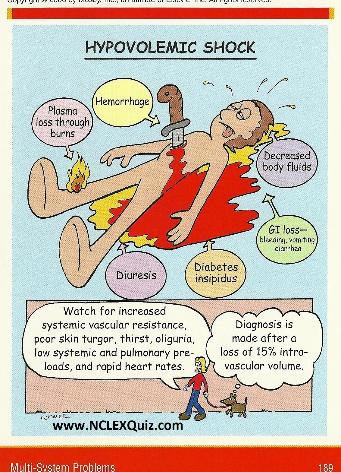
What causes hypovolemic shock?
The loss of intravascular fluid volume which causes hypovolemic shock can have a number of causes including dehydration from vomiting and diarrhea, hemorrhage, decreased intake of fluids, pathologic urinary losses (e.g. diabetic ketoacidosis, diabetes insipidus), and translocation of body fluids (e.g. burns, peritonitis, small bowel obstruction).
What fluids are used for hemorrhagic hypovolemic shock?
For hemorrhagic hypovolemic shock, boluses of isotonic crystalloid IV fluids are indicated, but the shock may not improve significantly. In this case, packed red blood cells (PRBCs) are indicated, and the standard dosing of PRBCs for refractory hemorrhagic hypovolemic shock is 10 mL/kg.
Why does the volume of blood decrease during stroke?
This reduction of the intravascular fluid volume causes a decrease in stroke volume because of the resulting decrease in preload. The decrease in preload impairs cardiac output which ultimately leads to inadequate delivery of oxygen and nutrients to the tissues and organs (shock).
What is the most common form of shock in children?
Hypovolemic shock is the most common form of shock that occurs in children. The most common cause of hypovolemic shock and infant deaths worldwide in the pediatric population is dehydration resulting from diarrhea. Remember: Heart Rate x Stroke Volume (preload, afterload, contractility) = Cardiac Output. When preload is decreased, there are three ...
What is hypovolemia in medical terms?
Hypovolemia is defined as a decrease in the blood volume resulting from loss of blood, plasma and/or plasma water, thereby causing a loss of intravascular content and resulting in a potential limitation of tissue perfusion .[1] . It is often seen in case of severe dehydration or blood loss owing to trauma or surgery.
What is the first step in hemorrhagic shock?
Although fluid resuscitation is the first step to restore tissue perfusion in severe hemorrhagic shock, it remains a matter of controversy for decades whether colloids or crystalloids, and more specifically, which colloid, should be used.
What should new generation fluids be developed with?
Based on patho-physiology of hypovolemia, new-generation fluids should be developed with a focus of research on improving oxygen-carrying capacity by using hemoglobin-based oxygen carriers and with an emphasis on limiting the proinflammatory effects of fluids. Acknowledgement.
What is volume expansion?
This effort involves the use of fluid resuscitation, vasopressors, and blood transfusion. Currently, volume expansion aims at increasing global blood flow, with the hope that such an increase will improve flow to the microcirculation increasing oxygen availability to the tissues.[1] .
Is colloidal fluid safe for perioperative use?
Despite little published evidence suggesting specific advantages over other intravenous fluids, and emerging evidence of harm in septic and critically ill patients, the colloidal fluids remain a popular choice for perioperative fluid therapy.
Is there a fluid for resuscitation?
With current evidence, no ideal resuscitation fluid exists. It is better to observe the patient's response to volume infusion than to follow blindly any specific rule as every patient responds differently to fluid therapy. Therapy should target physiologic goals of hemodynamic stabilization.
What happens when your preload decreases?
When our preload decreases, cardiac output also decreases and our body has mechanisms it uses to try to compensate. So you’ll see a lot of vasoconstriction in the body because it’s trying to push the blood back toward the heart – that means the pressure our heart has to pump against is increased – that’s our afterload.
Why does blood pressure decrease after stroke?
After a while, we’ll begin to see our Blood pressure decrease because the body can only compensate for so long . Ultimately, there’s just not enough circulating blood volume to serve the whole system, and it will start to shut down.
Why is hypovolemic shock important?
This is because hypovolemic shock can be caused by blood loss from traumatic injuries , internal bleeding, like a GI bleed or a surgical complication, and postpartum hemorrhage or fluid loss from burns, diarrhea and vomiting.
What is hypovolemic shock?
Pathophysiology. Hypovolemic shock is a loss of blood volume leading to decreased oxygenation of vital organs. The body’s compensatory mechanisms fail and organs begin to shut down.
How often should you give blood products during hypovolemic shock?
Usually, this would be every 15 minutes, times two, every 30 minutes times one in every hour after that. However, in hypovolemic shock, even blood products are given rapidly. Here is a look at the completed hypovolemic shock care plan. Let’s do a quick review. Hypovolemic shock is the loss of blood volume leading to decreased oxygenation of organs.
How fast can a catheter pump infuse fluid?
An infusion pump is only capable of infusing one liter an hour, so fluids should be given as soon as possible and as fast as possible to restore circulating blood volume.
Does monitoring vital signs help with shock?
Monitoring vital signs could help to prevent hypovolemic shock if caught early, but also help to determine the patient’s response to treatment.
What is hypovolemic shock?
A perilous plunge in perfusion. Hypovolemic shock occurs when circulatory volume drops significantly. As in other types of shock, systemic reduction in tissue perfusion leads to decreased oxygen delivery.
What are the complications of hypovolemic shock?
Hypovolemic shock also may cause other complications, including: systemic infection from use of a large-bore I.V. line for fluid resuscitation. transfusion reaction if blood transfusions are given. hypothermia, which may follow trauma, surgery, or infusion of massive amounts of I.V. fluids.
How does SVR affect blood volume?
Increased SVR in turn boosts cardiac output, increases tissue perfusion pressure, and triggers catecholamine release. Blood volume rises as interstitial fluid shifts to the intravascular spaces and the liver and spleen release stored red blood cells (RBCs).
What is the role of the renin-angiotensin-aldosterone system in the
These changes activate the renin-angiotensin-aldosterone system, which promotes sodium and water retention in an effort to raise systolic pressure. Urine output then declines. If blood loss continues, compensatory mechanisms maintain perfusion to vital tissues by shunting blood away from nonvital organs.
What does hematocrit mean in blood work?
Hemoglobin and hematocrit values may indicate bleeding ; during treatment, they provide a baseline for gauging the patient’s response to blood transfusions or other replacement fluids.
Why are children at greater risk for hypovolemia?
Children also are at greater risk due to their higher proportion of body water. Pathophysiologic process. In hypovolemia, decreased fluid volume reduces blood return to the heart, causing a decline in preload (the volume of blood remaining in the left ventricle at the end of diastole).
Does hypovolemia cause aspiration?
But be aware that such positioning heightens the risk of aspiration. Hypovolemia itself reduces gut perfusion; decreased blood flow to the stomach can increase the risk of gastric residuals, reflux, and aspiration. So if the patient’s receiving enteral nutrition, discontinue tube feedings temporarily.
