
Radiation therapy (also called radiotherapy) is a cancer treatment that uses high doses of radiation to kill cancer cells and shrink tumors. At low doses, radiation is used in x-rays to see inside your body, as with x-rays of your teeth or broken bones.
Are X-rays harmful for your body?
Radiation therapy in cancer treatment: X-rays and other types of high-energy radiation can be used to destroy cancerous tumors and cells by damaging their DNA. The radiation dose used for treating cancer is much higher than the radiation dose used for diagnostic imaging.
Is X ray bad for You?
X-rays use invisible electromagnetic energy beams to produce images of internal tissues, bones, and organs on film or digital media. Standard X-rays are performed for many reasons, including diagnosing tumors or bone injuries.
Why are X rays harmful?
Jan 08, 2019 · Radiation therapy (also called radiotherapy) is a cancer treatment that uses high doses of radiation to kill cancer cells and shrink tumors. At low doses, radiation is used in x-rays to see inside your body, as with x-rays of your teeth or broken bones.
What are the dangers of X rays?
Sep 02, 2020 · An X-ray is a common imaging test that’s been used for decades. It can help your doctor view the inside of your body without having to make an incision. This can help them diagnose, monitor, and...
Why are X-rays used for treatment?
When X-rays are used They're mainly used to look at the bones and joints, although they're sometimes used to detect problems affecting soft tissue, such as internal organs. Problems that may be detected during an X-ray include: bone fractures and breaks. tooth problems, such as loose teeth and dental abscesses.
Is an X-ray medical treatment?
An X-ray, also known as radiography, is a medical imaging technique. It uses tiny amounts of electromagnetic radiation to create images of structures inside the body.Mar 11, 2022
Are X-rays cancer treatment?
Radiation therapy (also called radiotherapy) is a cancer treatment that uses high doses of radiation to kill cancer cells and shrink tumors. At low doses, radiation is used in x-rays to see inside your body, as with x-rays of your teeth or broken bones.Jan 8, 2019
What disease can be treated with X-rays?
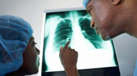
ChestLung infections or conditions. Evidence of pneumonia, tuberculosis or lung cancer can show up on chest X-rays.Breast cancer. Mammography is a special type of X-ray test used to examine breast tissue.Enlarged heart. This sign of congestive heart failure shows up clearly on X-rays.Blocked blood vessels.Feb 11, 2022
Can a chest xray show stomach problems?
Plain chest X-ray films can help diagnose and suspect volvulus in any patients presenting with an acute abdomen [5]. It characteristically demonstrates spherical stomach, double air-fluid level or the retrocardiac air-fluid level above the diaphragm on the upright chest films.Jul 16, 2020
How to do X-rays?
Although each facility may have specific protocols in place, generally, an X-ray procedure follows this process: 1 The patient will be asked to remove any clothing or jewelry which might interfere with the exposure of the body area to be examined. The patient will be given a gown to wear if clothing must be removed. 2 The patient is positioned on an X-ray table that carefully positions the part of the body that is to be X-rayed--between the X-ray machine and a cassette containing the X-ray film or specialized image plate. Some examinations may be performed with the patient in a sitting or standing position. 3 Body parts not being imaged may be covered with a lead apron (shield) to avoid exposure to the X-rays. 4 The X-ray beam will be aimed at the area to be imaged. 5 The patient must be very still or the image will be blurred. 6 The technologist will step behind a protective window and the image is taken. 7 Depending on the body part under study, various X-rays may be taken at different angles, such as the front and side view during a chest X-ray.
Why do you need a lead apron for X-rays?
Body parts not being imaged may be covered with a lead apron (shield) to avoid exposure to the X-rays. The X-ray beam will be aimed at the area to be imaged. The patient must be very still or the image will be blurred. The technologist will step behind a protective window and the image is taken.
What is the procedure for X-rays?
Although each facility may have specific protocols in place, generally, an X-ray procedure follows this process: The patient will be asked to remove any clothing or jewelry which might interfere with the exposure of the body area to be examined. The patient will be given a gown to wear if clothing must be removed.
What is the white line on an X-ray?
A bone or a tumor, which is more dense than soft tissue, allows few of the X-rays to pass through and appears white on the X-ray. When a break in a bone has occurred, the X-ray beam passes through the broken area and appears as a dark line in the white bone.
What are the parts of the body that allow X-rays to pass through?
The soft tissues in the body (such as blood, skin, fat, and muscle) allow most of the X-ray to pass through and appear dark gray on the film or digital media.
What type of image is made when X-rays pass through body structures?
X-rays pass through body structures onto specially-treated plates (similar to camera film) or digital media and a "negative" type picture is made (the more solid a structure is, the whiter it appears on the film).
Can X-rays be used during pregnancy?
X-ray technology is used in other types of diagnostic procedures, such as arteriograms, computed tomography (CT) scans, and fluoroscopy. Radiation during pregnancy may lead to birth defects. Always tell your radiologist or doctor if you suspect you may be pregnant.
What are the side effects of barium sulfate?
These include: 3 . A history of asthma, hay fever, or other allergies, which increases the risk of an allergic reaction to additives in the barium sulfate agent. Cystic fibrosis, which will increase the risk of blockage in the small bowel. Severe dehydration, which may cause severe constipation.
How long before barium X-ray?
If you will be having an X-ray using barium contrast dye, which is used most often to highlight structures in the digestive system, you will be told not to eat for at least three hours before your appointment. 3 People with diabetes usually are allowed to eat a light meal three hours before receiving barium. If the barium will be administered via an enema, you also may be asked to eat a special diet and take medication to cleanse your colon beforehand.
Why do we need X-rays?
X-rays often are done to view bones and teeth, making them useful for diagnosing breaks, fractures, and diseases such as arthritis.
What is radiograph used for?
As such, the image it creates, known as a radiograph, is useful for doctors interested in visualizing significant internal structures. Sometimes a contrast medium, a type of dye, is introduced into the body to help images show up in greater detail. The individual elements render in various shades of white and grey.
What is the best way to find evidence of lung cancer?
Finding evidence of pneumonia, tuberculosis, or lung cancer (chest X-rays) Examining breast tissue for signs of cancer using a special X-ray technique called mammography 1 . Looking for signs of heart failure or changes in blood flow to the lungs and heart.
Why do you need an X-ray before a dental exam?
Before the Test. Often, an X-ray will be done as part of a visit to your doctor or emergency room to diagnose symptoms or evaluate an injury. X-rays are also taken as part of certain routine exams, such as dental checkups.
How is X-ray technology used?
X-ray technology is used throughout the medical world for a multitude of purposes. Conventional X-ray images can be very useful to doctors in evaluating symptoms that originate inside the body as well as diagnosing injuries. Among the most common uses of conventional X-ray are:
What is intraoperative radiation therapy?
During surgery, so that it goes straight to the cancer without passing through the skin. Radiation therapy used this way is called intraoperative radiation.
How does radiation help cancer?
When radiation is combined with surgery, it can be given: 1 Before surgery, to shrink the size of the cancer so it can be removed by surgery and be less likely to return. 2 During surgery, so that it goes straight to the cancer without passing through the skin. Radiation therapy used this way is called intraoperative radiation. With this technique, doctors can more easily protect nearby normal tissues from radiation. 3 After surgery to kill any cancer cells that remain.
What is brachytherapy with liquid source?
Learn more about brachytherapy. Internal radiation therapy with a liquid source is called systemic therapy. Systemic means that the treatment travels in the blood to tissues throughout your body, seeking out and killing cancer cells.
What is the best radiation treatment for thyroid cancer?
A systemic radiation therapy called radioactive iodine, or I-131, is most often used to treat certain types of thyroid cancer.
What is the treatment for cancer that has spread to the bone called?
Pain from cancer that has spread to the bone can be treated with systemic radiation therapy drugs called radiopharmaceuticals.
Why do people with cancer need radiation?
Why People with Cancer Receive Radiation Therapy. Radiation therapy is used to treat cancer and ease cancer symptoms . When used to treat cancer, radiation therapy can cure cancer, prevent it from returning, or stop or slow its growth. When treatments are used to ease symptoms, they are known as palliative treatments.
What is external beam radiation therapy?
External Beam Radiation Therapy. External beam radiation therapy comes from a machine that aims radiation at your cancer. The machine is large and may be noisy. It does not touch you, but can move around you, sending radiation to a part of your body from many directions.
Why do doctors use X-rays?
It can help your doctor view the inside of your body without having to make an incision. This can help them diagnose, monitor, and treat many medical conditions . Different types of X-rays are used for different purposes. For example, your doctor may order a mammogram to examine your breasts. Or they may order an X-ray with a barium enema ...
How to order an X-ray?
Your doctor may order an X-ray to: 1 examine an area where you’re experiencing pain or discomfort 2 monitor the progression of a diagnosed disease, such as osteoporosis 3 check how well a prescribed treatment is working
What conditions can you see on an X-ray?
Conditions that may call for an X-ray include: bone cancer. breast tumors. enlarged heart. blocked blood vessels. conditions affecting your lungs. digestive problems.
Can dye cause anaphylactic shock?
itching. nausea. lightheadedness. a metallic taste in your mouth. In very rare cases, the dye can cause a severe reaction, such as anaphylactic shock, very low blood pressure, or cardiac arrest. If you suspect you’re having a severe reaction, contact your doctor immediately.
Can you have an MRI with a broken bone?
They may suggest a different imaging method, such as an MRI. If you’re having an X-ray done to help diagnose or manage a painful condition, such as a broken bone, you may experience pain or discomfort during the test. You will need to hold your body in certain positions while the images are being taken.
Can you get an X-ray with a barium enema?
Or they may order an X-ray with a barium enema to get a closer look at your gastrointestinal tract. There are some risks involved in getting an X-ray. But for most people, the potential benefits outweigh the risks. Talk to your doctor to learn more about what is right for you.
Can you remove jewelry before an xray?
They may also ask you to remove any jewelry or other metallic items from your body before your X-ray is taken. Always tell your doctor or radiologist if you have metal implants from prior surgeries. These implants can block X-rays from passing through your body and creating a clear image.
How is radiation given?
Radiation therapy can be given in 3 ways: 1 External radiation (or external beam radiation): uses a machine that directs high-energy rays from outside the body into the tumor. It’s done during outpatient visits to a hospital or treatment center. It's usually given over many weeks and sometimes will be given twice a day for several weeks. A person receiving external radiation is not radioactive and does not have to follow special safety precautions at home. 2 Internal radiation: Internal radiation is also called brachytherapy. A radioactive source is put inside the body into or near the tumor. With some types of brachytherapy, radiation might be placed and left in the body to work. Sometimes it is placed in the body for a period of time and then removed. This is decided based on the type of cancer. Special safety precautions are needed for this type of radiation for a period of time. But it's important to know if the internal radiation is left in the body, after a while it eventually is no longer radioactive. 3 Systemic radiation: Radioactive drugs given by mouth or put into a vein are used to treat certain types of cancer. These drugs then travel throughout the body. You might have to follow special precautions at home for a period of time after these drugs are given.
What doctor is trained to treat cancer?
Radiation oncologist: This doctor is specially trained to treat cancer with radiation. This person oversees your radiation treatment plan. Radiation physicist: This is the person who makes sure the radiation equipment is working as it should and that it gives you the exact dose prescribed by your radiation oncologist.
What is the treatment for cancer that has returned?
To treat cancer that has returned (recurred) If a person's cancer has returned (recurred), radiation might be used to treat the cancer or to treat symptoms caused by advanced cancer. Whether radiation will be used after recurrence depends on many factors.
How does radiation help cancer cells?
But cancer cells grow and divide faster than most normal cells. Radiation works by making small breaks in the DNA inside cells. These breaks keep cancer cells from growing and dividing and cause them to die.
Why do people get radiation to their head?
This is done to help prevent cancer from spreading to the head even before it can.
How does cancer spread?
Cancer can spread from where it started to other body parts. Doctors often assume that a few cancer cells might already have spread even when they can’t be seen on imaging scans like CT scans or MRIs. In some cases, the area where the cancer most often spreads to may be treated with radiation to kill any cancer cells before they grow into tumors. For instance, people with certain kinds of lung cancer may get radiation to the head, even when there is no cancer known to be there, because their type of lung cancer often spreads to the brain. This is done to help prevent cancer from spreading to the head even before it can. Sometimes, radiation to prevent future cancer can be given at the same time that radiation is given to treat existing cancer, especially if the area the cancer might spread to is close to the tumor itself.
How many people with cancer get radiation?
More than half of people with cancer get radiation therapy. Sometimes, radiation therapy is the only cancer treatment needed and sometimes it's used with other types of treatment. The decision to use radiation therapy depends on the type and stage of cancer, and other health problems a patient might have.
What is the procedure called when you have a contrast dye injected into your spinal canal?
CT scan. When a CT is used to image the spine, you may have a contrast dye injected into your spinal canal before the X-rays are taken — a procedure called a CT myelogram.
What is an EMG test?
Electromyography (EMG). This test measures the electrical impulses produced by the nerves and the responses of your muscles. This test can confirm nerve compression caused by herniated disks or narrowing of your spinal canal (spinal stenosis).
What is the best treatment for low back pain?
Alternative therapies commonly used for low back pain include: Acupuncture. In acupuncture, the practitioner inserts hair-thin needles into your skin at specific points on your body. Some studies have suggested that acupuncture can help back pain, while others have found no benefit.
What is the procedure to see if you have a bone spur?
X-ray. An X-ray of your spine may reveal an overgrowth of bone (bone spur) that may be pressing on a nerve. MRI. This procedure uses a powerful magnet and radio waves to produce cross-sectional images of your back. An MRI produces detailed images of bone and soft tissues such as herniated disks.
What can a physical therapist do for back pain?
This typically includes exercises to correct your posture, strengthen the muscles supporting your back and improve your flexibility.
How to get rid of nerve compression in lower back?
Stretching. Stretching exercises for your low back can help you feel better and might help relieve nerve root compression. Avoid jerking, bouncing or twisting during the stretch, and try to hold the stretch for at least 30 seconds. Over-the-counter medications.
How to get rid of a swollen ear?
Initially, you might get relief from a cold pack placed on the painful area for up to 20 minutes several times a day. Use an ice pack or a package of frozen peas wrapped in a clean towel. Hot packs. After two to three days, apply heat to the areas that hurt.
What is the test for pneumonia?
This measures the oxygen level in your blood. Pneumonia can prevent your lungs from moving enough oxygen into your bloodstream. Sputum test. A sample of fluid from your lungs (sputum) is taken after a deep cough and analyzed to help pinpoint the cause of the infection.
What is the best medicine for pneumonia?
It may take time to identify the type of bacteria causing your pneumonia and to choose the best antibiotic to treat it. If your symptoms don't improve, your doctor may recommend a different antibiotic. Cough medicine.
What to do if pneumonia isn't clearing?
If your pneumonia isn't clearing as quickly as expected, your doctor may recommend a chest CT scan to obtain a more detailed image of your lungs. Pleural fluid culture. A fluid sample is taken by putting a needle between your ribs from the pleural area and analyzed to help determine the type of infection.
How to get rid of pneumonia?
Get plenty of rest. Don't go back to school or work until after your temperature returns to normal and you stop coughing up mucus. Even when you start to feel better, be careful not to overdo it. Because pneumonia can recur, it's better not to jump back into your routine until you are fully recovered.
What tests are done to determine if you have pneumonia?
If pneumonia is suspected, your doctor may recommend the following tests: Blood tests . Blood tests are used to confirm an infection and to try to identify the type of organism causing the infection. However, precise identification isn't always possible. Chest X-ray.
What does chest X-ray show?
Diagnosis. This chest X-ray shows an area of lung inflammation indicating the presence of pneumonia. Your doctor will start by asking about your medical history and doing a physical exam, including listening to your lungs with a stethoscope to check for abnormal bubbling or crackling sounds that suggest pneumonia.
How fast can you breathe in a minute?
Your breathing is rapid (30 breaths or more a minute) You need breathing assistance. Your temperature is below normal. Your heart rate is below 50 or above 100. You may be admitted to the intensive care unit if you need to be placed on a breathing machine (ventilator) or if your symptoms are severe.
How to treat bursa inflammation?
Medication. If the inflammation in your bursa is caused by an infection, your doctor might prescribe an antibiotic. Therapy. Physical therapy or exercises can strengthen the muscles in the affected area to ease pain and prevent recurrence. Injections.
How to relieve pain from bursitis?
Measures you can take to relieve the pain of bursitis include: Rest and don't overuse the affected area. Apply ice to reduce swelling for the first 48 hours after symptoms occur. Apply dry or moist heat, such as a heating pad or taking a warm bath.
What tests can be done to diagnose bursitis?
Testing, if needed, might include: Imaging tests. X-ray images can't positively establish the diagnosis of bursitis, but they can help to exclude other causes of your discomfort. Ultrasound or MRI might be used if your bursitis can't easily be diagnosed by a physical exam alone. Lab tests.
How to relieve pain from a swollen knee?
Take an over-the-counter medication, such as ibuprofen (Advil, Motrin IB, others) or naproxen sodium ( Aleve, others), to relieve pain and reduce inflammation. Some are available in a form you apply to the skin. Cushion your knees if you sleep on your side by placing a small pillow between your legs.
Can a doctor inject a corticosteroid into a bursa?
Injection of a corticosteroid medication into your bursa can relieve the pain and inflammation of bursitis. In some cases, your doctor might use ultrasound to guide the injection into the affected bursa. The ultrasound's hand-held transducer provides a live-action display your doctor can view on a monitor during the procedure.
Does ultrasound help with bursitis?
The ultrasound's hand-held transducer provides a live-action display your doctor can view on a monitor during the procedure . Bursitis generally gets better on its own. Conservative measures, such as rest, ice and taking a pain reliever, can relieve discomfort.

How It Works
When It's Used
Risks
Contraindications
How to Prepare
During The Test
After The Test
Interpreting Results
Summary
- X-rays are imagery tests that use small amounts of electromagnetic radiation to obtain images of the inside structures of your body. In addition to conventional X-rays, several other specialized forms of X-rays capture images in more precise ways. Sometimes a contrast agent can help healthcare providers see things better. These dyes might be given ...
A Word from Verywell