How Is Adenomyosis Treated?
- Anti-inflammatory medications. Your doctor may prescribe nonsteroidal anti-inflammatory drugs (NSAIDs) to relieve mild...
- Hormone therapy.
- Uterine artery embolization.
- Endometrial ablation.
What is adenomyosis and how can it be treated?
Adenomyosis treatment is focused primarily on symptom management, as the cause is not well understood. There are nutrition and lifestyle practices that can help reduce pain, improve hormonal health, lower inflammation, and improve health outcomes. In some cases, surgical intervention may be necessary.
Is there any cure for adenomyosis?
Vitamin B complex and vitamin E play a significant role in the treatment of conditions like adenomyosis. Studies have found that vitamins B1, B6, and E can help reduce menstrual pain (9), (10). You may get the required amounts of these vitamins by consuming eggs, milk, cheese, fish, poultry, almonds, spinach, and kale.
Is dienogest effective for treating symptomatic adenomyosis?
The combination of MEA and postoperative dienogest is useful for treating uterine adenomyosis with menorrhagia and dysmenorrhea. Combination of microwave endometrial ablation and postoperative dienogest administration is effective for treating symptomatic adenomyosis
What techniques are used to diagnose adenomyosis?
These include:
- oral contraceptive pills
- high-dose progestins
- a levonorgestrel-releasing intrauterine device
- danazol
- gonadotropin-releasing hormone (GnRH) agonists and antagonists, like elagolix
- endometrial ablation, which is an outpatient procedure that uses a laser or other ablation techniques to destroy the lining of the uterus

Can adenomyomatosis be cured?
Can Adenomyosis Be Cured? The only definitive cure for adenomyosis is a hysterectomy, or the removal of the uterus. This is often the treatment of choice for women with significant symptoms.
How serious is gallbladder adenomyomatosis?
Originally recognized as a precancerous lesion, adenomyomatosis is currently recognized by recent studies as a benign alteration of the gallbladder that is often associated with cholecystitis and cholecystolithiasis. Gallbladder carcinoma is an extremely malignant disease with a 5-year survival rate of less than 5%.
Can adenomyosis be treatment without surgery?
Adenomyosis cannot be fully treated without surgery. However, mild symptoms may be temporarily managed with some nonsurgical options. Because the condition gets worse over time, surgery will likely become necessary. Adenomyosis causes thickening of the uterine wall that ultimately leads to an enlarged uterus.
Does focal adenomyomatosis in the gallbladder require surgery?
At present, the main treatment for gallbladder adenomyomatosis is surgery, but it is very important to screen patients and make clear the operation indications, taking the complications after cholecystectomy into consideration.
What are the symptoms of gallbladder adenomyomatosis?
The most common presentation of GAM is pain in the upper right quadrant of the abdomen, which is similar to gallstone pain with or without cholecystitis. This pain is intermittent and mostly self-limiting [5,6]. It is possible that GAM symptoms are secondary to gallstones and inflammation.
How common is gallbladder adenomyomatosis?
1. Gallbladder adenomyomatosis is a common benign lesion (1–9% of the patients).
What happens if adenomyosis is left untreated?
Adenomyosis Gets Worse Over Time If left untreated it may lead to infertility or other problems such as pelvic organ prolapse. As women continue to live longer lives reproductive issues like adenomyosis have increased in prevalence over the last 30 years.
What foods to avoid if you have adenomyosis?
Foods to avoid on an adenomyosis diet include:Wheat and gluten.Artificial sugars.Dairy.Bananas.Yeast-based products including alcohol, tea, and coffee.Chasteberry (Vitex agnus-castus) and red raspberry leaf/raspberry teas.
What vitamins should I take for adenomyosis?
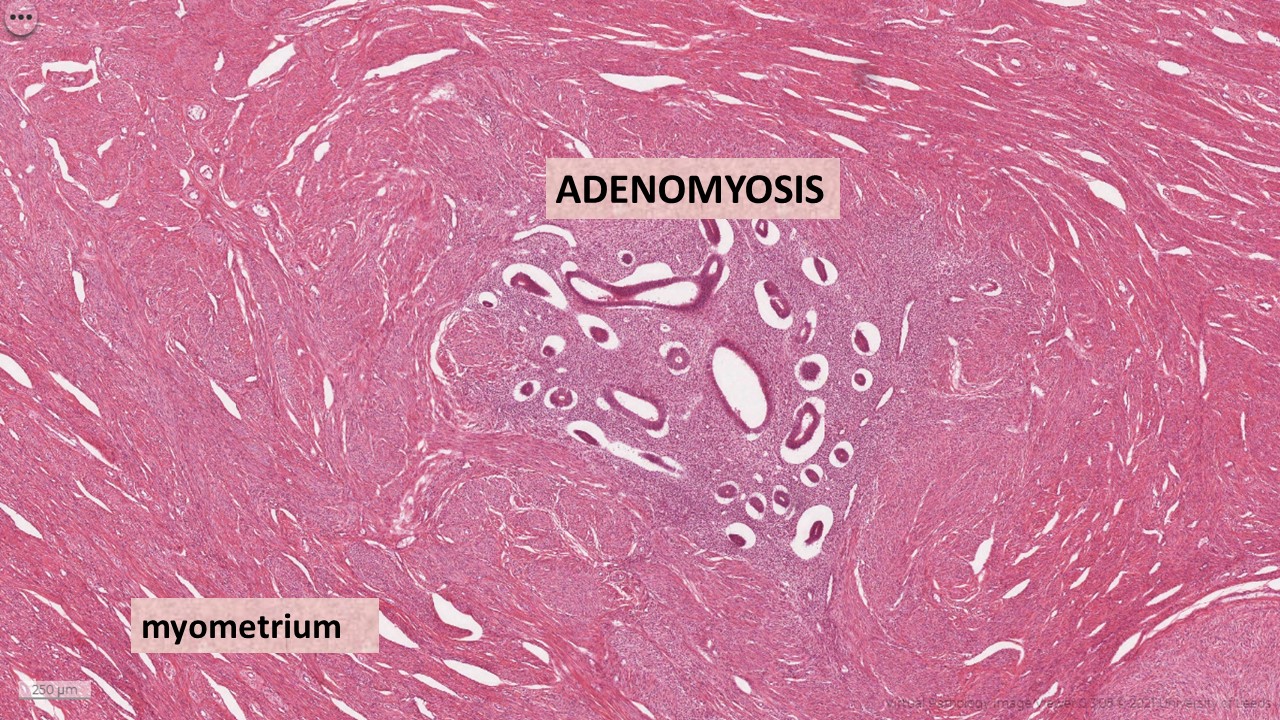
Adenomyosis is a gynaecological condition that affects the uterus. Cells that normally form the lining of the uterus (the endometrial tissue) also grow within the muscle walls of the uterus (the myometrium)....Key antioxidants include:Vitamin A.Vitamin C.Vitamin E.Zinc.Copper.
What does adenomyomatosis mean?
Adenomyomatosis is a benign condition that can present with right upper quadrant pain and is characterized by hyperplasia of the gallbladder wall mucosa and muscularis propria, with pathognomonic epithelial invaginations forming cystic pockets (Rokitansky-Aschoff sinuses).
Does adenomyomatosis enhance?
Abnormal gallbladder wall thickening and enhancement are common but nonspecific CT features of adenomyomatosis.
What is focal adenomyomatosis at the gallbladder fundus?
Adenomyomatosis is a common tumorlike lesion of the gallbladder with no malignant potential and may involve the gallbladder in a focal, segmental, or diffuse form. The focal type is the most common and usually involves the gallbladder fundus.
What can you tell me about a condition called gallbladder adenomyomatosis?
A benign finding: This is a benign finding in many gallbladders. It is associated with cholesterol crystals that build up in a thickened gallbladder wall. Often associa... Read More
I just was diagnosed with adenomyomatosis of gallbladder, i also have 2 small polyps . should i get removed, should i fear of it becoming cancerous!
Up to you: Smaller polyps tend to be benign , size 2 cm or more have higher incidence of malignancy , the one you describing could be observed but need clos... Read More
Gallbladder without gallstones but with a few small hyperechoic nodules along its inner wall largest measuring 0.8 x 0.9 x 0.6 cm compatible with small polyps versus adenomyomatosis. no sxs. treat?
Up to you: General recommendation for polyps is observation and repeat ultrasound in 6-12 months if less than 1cm. If you're having pain, and you think it could ... Read More
My ultrasound reported: gallbladder exhibits features of adenomyomatosis. there is no cholelithiasis or billiard tract dilatation. what it could be?
Hyperplastic gb: Adenomyomatosis denotes 3 hyperplastic changes in the gallbladder--overgrowth of the mucous membrane, thickening of the muscle layer, & intramural... Read More
What to do if i have been told i have adenomyomatosis of the gall bladder has anyone else heard of this?
Polyps?: If you have adenomatous changes or polyps of the gall bladder you should be evaluated by a surgeon for removal of the gall bladder.
I have a hyperechoic nodule on the right hepatic lobe measuring 0.750.820.39 cm that is stable.focal fat sparing in the periportal region noted. gall bladder adenomyomatosis detected. should i go for a further scan or it is just normal? this is my ultra
Maybe: You should discuss this with your doctor. Do you have underlying liver disease and would be at risk of a problem in the liver? Liver cysts are usuall... Read More
What is an extended gallbladder and what is the treatment?
DIStended?: The gallbladder's function is to store bile. A distended gallbladder is simply one filled with bile. We can see distention in many different condition... Read More
How to diagnose adenomyosis?
Until recently, the only definitive way to diagnose adenomyosis was to perform a hysterectomy and examine the uterine tissue under a microscope. However, imaging technology has made it possible for doctors to recognize adenomyosis without surgery.
What hormones cause adenomyosis?
Though the cause of adenomyosis isn't known, studies have suggested that various hormones -- including estrogen, progesterone, prolactin, and follicle stimulating hormone -- may trigger the condition.
Can ultrasound diagnose adenomyosis?
An ultrasound can allow a doctor to see the uterus, its lining, and its muscular wall. Though ultrasound cannot definitively diagnose adenomyosis, it can help to rule out other conditions with similar symptoms. Another technique sometimes used to help evaluate the symptoms associated with adenomyosis is sonohysterography.
Is adenomyosis a benign condition?
The condition can be located throughout the entire uterus or localized in one spot. Though adenomyosis is considered a benign ( not life-threatening) condition, the frequent pain and heavy bleeding associated with it can have a negative impact on a woman's quality of life.
Is adenomyosis the same as fibroids?
However, the two conditions are not the same. While fibroids are benign tumors growing in or on the uterine wall, adenomyosis is less of a defined mass of cells within the uterine wall. An accurate diagnosis is key in choosing the right treatment.
How to relieve pain from adenomyosis?
To ease pelvic pain and cramping related to adenomyosis, try these tips: Soak in a warm bath. Use a heating pad on your abdomen. Take an over-the-counter anti-inflammatory medication, such as ibuprofen (Advil, Motrin IB, others).
How to treat adenomyosis after menopause?
Treatment options for adenomyosis include: Anti-inflammatory drugs. Your doctor might recommend anti-inflammatory medications , such as ibuprofen (Advil, Motrin IB, others), to control the pain.
Can endometrial biopsy confirm adenomyosis?
But an endometrial biopsy won 't help your doctor confirm a diagnosis of adenomyosis. Pelvic imaging such as ultrasound and MRI can detect signs of adenomyosis, but the only way to confirm it is to examine the uterus after hysterectomy.
Can an MRI confirm adenomyosis?
But an endometrial biopsy won't help your doctor confirm a diagnosis of adenomyosis.
What is adenomyomatosis?
Adenomyomatosis is a benign condition that is pathologically characterized by hyperplasia of the gallbladder wall mucosa and muscularis propria, with pathognomonic epithelial invaginations forming cystic pockets (Rokitansky-Aschoff sinuses).
Can adenomyomatosis be diagnosed with gallbladder cancer?
Sometimes, mainly when the characteristic imaging findings are not present, ade nomyomatosis can be challenging to distinguish from gallbladder cancer based on the diagnostic imaging finding s. Adenomyomatosis is often asymptomatic and incidentally detected, requiring no specific treatment.
Is adenomyomatosis a benign condition?
Adenomyomatosis is a benign condition that is pathologically char …. Adenomyomatosis, also known as adenomyoma or adenomyomatous hyperplasia of the gallbladder, is one of the two hyperplastic cholecystoses. The other hyperplastic cholecystosis is cholesterolosis, also known as "strawberry gallbladder.". Adenomyomatosis is a benign condition that is ...
What is focal adenomyomatosis?
This can lead to focal cholelithiasis or cholecystitis involving only the fundus of the gallbladder. Focal adenomyomatosis manifesting as a mass is sometimes referred to as an adenomyoma.
What is adenomyomatous hyperplasia?
Adenomyomatosis of the gallbladder, also referred to as adenomyomatous hyperplasia and intramural diverticulosis, is an acquired, hyperplastic lesion of the gallbladder characterized by excessive proliferation of surface epithelium with invaginations into the thickened, hypertrophied muscularis propria. 215 The intramural diverticula formed by epithelial invaginations into the muscularis are referred to as Rokitansky-Aschoff sinuses (RAS) and dilated RAS are a prominent feature of this disorder. The pathophysiology of this disorder is not entirely certain. This is not usually associated with cholesterolosis, indicating a separate pathophysiology. Pathogenesis has been postulated to result from mechanical obstruction of the gallbladder (from stones, cystic duct kinking or congenital septum), chronic inflammation and anomalous pancreaticobiliary ductal union. 215 The functional obstruction to bile outflow causes increased pressure within the gallbladder lumen and results in invagination of the mucosa through the muscularis, forming the dilated RAS. The reported incidence of adenomyomatosis in cholecystectomy specimens is up to 8%. 219 The association of this disorder with clinical findings is controversial The disorder may be asymptomatic or associated with symptoms of chronic cholecystitis. Over 90% of cases are associated with gallstones which may be responsible for biliary symptoms. 2 Adenocarcinoma of the gallbladder has been found in association with adenomyomatosis, however, a causal link has not been established. 215 In a surgical series of 3,000 resected gallbladders, there was a higher frequency of gallbladder carcinoma (6.4%) in gallbladders with segmental adenomyomatosis than those without. 219
Can focal adenomyomatosis be missed?
Focal adenomyomatosis is more difficult to diagnose and can be missed ; this is supported by the pathological examination of gallbladders in which focal changes are seen more frequently than shown on imaging studies. Ultrasound scan shows gallbladder wall thickening and occasionally may show sinus formation.
Is adenomyomatosis a focal or segmental?
Adenomyomatosis may be generalized, segmental or focal and the focal form is most common. 220 Diffuse adenomyomatosis causes thickening and irregularity of the mucosa and muscular layer with associated RAS, which on gross inspection appear as collections of bile in the gallbladder wall.
Is adenomyomatosis a form of cholecystosis?
Adenomyomatosis is one of two forms of hyperplastic cholecystoses (cholesterolosis is the other). Like cholesterolosis, the etiology is unknown. Pathologically, adenomyomatosis is characterized by mucosal hyperplasia and thickening of the muscular layer of the gallbladder. Mucosal herniations into the muscular layer are called Rokitansky-Aschoff ...
Is cholecystectomy asymptomatic?
Many patients are asymptomatic. Treatment of symptomatic patients is controversial. There is evidence to suggest, however, that symptomatic patients who receive cholecystectomy frequently experience abatement of symptoms. Cholesterolosis has been described in the previous section.
Is gallbladder adenomyomatosis benign?
Gallbladder ade nomyomatosis is a benign condition occurring either focally (most commonly at the fundus), diffusely, or segmentally. There is hyperplasia of epithelial and muscular elements resulting in mucosal outpouchings into a thickened wall, forming intramural diverticula (also known as Rokitansky-Aschoff sinuses) (Fig. 3-54 ).
How to diagnose adenomyosis?
Healthcare providers often diagnose adenomyosis based on symptoms and one or more of these tests: Pelvic exam: During a pelvic exam, your provider may notice that your uterus has gotten larger, softer, or more painful when palpated. Ultrasound: A transvaginal ultrasound uses sound waves to produce images of pelvic organs.
What is adenomyosis in women?
What is adenomyosis? Adenomyosis (add-en-o-my-OH-sis) is a condition of the female reproductive system. It causes the uterus to thicken and enlarge. Endometrial tissue lines the inside of the uterine wall (endometrium).
Can adenomyosis cause miscarriage?
Infertility treatments may help. Once pregnant, there is an increased risk of: Miscarriage (loss of pregnancy before a baby fully develops).
Can adenomyosis be seen in MRI?
Imaging scans: Magnetic resonance imaging (MRI) scans can show uterine enlargement and thickening of certain areas of the uterus which can be indicative of adenomyosis.