Available treatments include medical suppression and catheter ablation. Beta-blockers are the mainstay of medical suppression of PVCs. Verapamil and diltiazem are highly effective in treating fascicular PVCs.
Full Answer
What is a fusion beat in cardiology?
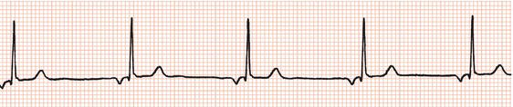
Fusion Beats. A fusion beat occurs when a supraventricular and a ventricular impulse coincide to produce a hybrid complex. It indicates that there are two foci of pacemaker cells firing simultaneously: a supraventricular pacemaker (e.g. the sinus node) and a competing ventricular pacemaker (source of ventricular ectopics). The fusion beats are...
What are the conditions for ventricular escape rhythm?
Conditions leading to the emergence of a junctional or ventricular escape rhythm include: Sinus pause / arrest (there is a single P wave visible on the 6-second rhythm strip). Broad complex escape rhythm with a LBBB morphology at a rate of 25 bpm.
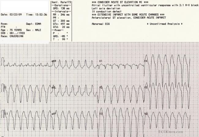
What are premature ventricular complexes (PVC)?
Premature ventricular complexes are the most common arrhythmia observed in patients without structural heart disease 1. PVCs are characterized by the premature occurrence of a wide QRS complex that is bizarre in shape.
How can I control my ventricular premature complexes?
Your doctor may also recommend that you find ways to control your stress and anxiety levels. If your ventricular premature complexes are caused by an underlying health condition, your doctor may recommend certain medications to treat it.
What is the treatment for ventricular escape rhythm?
Ouabain. Ouabain infusion decreases ventricular escape time and increases ventricular escape rhythm.
Are fusion complexes serious?
Fusion beats are typically benign but can be helpful diagnostically, such as in cases of ventricular tachycardia.
What is ventricular escape complex?
Definition. Ventricular Escape Rhythm: A ventricular rhythm with a rate of 20-40 bpm. QRS complexes are broad (≥ 120 ms) and may have a LBBB or RBBB morphology. Also known as Idioventricular escape rhythm.
Is ventricular escape rhythm shockable?
Abstract. Sudden cardiac death (SCD) is caused by lethal arrhythmia. Ventricular fibrillation (VF) and ventricular tachycardia (VT) are amenable to defibrillation or electrical shock therapy (“shockable” arrhythmia) that can abolish the VF/VT and restore normal electrical and mechanical heart function.
What causes fusion complexes on ECG?
A fusion beat occurs when a supraventricular and a ventricular impulse coincide to produce a hybrid complex. It indicates that there are two foci of pacemaker cells firing simultaneously: a supraventricular pacemaker (e.g. the sinus node) and a competing ventricular pacemaker (source of ventricular ectopics).
Are fusion beats normal?
Fusion beats are common and are characterized by a short PR interval and an intermediate QRS morphology (similar in shape to ectopic ventricular complex and the normal QRS). E. Normal, upright QRS complexes may occur if the arrhythmia originates near the septum, but the QRS duration is prolonged.
What symptoms might occur in a patient with junctional escape rhythm?
Junctional rhythm can cause symptoms due to bradycardia and/or loss of AV synchrony. These symptoms (which can be vague and easily missed) include lightheadedness, palpitations, effort intolerance, chest heaviness, neck tightness or pounding, shortness of breath, and weakness.
What is the ventricular escape rate?
The ventricular escape beats typically occur at a rate of about 40 beats/min.
What is the initial treatment for 3rd degree heart block?
Transcutaneous pacing is the treatment of choice for any symptomatic patient. All patients who have third-degree atrioventricular (AV) block (complete heart block) associated with repeated pauses, an inadequate escape rhythm, or a block below the AV node (AVN) should be stabilized with temporary pacing.
What does a ventricular escape beat look like?
1:063:00Ventricular Escape Beat ECG - EMTprep.com - YouTubeYouTubeStart of suggested clipEnd of suggested clipWith the ventricular escape beats QRS width on a ventricular escape beat is going to be wide. AndMoreWith the ventricular escape beats QRS width on a ventricular escape beat is going to be wide. And bizarre-looking and there are no dropped beats.
What are the 5 lethal cardiac rhythms?
You will learn about Premature Ventricular Contractions, Ventricular Tachycardia, Ventricular Fibrillation, Pulseless Electrical Activity, Agonal Rhythms, and Asystole. You will learn how to detect the warning signs of these rhythms, how to quickly interpret the rhythm, and to prioritize your nursing interventions.
What is the rate of ventricular escape?
Ventricular Escape Rhythm: A ventricular rhythm with a rate of 20-40 bpm.
What is the rate of a broad complex escape rhythm?
Broad complex escape rhythm with a LBBB morphology at a rate of 25 bpm.
Diagnostic Considerations
Differentiating VPCs from other arrhythmias can be challenging, because several arrhythmias may mimic VPCs, as discussed below.
Aberrant premature atrial contractions
The presence of ectopic P waves, usually absence of full compensatory pause (R-R interval containing the premature contraction is < 2 times the R-R interval of basic rhythm), and a relatively narrow QRS complex morphology help differentiate atrial premature complexes from VPCs.
Fusion beat
Simultaneous activation of the ventricle by 2 sources can lead to a beat with characteristics between the conducted sinus beat and the ectopic beat.
Premature junctional contractions
The origin of this arrhythmia is automaticity or reentry in AV junctional tissues. The P waves usually are inverted because of retrograde atrial depolarization. If the ectopic beat originates in high nodal tissue, the QRS complex can be narrow.
Idioventricular escape rhythms
A very slow pacemaker in the ventricle takes over when sinoatrial node and AV junctional pacemakers fail to function. The rate usually is less than 45 beats per minute, which helps to differentiate it from other arrhythmias.
Ventricular tachycardia
When 3 or more consecutive ventricular contractions occur, they are called VT. VT that persists for 30 seconds or causes hemodynamic collapse is called sustained VT.
Parasystole
Parasystole occurs when a protected focus discharges independently of the dominant pacemaker. The characteristics of parasystole include wide QRS complexes with a varying coupling interval between the ectopic (parasystolic) and the dominant (usually sinus) complex, fusion beats, and variable coupling interval.
What is aberrant ventricular conduction?
Aberrant Ventricular Conduction: defined as the intermittent abnormal intraventricular conduction of a supraventricular impulse. The phenomenon comes about because of unequal refractoriness of the bundle branches and critical prematurity of a supraventricular impulse (see diagram of Three Fates of PACs ). With such critical prematurity, the supraventricular impulse encounters one bundle branch (or fascicle) which is responsive, and the other which is refractory, and is consequently conducted with a bundle branch block or fascicular block pattern.
Why is it called isochronic ventricular rhythm?
Sometimes called isochronic ventricular rhythm because the ventricular rate is close to underlying sinus rate
Why is timing important for a premature wide QRS complex?
The timing of the premature wide QRS complex is also important because aberrantly conducted QRS complexes only occur early in the cardiac cycle during the refractory period of one of the conduction branches. Therefore, late premature wide QRS complexes (after the T wave, for example) are most often ventricular ectopic in origin.
What happens after a PVC?
The events following a PVC are of interest. Usually a PVC is followed by a complete compensatory pause because the sinus node timing is not interrupted ; one sinus P wave isn't able to reach the ventricles because they are still refractory from the PVC; the following sinus impulse occurs on time based on the sinus rate.
Can a PVC capture the atrium?
Finally a PVC may retrogradely capture the atrium, reset the sinus node, and be followed by an incomplete pause. Often the retrograde P wave can be seen on the ECG, hiding in the ST-T wave of the PVC.
Do all PVCs have a pause?
Not all PVCs are followed by a pause. If a PVC occurs early enough (especially if the heart rate is slow), it may appear sandwiched in between two normal beats. This is called an interpolated PVC. The sinus impulse following the PVC may be conducted with a longer PR interval because of retrograde concealed conduction by the PVC into the AV junction slowing subsequent conduction of the sinus impulse. test
Is a wide QRS tachycardia ventricular?
Differential Diagnosis: just as for single premature funny-looking beats, not all wide QRS tachycardias are ventricular in origin (i.e., they may be supraventricular tachycardias with bundle branch block or WPW preexcitation)!
What is fusion beat?
Fusion beats or fusion complexes occur when a supraventricular impulse reaches the ventricles during a ventricular beat and they coincide to produce a hybrid complex. The morphology and duration of fusion beats are usually intermediate between the morphologies and durations of two QRS complexes.
What is a premature ventricular complex?
Premature ventricular complexes reflect activation of the ventricles from a site below the AV node.
What pattern does a left ventricular ectopic pacemaker have?
As a broad general rule, the right ventricular ectopic pacemaker generates a ventricular complex with left bundle branch block (LBBB) pattern, and the left ventricular ectopic pacemaker generates a ventricular complex with right bundle branch block (RBBB) pattern 2.
Why do PVCs occur?
PVCs caused by triggered activity are often seen in patients with ventricular arrhythmias due to digoxin toxicity and reperfusion therapy after an acute myocardial infarction. The mechanism of PVCs in patients without structural heart disease is thought to be enhanced automaticity versus triggered activity.
How many sinus beats does PVC occur?
Trigeminy: PVC occurring every third beat (two sinus beats followed by a PVC).
Where is the most common site of origin for PVC in the absence of structural heart disease?
The right or left ventricular outflow tracts and aortic cusp are the most common sites of origin for PVC in the absence of structural heart disease.
Is ablation a reversal of ventricular dysfunction?
In addition to an excellent safety profile, ablation has been associated with reversal of ventricular dysfunction, but is typically reserved for drug-intolerant or medically refractory patients with a high PVC burden 4.