Treatment may include: Monitoring with MRI or CT. These tests are done to check the size and rate of growth of the aneurysm. Managing risk factors.
What does uncoiling of thoracic aorta mean?
Answered in 12 minutes by: This uncoiling usually refers to the enlargement and straightening of the thoracic aorta on the imaging test. Osteophytes throughout the dorsal spine are what are more commonly known as "bone spurs", and grow on the spinal column after years of degenerative changes.
What is the treatment for Thoracic Aneurysm?
Once formed, an aneurysm will gradually increase in size and get progressively weaker. Treatment for a thoracic aneurysm may include surgical repair or removal of the aneurysm, or inserting a metal mesh coil (stent) to support the blood vessel and prevent rupture.
What does it mean when your thoracic aorta is slightly enlarged?
It usually refers to a mild enlargement and straightening of the thoracic aorta on the imaging test. It is an indication of normal aging, atherosclerosis and hypertension. It is not a sign of anything serious unless there are other findings such as an aneurysm.
Does calcification of the aorta require surgery?
Aortic valve sclerosis — thickening and stiffness of the valve and mild aortic calcification — usually doesn't cause significant heart problems, but requires regular checkups to make sure your condition isn't worsening. If the valve becomes severely narrowed (stenotic), aortic valve replacement surgery may be necessary.

What does mild uncoiling of the thoracic aorta mean?
“Uncoiled Aorta” reflects a change of the silhouette of the thoracic aorta as seen on the X ray. It usually refers to a mild enlargement and straightening of the thoracic aorta on the imaging test. It is an indication of normal aging, atherosclerosis and hypertension.
Is unfolding of aorta serious?
Unfolding is often associated with aortic calcification which implies aortic degeneration and hypertension. Aortic unfolding, though not serious, should be differentiated from the more severe dissection of the aorta.
How is thoracic aortic aneurysm treated?
Treatment for a thoracic aneurysm may include surgical repair or removal of the aneurysm, or inserting a metal mesh coil (stent) to support the blood vessel and prevent rupture. "Thoracic" refers to the part of the aorta that runs through the chest (thoracic aortic aneurysm).
How serious is a thoracic aortic aneurysm?
A thoracic aortic aneurysm is a serious health risk because, depending on its location and size, it may rupture or dissect (tear), causing life-threatening internal bleeding. When detected in time, a thoracic aortic aneurysm can often be repaired with surgery or other less invasive techniques.
Can unfolded aorta be treated?
Most people with a thoracic aortic aneurysm have open-chest surgery, but sometimes a less-invasive procedure called endovascular surgery can be done. The type of surgery done depends on the specific health condition and the location of the thoracic aortic aneurysm. Open-chest surgery.
What are the symptoms of aorta problems?
Signs and symptoms that a thoracic aortic aneurysm has ruptured or dissected include:Sharp, sudden pain in the upper back that spreads downward.Pain in the chest, jaw, neck or arms.Difficulty breathing.Low blood pressure.Loss of consciousness.Shortness of breath.Trouble swallowing.
How long can you live with a thoracic aortic aneurysm?
Median survival was 6.6 years. The leading cause of death in this cohort was rupture of the thoracic aortic aneurysm, which accounted for 30% of the deaths. Cardiac events accounted for another 25%, along with pulmonary causes in 15%, cancer in 10%, stroke in 4%, and various other causes of death in 16%.
At what size should an aortic aneurysm be repaired?
5.5 centimetersIf the aneurysm is more than 5.5 centimeters in size, or if it's rapidly getting larger, your doctor may recommend surgery to repair the aneurysm.
What is the success rate of thoracic aortic aneurysm surgery?
Discussion. The present population-based study of primary open thoracic aortic surgery, using data from 1993 to 2010, demonstrated an overall survival rate of 86.6% at 1 year, which declined to 44.7% at 15 years.
What is the most common cause of thoracic aortic aneurysm?
Causes. The most common cause of a thoracic aortic aneurysm is hardening of the arteries. This condition is more common in people with high cholesterol, long-term high blood pressure, or who smoke.
Can aneurysms be treated with medication?
Aneurysms can be treated with medicine to slow their growth or with surgery to repair them if they are found before they rupture. An aneurysm occurs when part of an artery wall weakens.
What should you not do with an aortic aneurysm?
DON'T:Push, pull, bear down or lift anything heavier than 30 pounds (or 10 pounds for patients recovering from surgery).Get a tattoo or body piercing.Smoke (or be exposed to secondhand smoke) or use any other tobacco products.Shovel snow, chop wood, dig earth or use a sledgehammer or snow blower.Take illicit drugs.More items...•
What causes a thoracic aortic aneurysm?
Thoracic Aortic Aneurysm. (Click to Enlarge) Different disease processes can cause thoracic aortic aneurysms including: Degenerative disease that causes breakdown of the tissue of the aortic wall. Genetic disorders. Family history.
How to control aneurysms?
Managing risk factors. Steps, such as quitting smoking, controlling blood sugar if you have diabetes, losing weight if overweight, and eating a healthy diet may help control the progression of the aneurysm. Medicine. Used to control factors such as high cholesterol or high blood pressure.
How many layers are there in the aortic wall?
The aortic wall is made up of 3 layers of tissue. When a tear occurs in the innermost layer of the aortic wall, blood is then channeled into the wall of the aorta separating the layers of tissues. This generates a weakening in the aortic wall with a potential for rupture.
How to tell if you have a thoracic aneurysm?
Symptoms of a thoracic aneurysm may include: Pain in the jaw, neck, or upper back. Pain in the chest or back. Wheezing, coughing, or shortness of breath as a result of pressure on the trachea (windpipe) Hoarseness as a result of pressure on the vocal cords. Trouble swallowing due to pressure on the esophagus.
What are the diseases of the aorta?
Connective tissue disorders, such as Marfan disease, Ehlers-Danlos syndrome, and Turner syndrome. Cystic medial disease (a degenerative disease of the aortic wall) Aortitis (inflammation of the aorta) Atherosclerosis. Bicuspid aortic valve (only 2 flaps in the aortic valve, rather than the normal 3) Trauma.
What is the purpose of X-rays on the chest?
Chest X-ray. This test uses invisible electromagnetic energy beams to make images of internal tissues, bones, and organs onto film. Arteriogram (angiogram). This is an X-ray image of the blood vessels that is used to assess conditions such as aneurysm, narrowing of the blood vessel, or blockages.
Can a thoracic aneurysm cause pain?
Thoracic aortic aneurysms may not cause symptoms. When symptoms do occur, they may be related to the location, size, and how fast the aneurysm is growing. Sudden, severe pain associated with a thoracic aneurysm may be a sign of a life-threatening medical emergency. Symptoms of a thoracic aneurysm may include:
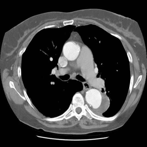
What is porcelain aorta?
In severe cases of TAC, patients may be labeled as having a porcelain aorta, which is defined practically as severe calcification that prevents safe aortic cross-clamping or cannulation (55). Because of a lack of standardization, the term has been used inconsistently, and traditionally, various assessments aided in diagnosis included chest x-ray, fluoroscopy, and manual palpation (56). More recently, CT has been used for pre-procedural planning and has facilitated a more standard definition by delineating the location and circumferential extent of atherosclerosis ( Figure 9) (57). Clinical trials in aortic stenosis (AS) have also been instrumental in this standardization. According to the Valve Academic Research Consortium-2 consensus, a porcelain aorta is defined as “heavy circumferential calcification or severe atheromatous plaque of the entire ascending aorta such that cross-clamping is not feasible” (58).
What is TAC in medical terms?
Thoracic aortic calcification (TAC) is associated with adverse cardiovascular outcomes, and for the cardiovascular imager, is predominantly encountered in 4 settings: 1) incidentally, for example, during a coronary artery calcium scan; 2) as part of dedicated screening; 3) in the evaluation of an embolic event; or 4) in procedural planning.
What are the advantages of echocardiography?
Compared with CT, the advantages of echocardiography include the lack of ionizing radiation and improved temporal resolution, which facilitates assessment of plaque mobility. In addition, unlike noncontrast CT, echocardiography more readily visualizes noncalcified and calcified atheroma. However, CT often has improved spatial resolution, and severe calcification can also confound the echocardiographic assessment due to reverberation artifact and acoustic shadowing (24). Moreover, in distinction to the Agatston method for CT (8), ultrasound physics are not readily amenable to quantification of calcification. Standard echocardiography also does not image the entire thoracic aorta. Despite these limitations, echocardiographic assessment for thoracic aortic atheroma has been used to aid in the diagnosis of stroke, inform the probability of concomitant coronary artery disease (CAD), and risk stratify for cardiovascular events 25, 26, 27.
What is a TAC?
A common imaging finding, thoracic aortic calcification (TAC) reflects systemic atherosclerosis and its attendant cardiovascular morbidity and mortality risks . Typically, TAC is encountered in 4 contexts: 1) incidentally, for example, as part of a coronary artery calcium (CAC) scan or any chest computed tomography (CT) study; 2) as part of a dedicated screening assessment in an asymptomatic patient; 3) in the evaluation of a patient with an embolic event; or 4) as a pre-procedural assessment in a patient with severe coronary or valvular heart disease. Because these assessments are often performed with CT or echocardiography, this review primarily discusses these modalities. Thoracic aortic pathology is also frequently assessed with magnetic resonance imaging (MRI), but due to signal void, calcification is not imaged. In addition, although regression of thoracic atheroma can be measured with MRI, the technique remains a research application, and is discussed elsewhere 1, 2, 3. Historically, fluoroscopy and chest x-rays have also been used for diagnosis and prognosis, but because of limited accuracy and lack of treatment implications, neither is recommended for evaluating TAC 4, 5, 6, 7, 8.
Where does TAC occur?
Within atherosclerosis, TAC is common, variable in extent, and begins in the intima with a patchy distribution. In metabolic disorders, aortitis, and radiation-associated cardiovascular disease, calcification preferentially involves the media and is often more concentric.
Can aortic calcification be measured with MRI?
Thoracic aortic pathology is also frequently assessed with magnetic resonance imaging (MRI), but due to signal void, calcification is not imaged. In addition, although regression of thoracic atheroma can be measured with MRI, the technique remains a research application, and is discussed elsewhere 1, 2, 3.
Does atherosclerosis cause plaque ulceration?
Atherosclerosis leads not only to calcification through cellular osteogenic differentiation, but can also stiffen the aorta, and depending on the relationship to neighboring lipid pools, may increase local wall stress and contribute to plaque ulceration (13).
What is aortic unfolding?
Aortic unfolding is an abnormality visible on a chest X-ray, that shows widening of the mediastinum which may mimic the appearance of a thoracic aortic aneurysm. With aging, the ascending portion of the thoracic aorta increases in length by approximately 12% per decade, whereas the diameter increases by just 3% per decade.
Is unfolding of the aorta serious?
Unfolding is often associated with aortic calcification which implies aortic degeneration and hypertension. Aortic unfolding, though not serious, should be differentiated from the more severe dissection of the aorta.
What is a calcification of the aortic valve?
Aortic valve calcification is a condition in which calcium deposits form on the aortic valve in the heart. These deposits can cause narrowing at the opening of the aortic valve. This narrowing can become severe enough to reduce blood flow through the aortic valve — a condition called aortic valve stenosis. Aortic valve calcification may be an early ...
Can aortic valve sclerosis cause heart problems?
Aortic valve sclerosis — thickening and stiffness of the valve and mild aortic calcification — usually doesn't cause significant heart problems, but requires regular checkups to make sure your condition isn't worsening. If the valve becomes severely narrowed (stenotic), aortic valve replacement surgery may be necessary.
Can aortic valve calcification be a sign of heart disease?
Aortic valve calcification may be an early sign that you have heart disease, even if you don't have any other heart disease symptoms. Calcification and stenosis generally affects people older than age 65. When it occurs in younger people, it's often caused by: