What is the prognosis for someone with retinal vein occlusion?
What Is Retinal Vein Occlusion?
- Symptoms. You may not always know that you’re going to have retinal vein occlusion. Almost always, it happens in only one eye.
- Diagnosis. Your doctor will check your eyes and ask about your medical history. ...
- Treatments. There’s no cure for retinal vein occlusion. ...
- Prevention. Usually, an underlying medical condition brings on a retinal vein occlusion. ...
What is the treatment to cure bleeding behind retina?
- Injections of VEGF inhibitors into the eye
- Removing all or part of the vitreous (vitrectomy).
- Surgical reattachment of the retina (for retinal detachment).
- Injections of corticosteroids into the eye
What are the best treatments for retinal tears?
Surgery for Retinal Detachment
- Scleral Buckle. Scleral buckle is a common surgery used to treat retinal detachment. ...
- Vitrectomy. During a vitrectomy, your doctor makes an incision in the sclera of the eye and inserts an instrument to remove the vitreous gel.
- Pneumatic Retinopexy. For certain locations of retinal detachment, our ophthalmologists may perform a pneumatic retinopexy.
Are there any cures for retinal detachment?
There are no non-surgical treatments for retinal tears or retinal detachment. Fortunately, however, many retinal detachment surgeries can be performed on an outpatient basis, with no need for general anesthesia. Depending on the extent of your injury, you are likely to need one of these surgeries performed:

Can retinal vein occlusion be treated?
Treatment for retinal vein occlusions depends on their type and severity, and can include: laser treatment, injections or a vitrectomy. A BRVO may not require any treatment and may heal itself given time. A CRVO, on the other hand, may require immediate treatment.
How serious is retinal vein occlusion?
Sometimes there is a complete loss of vision almost immediately. If these symptoms occur, it is important to schedule an appointment with your doctor as soon as possible. Retinal vein occlusion often causes permanent damage to the retina and loss of vision. It can also lead to other eye problems.
How do you fix retinal vein occlusion?
There's no cure for retinal vein occlusion. Your doctor can't unblock the retinal veins. What they can do is treat any complications and protect your vision.
Can you recover from retinal vein occlusion?
Each case of retinal vein occlusion is unique. The mild cases of vein occlusion may get better without treatment but only 1o to 20% of cases with severe occlusion may recover some vision. The majority of patients with CRVO do not recover vision and often get worse if left untreated for several months.
Can you go blind with BRVO?
Symptoms of Branch Retinal Vein Occlusion (BRVO) This swelling, known as macular edema, may require treatment. If left untreated, branch retinal vein occlusion can lead to complete and permanent vision loss / blindness in the affected eye.
How long does BRVO take to resolve?
The median time to resolution of BRVO, consisting of resolution of both macular edema and retinal hemorrhages, is 21 months in major BRVO and 18 months in macular BRVO.
Does BRVO go away?
While BRVO cannot be cured, there are effective treatments that can help patients maintain or improve their visual outcome by reducing the associated macular edema. Treatment options include intravitreal injection (injection of medicine into the eye) and laser.
What is the most common cause of retinal artery occlusion?
What causes central retinal artery occlusion (CRAO)?Cholesterol is the most common type, but it can also be from calcium, bacteria, or talc from intravenous drug use.This is associated with poorer visual acuity and higher overall morbidity and mortality.More items...•
How common is retinal vein occlusion?
Retinal vein occlusion (RVO) is the second most common sight-threatening retinal vascular disorder after diabetic retinopathy.
How do you increase blood flow to the retina?
TreatmentsEye massage. Your doctor will massage your closed eyelid with a finger to dislodge the clot.Carbon dioxide-oxygen. You breathe in a mixture of carbon dioxide and oxygen to increase blood flow to the retina. ... Paracentesis. ... Medications.
What Is Retinal Vein Occlusion?
The eye has only one vein with multiple branches, and when that vein or one of the branches is blocked, blood flow backs up and stagnates. Without...
Symptoms of Retinal Vein Occlusions
The symptoms of a retinal vein occlusion can be easy to miss at first because in some cases there may be no symptoms. Symptoms usually appear only...
What Can Cause A Retinal Vein Occlusion?
A retinal vein occlusion can happen to anyone, but it is more common in people who are over the age of 65 or who have certain medical conditions (o...
What Are The Various Types of Retinal Vein Occlusion?
Health professionals subdivide retinal vein occlusion into ischemic and nonischemic forms, but this classification is still controversial. Ischemic...
How Is Retinal Vein Occlusion Treated?
In many cases, a retinal vein occlusion is an emergency situation. Consultation with a retinal specialist is typically necessary for proper diagnos...
What Is The Usual Prognosis For Retinal Vein Occlusion?
In some cases, vision may improve spontaneously orafter treatment, but frequently a retinal vein occlusion does lasting damage. The degree to which...
Why do you need a return visit for retinal vein occlusion?
Return visits are recommended to monitor your disease progress. It is important to detect changes in your condition and formulate treatment plans as needed. It is also important to inform your primary care doctor of your retinal vein occlusion, so he or she can evaluate and treat any underlying systemic illnesses.
How do you know if you have a retinal vein occlusion?
The symptoms of retinal vein occlusion range from subtle to very obvious. There is painless blurring or loss of vision. It almost always happens in just one eye. At first, the blurring or loss of vision might be slight, but it gets worse over the next few hours or days.
What causes blurred vision and loss of vision?
Blood and fluid leaking into the macula cause swelling, a condition called macular edema, which causes blurring and/or loss of vision. Neovascularization: RVO can cause the retina to develop new, abnormal blood vessels, a condition called neovascularization.
Why is blood flow blocked in the retina?
When the flow of blood from the retina is blocked, it is often because a blot clot is blocking the retinal vein. This condition is called retinal vein occlusion (RVO). Nerve cells need a constant supply of blood to deliver oxygen and nutrients. Blood vessels provide this supply.
What are the different types of RVO?
There are two types of RVO: 1 Central retinal vein occlusion (CRVO) is the blockage of the main retinal vein. 2 Branch retinal vein occlusion (BRVO) is the blockage of one of the smaller branch veins.
What happens when a retinal vein is blocked?
When a retinal vein is blocked, it cannot drain blood from the retina. This leads to hemorrhages (bleeding) and leakage of fluid from the blocked blood vessels. There are two types of RVO: Central retinal vein occlusion (CRVO) is the blockage of the main retinal vein. Branch retinal vein occlusion ...
Why do I have a retinopathy in my eye?
Retinal vein occlusion happens when a blood clot blocks the vein. Sometimes it happens because the veins of the eye are too narrow. It is more likely to occur in people with diabetes, and possibly high blood pressure, high cholesterol levels, or other health problems that affect blood flow.
What is the test for retinal blockage?
They'll put drops in your eyes to open up your pupils. They’ll use a tool called an ophthalmoscope to check your retina for signs of blockage or bleeding. Your doctor may also order a test called a fluorescein angiography.
How long does it take for eyesight to improve?
Most people’s eyesight will get better after a few months. But some may not see any improvements. Prevention. Usually, an underlying medical condition brings on a retinal vein occlusion. So it’s important to keep your blood pressure, cholesterol, and blood sugar under control.
What causes blurry vision?
Eye Stroke. Retinal Vein Occlusion. Glaucoma Treatment. Your retina is a thin layer of tissue that lines the back of your eyeball. It turns light into signals to the brain, which interprets them as sight. When a vein in the retina becomes blocked, it’s called retinal vein occlusion. This can give you blurry vision or even sudden permanent blindness ...
Can a doctor see if you have a vein occlusion?
You may get drops to dilate your pupils and then a machine scans your eyes with rays of light to make a detailed image of your retina. There’s no cure for retinal vein occlusion.
Can a blood clot cause bleeding in the eye?
That raises pressure inside your eye, which can cause bleeding, swelling, and fluid leaks. Retinal vein occlusions can harm your eye in minutes. Usually, a blood clot blocks the vein. Sometimes, a nearby artery can be a problem. In the retina, arteries and veins cross over each other.
Can you have a retinal vein occlusion in one eye?
You may not always know that you’re going to have retinal vein occlusion. Almost always, it happens in only one eye. Some people -- especially those with blockage in smaller blood vessels -- have no symptoms.
What is the treatment for occlusion of the veins?
More controversial methods for treating vein occlusions may include heparin (dalteparin), vitrectomy (removing the vitreous jelly from the back of the eye), radial optic neurotomy (incisions in the sheath of the optic nerve), or hyperbaric oxygen.
How do you know if you have a retinal vein occlusion?
Symptoms of a retinal vein occlusion can include: Pain in the eye from increased eye pressure brought about by secondary glaucoma. Blurred vision. Loss of side vision. Visual distortions. Symptoms that worsen in hours or days.
What are the complications of ischemic retinal vein occlusion?
Complications that may occur with ischemic retinal vein occlusion include secondary glaucoma (high intraocular eye pressure) and macular edema (swelling in the retina). Symptoms such as blurry vision, eye pain, or visual disturbances should be reported to a physician right away.
What causes partial vision loss?
It is caused by a blockage in the primary vein that drains blood from the retina, or a smaller branch of this vein. Different eye care professionals treat this condition differently, but some medications ...
What are the health problems associated with retinal vein occlusion?
Some of the other health conditions associated with a retinal vein occlusion include trauma to the eye, diabetes, secondary glaucoma, and high cholesterol.
What causes blurry vision in the central vision?
Often, the blockage is associated with swelling of the retina in the central, or “macular” region (macular edema), which can cause blurring of the central vision .
What happens if your eye has only one vein?
The eye has only one vein with multiple branches, and when that vein or one of the branches is blocked , blood flow backs up and stagnates. Without regular blood flow, the cells in the retina may start to die. A retinal vein occlusion will impair sight in the affected eye and can eventually cause permanent damage.
What is retinal vein occlusion?
Retinal vein occlusion usually occurs when one of the following occurs: A retinal vein is 'pinched off' through the pressure of an artery lying on top of the vein. A retinal vein is blocked with a blood clot or fragment of fatty deposit (atherosclerotic plaque) in the wall of the artery.
Why do people with retinal vein occlusion have permanent visual problems?
This is swelling of the macula at the centre of the retina. It is the main reason why someone with retinal vein occlusion may develop permanent visual problems. Neovascularisation. This is abnormal new blood vessel formation at the back of the eye.
What happens if only one of the four branch veins is blocked?
If only one of the four branch veins is blocked, the other three will still drain blood away from the macula. There are two main types of retinal vein occlusion: Branch retinal vein occlusion - the blockage occurs somewhere along the course of one of the four retinal veins. (One retinal vein drains each quarter of the eye.)
What causes vision loss in the retina?
Retinal vein occlusion can cause very profound visual loss. This is more commonly seen in central retinal vein occlusion, which affects the whole of the retina, (including the macula where central vision is formed).
Why do retinal veins become stiff?
If the retinal arteries are narrowed due to atherosclerosis, they can become stiff and rigid. It is thought they then press on nearby veins and disturb the blood flow in them. This means that a clot is more likely to form in the vein, leading to retinal vein occlusion.
What is the name of the membrane that lines the back of the eye?
The retina is a thin, light-sensitive membrane that lines the back of your eye. An occlusion is a medical term for blockage so retinal vein occlusion means the retinal vein is blocked. This stops blood draining away from the retina and blood 'backs up' behind the blockage.
Why does my vein plug?
It happens because the vein is blocked. This blockage may occur either because of pressure on the outside of the vein (usually from a retinal artery) which squashes or kinks it, or because of sludging of fatty deposits or clotting of blood inside the vein, forming a plug.
WHAT IS A RETINAL VEIN OCCLUSION?
Veins take blood back to the heart whereas arteries take blood from the heart to the different parts of your body including the eyes. A retinal vein occlusion happens when you have a blockage of either the main vein of the retina or one of its branches. It is considered a hemorrhagic stroke of the retina as the veins start to bleed into the retina.
WHAT ARE THE RISK FACTORS FOR A RETINAL VEIN OCCLUSION?
A retinal vein occlusion most commonly occurs secondary to the changes that happen in the blood vessel walls of the arteries. As the blood vessel wall of the arteries thickens, it presses on the veins which causes changes in the blood flow within your veins which lead to blood clots (thrombosis) that block the venous flow.
WHAT ARE THE TREATMENTS FOR A RETINAL VEIN OCCLUSION?
The treatment is dependent on a detailed evaluation by your eye specialist and on the results of the recommended testing, as a wide variety of diseases can cause a retinal vein occlusion.
What is it called when the macula is occluding?
It can also result in the macula receiving inadequate blood flow. This is called macular edema. The symptoms commonly associated with branch retinal vein occlusion are due to this effect on the macula. This is also why it’s important to see a doctor if you think you might have this condition.
Why is it important to see a doctor for macula?
This is also why it’s important to see a doctor if you think you might have this condition. Poor circulation and minute amounts of swelling may still do some permanent damage. You could potentially lose much more vision in the affected eye without treatment as the macula is further affected.
What does BRVO mean in the eye?
Whether it’s due to a blood clot, narrowed blood vessels, or a thickened artery crossing over a branch vein, BRVO happens when something begins to block a branch retinal vein. Blood and other fluid can then lead out of the blocked vein, which can swell the macula (an area of the eye very important for detailed vision).
How to control BRVO?
( Learn More) Proper diet, exercise, and weight maintenance can help, as does avoiding behaviors with a negative impact on circulation, such as smoking.
What test is used to determine if you have BRVO?
Abnormal growth of new blood vessels in the retina. Once a doctor has determined if you likely have BRVO, an optical coherence tomography (OCT) test may be performed. This noninvasive imaging test uses light to make an 3D image of your eye that will be useful in your evaluation and treatment.
Can branch retinal vein occlusion cause blindness?
The condition can worsen over time and lead to blindness in the affected eye. Getting help sooner rather than later increases the chance that some of your sight may be restored. ( Learn More) The prognosis for recovery from branch retinal vein occlusion is mixed.
Can BRVO cause vision problems?
This is why any visual floaters, as discussed above, should not be ignored. Left untreated, neovascularization can lead to a host of vision problems.
Symptoms
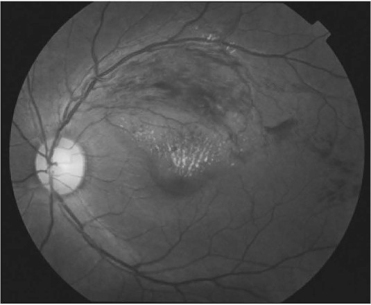
The most common symptom of branch retinal vein occlusion (BRVO) is vision loss or blurry vision in part or all of one eye. It can happen suddenly or become worse over several hours or days. Sometimes, you can lose all vision suddenly.
Who is at risk for BRVO?
Branch retinal vein occlusion (BRVO) usually happens in people who are aged 50 and older.
Diagnosis
Your ophthalmologist will widen (dilate) your pupils with eye drops and check your retina.
Treatment
With branch retinal vein occlusion (BRVO), vision usually worsens due to swelling of the macula. The main goal of treatment is to dry up the retina. In most cases, medication or laser help reduce fluid and swelling.
