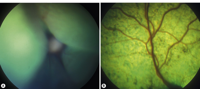
Treatment will depend upon the cause of your cat's retinal detachment. Medications can lower blood pressure to treat most cases of feline retinal detachment. Surgery may be needed to repair the damage.
What to do if your cat has a retinal detachment?
Home care 1 Administer medications as directed by your veterinarian. 2 Keep your cat indoors and restrict activity during recovery. 3 Regular follow-ups to monitor your cat’s progress and blood pressure. 4 Restrict activity until the retina has re-attached.
What is the treatment for retinal detachment?
Retinal detachment. Pneumatic retinopexy. Pneumatic retinopexy After sealing a retinal tear with cryopexy, a gas bubble is injected into the vitreous. The bubble applies gentle pressure, helping a detached section of the retina to reattach to the eyeball.
What causes retinal detachment in cats?
Feline retinal detachment usually occurs as a result of fluid build up under the retina. It's often a symptom of a more serious underlying illness. The most common cause of feline retinal detachment is high blood pressure. High blood pressure causes bleeding in the blood vessels beneath the retina.
What is the prognosis for a cat with a detached retina?
Depending on the severity of the detachment, your cat has a good prognosis for long term recovery after suffering retinal detachment. In most cases, your cat will have partial to full recovery of vision within several months.

Can retinal detachment be fixed in cats?
Retinal detachment is most frequently associated with high blood pressure, an overly active thyroid gland, or kidney disease. In some instances, prompt and proper veterinary treatment can restore partial vision to a cat with a retinal detachment, but in most cases, permanent blindness will result.
How long does it take for the retina to reattach in cats?
Retinal reattachment studies in the cat 10 and primate 12 have shown that the morphology of the RPE–retina interface does not return to normal, even after recovery periods of up to 6 months.
What is the immediate treatment for retinal detachment?
If your retina has detached, you'll need surgery to repair it, preferably within days of a diagnosis. The type of surgery your surgeon recommends will depend on several factors, including how severe the detachment is. Injecting air or gas into your eye.
Can retinal detachment be fixed on its own?
A detached retina won't heal on its own. It's important to get medical care as soon as possible so you have the best odds of keeping your vision. Any surgical procedure has some risks.
How long before retinal detachment causes blindness?
A retinal detachment may cause permanent blindness over a matter of days and should be considered an eye emergency until evaluated by a retina specialist. Most retinal detachments occur suddenly and can threaten the central vision within hours or days.
Can blind cats see light?
Some cats may still see light, in that case turning on a light when you come into the room can help as well.
What happens if retinal detachment is not treated?
If the retinal detachment isn't treated right away, more of the retina can detach — which increases the risk of permanent vision loss or blindness.
How much does a retinal detachment surgery cost?
Without insurance, the cost ranges from $5,000 to $10,000 per eye, depending on where the procedure is performed, the severity of detachment, and the expertise of your doctor. Keep in mind that surgery is the only form of treatment, making it necessary to prevent permanent vision loss.
What are the warning signs of a detached retina?
Detached retina (retinal detachment)dots or lines (floaters) suddenly appear in your vision or suddenly increase in number.you get flashes of light in your vision.you have a dark "curtain" or shadow moving across your vision.your vision gets suddenly blurred.
Is detached retina an emergency?
Seek immediate medical attention if you are experiencing the signs or symptoms of retinal detachment. Retinal detachment is a medical emergency in which you can permanently lose your vision.
Is retinal detachment painful?
There's no pain associated with retinal detachment, but there are usually symptoms before your retina becomes detached. Primary symptoms include: blurred vision. partial vision loss, which makes it seem as if a curtain has been pulled across your field of vision, with a dark shadowing effect.
What is the most common cause of retinal detachment?
Aging is the most common cause of rhegmatogenous retinal detachment. As you get older, the vitreous in your eye may change in texture and may shrink. Sometimes, as it shrinks, the vitreous can pull on your retina and tear it.
What is the best way to diagnose retinal detachment in cats?
A thorough ophthalmic examination is indicated. Some retinal detachments are easily identified, while others can be difficult to see. Your veterinarian may refer your cat to a veterinary ophthalmologist for further evaluation using specialized instrumentation.
Why does my cat have a retinal detachment?
It is a disease of older cats. High blood pressure results in fluid leakage and bleeding from blood vessels of the retina and under the retina. As fluid accumulates under the retina, the retina is pushed away from the underlying pigmented epithelium and a detachment develops.
How long does it take for a cat to go blind?
If only one eye is affected, the animal’s behavior may be normal. The onset of blindness can be gradual or rapid. In cats with detachments due to hypertension, the onset of blindness is usually very rapid (within 1 to 3 days) and involves both eyes.
How to keep a cat from changing the furniture?
Avoid changing the location of the furniture and leaving chairs or other objects out of place in the house. Your cat will memorize a familiar (stable) environment in a relatively short time.
Is retinal detachment uncommon?
Fortunately detachment of the retina is an uncommon complication of these conditions.
Can a cat go blind from a detached retina?
Therapy must be instituted as early in the disease process as possible, or the detached retina will deteriorate and the cat will be permanently blind. Treatment is usually directed at the underlying cause of the retinal detachment. The detachment itself is very difficult to treat. Depending on the physical condition of the patient, treatment options may include outpatient care or may necessitate hospitalization.
Can cats go blind?
They may be blind, however. In the event that vision cannot be saved, understand that such vision loss is not life threatening and the vast majority of cats adjust very well to their blindness.
What do you need to know about a cat's retinal detachment?
Your vet will need a complete medical history and physical exam to determine the extent of your cat's retinal detachment. A veterinary eye exam will be in order, and your cat may need to see a veterinary ophthalmologist. A range of tests, including blood tests, hormone level tests, fecal exams, urinalysis, X-rays and ultrasounds may be used ...
What tests can be done to determine if a cat has a detached retina?
A range of tests, including blood tests, hormone level tests, fecal exams, urinalysis, X-rays and ultrasounds may be used to determine the underlying cause of your cat's detached retina. Treatment will depend upon the cause of your cat's retinal detachment.
How to tell if a cat's retina is detached?
Other symptoms of detached retina in cats include dilated pupils. Your cat's pupil will slowly lose its ability to react reflexively to light as your cat's retina becomes further separated from the tissues of the inner eye. As your cat's retina becomes more detached, his pupil will dilate more slowly. When your cat loses sight altogether, the pupil will cease to dilate at all.
What is the retina in cats?
... Detached retina in cats occurs when the innermost layer of tissue in the back of the eye, or retina, detaches from the epithelium and choroid, the outermost layers.
Why does my cat have retinal detachment?
Causes of Retinal Detachment in Cats. The most common cause of feline retinal detachment is high blood pressure. High blood pressure causes bleeding in the blood vessels beneath the retina. As blood accumulates under the retina, it's pushed away from the other layers of tissue at the back of the eye. Hyperthyroidism and chronic kidney disease are ...
What are the symptoms of a cat's retina?
Symptoms of Feline Retinal Detachment. Blindness or vision loss is the most obvious symptom of detached retina in cats. The more the retina separates from the tissues of the inner eye below it, the more vision loss can occur.
Can you treat retinal detachment in cats?
Surgery may be needed to repair the damage. Some cases of detached retina in cats can't be treated. Related Links:
How to restore vision in cats?
Since high blood pressure is one cause of retinal detachment, your vet will lower your cat’s blood pressure to try to restore its vision. However, if the retinas have been detached for more than 2 days, it’s unlikely that your cat’s vision will be restored. Alternatively, your vet may recommend surgical treatments which use lasers to reattach the retina to the back of the eye. For more advice from our Veterinary co-author, including how to support your visually impaired cat at home, keep reading!
How to reattach retina to back of eye?
Consider surgery. For retinal detachment, a few surgical treatments (laser retinopexy, retinal cryopexy) are available to reattach the retina to the back of the eye. These treatments use lasers to create scar tissue on the retina; this scar tissue would act like a seal between the retina and the back of the eye. These surgical methods, unfortunately, are not always successful.
How to restore vision to a cat with high blood pressure?
Lower your cat’s blood pressure. If your cat’s retinal detachment and subsequent blindness are due to high blood pressure, then your vet will need to lower her blood pressure quickly . Lowering her blood pressure could restore her vision. However, if her retinas have been detached more than 1 or 2 days, it is unlikely that her vision will be restored.
How to tell if a cat has PRA?
Watch your cat’s movements in dark areas. The retina is made up of structures called photoreceptors—rods and cones—that allow your cat to see light and color. With PRA, the rods and cones start breaking down. Without the rods, your cat would not be able to see very well in dark areas, like a hallway. You may notice your cat avoiding dark areas of your home.#N#Inability to see in the dark is known as night blindness. It is an early sign of PRA and eventually progresses to total blindness.
Why does my cat have vision loss?
Observe how your cat navigates her environment. With retinal detachment, vision loss occurs because the retina has separated from the back of the eye. If retinal detachment has caused complete blindness in both of your cat's eyes, she’ll probably bump into walls and furniture as she walks. If she has partial retinal detachment (causing partial blindness), or is blind in only one eye, she may be able to compensate and navigate your home fairly well.
What to do if my cat doesn't have taurine?
If your cat’s current diet does not have enough taurine, talk to your vet about high-quality taurine supplements.
Why are my cat's eyes bigger than normal?
Look at your cat’s pupils. With PRA and retinal detachment, your cat’s pupils will be larger than normal. Looking through her pupils, you may be able to see a reflection of light from the backs of her eyes. Her pupils may be larger because they trying to take in more light.
Introduction
Complete or partial detachment of the neurosensory retina (nsr) from the retinal pigment epithelium (rpe). Usually bilateral.
Pathophysiology
Primary (spontaneous or traumatic) - secondary to systemic or ocular disease. Partial (focal or multifocal) - total.
Timecourse
Usually 'sudden' in onset, ie sudden objective signs but this relates to the second eye affected - the first may have a chronically detached (and thus permanently damaged) retina.
Why does my cat have a retinal detachment?
Hypertension (high blood pressure) is one of the most common causes of retinaldetachment in cats. High blood pressure causes fluid to leak from the bloodvessels behind the retina which over time causes it to separate from the underlyinglayer it is attached to (serous retinal detachment). Hypertension may be primary orsecondary (see below).
What is the goal of retina surgery?
The goal of treatment is address the underlying issue and repair the retina (if possible).Your veterinarian may refer you to a specialist eye veterinarian (ophthalmologist) toperform the surgery. There are several surgical options:
What does a veterinarian do for a cat?
Your veterinarian will perform a complete physical examination of your cat including athorough ocular examination. He will obtain a medical history from you including othersymptoms you may have noticed, known medical disorders, the age of your cat, anymedications he may be taking or exposure to toxins.
How long does it take for a cat's retina to be detached?
The prognosis is poor if the retina has been detached for more than 24 hours, in which case it may be necessary to remove the eye ( enunciation ). This is an unfortunate outcome; however, cats can adapt very well to the loss of their sight.
What is retinal detachment?
A retinal detachment (RD) is a common, severe and sight-threatening disorder that occurs when the retina (a thin layer of tissue at the back of the eye) lifts or pulls away from the retinal pigment epithelium which provides nourishment and oxygen. The retina is the thin, transparent layer of light-sensitive tissue that lines the rear (posterior) ...
What causes exudative retinal detachment?
The most common causes of exudative retinal detachment are inflammatory conditions. Traction retinal detachment occurs when the retina is pulled off the retinal pigment epithelium due to tractional forces.
What is the term for a tear in the retina?
There are three types of retinal detachment, including: Rhegmatogenous retinal detachment (RRD) occurs when there is a tear in the retina which leads to vitreous humour seeping through the tear and behind the retina, separating it from the underlying retinal pigment epithelium. Exudative (serous) retinal detachment is due to a build-up ...
How many layers of the retina are there?
The retina has nine neurosensory layers including rods and cones (responsible for vision) which are located in the innermost layer and the retinal pigment epithelium (RPE) behind the photosensitive layer. In the centre of the retina is the optic nerve, which travels from the retina to the brain where it transmits visual information.
How is a tear repaired?
The tear is then repaired by cryosurgery or laser.
What is the condition called when the eye is damaged by a previous eye operation?
This condition is known as diabetic retinopathy. Trauma: Blunt force, penetrating injury or surgical trauma from a previous eye operation. Cancers: Lymphoma, multiple myeloma (cancer of the plasma cells), tumours of the eye (most commonly melanoma or ciliary body adenocarcinoma) and aggressive metastatic cancer which can infiltrate ...
What is the retina in a dog?
Overview#N#The retina is the light-sensitive tissue that lines the inner surface of the eye. When it becomes detached from the tissue supporting it, a very serious situation exists. It is extremely important to get your pet to the veterinarian immediately if you suspect he is having vision problems.
Why do you do electrolyte tests on pets?
Electrolyte tests to ensure your pet isn’t suffering from an electrolyte imbalance
How to repair a detached retina?
The type of surgery your surgeon recommends will depend on several factors, including how severe the detachment is. Injecting air or gas into your eye.
How to prevent retinal detachment?
When a retinal tear or hole hasn't yet progressed to detachment, your eye surgeon may suggest one of the following procedures to prevent retinal detachment and preserve vision. Laser surgery (photocoagulation). The surgeon directs a laser beam into the eye through the pupil. The laser makes burns around the retinal tear, ...
What is the procedure called when you inject air into your eye?
Injecting air or gas into your eye. In this procedure, called pneumatic retinopexy (RET-ih-no-pek-see), the surgeon injects a bubble of air or gas into the center part of the eye (the vitreous cavity). If positioned properly, the bubble pushes the area of the retina containing the hole or holes against the wall of the eye, stopping the flow of fluid into the space behind the retina. Your doctor also uses cryopexy during the procedure to repair the retinal break.
What is the procedure called when you put silicone on your eye?
This procedure, called scleral (SKLAIR-ul) buckling, involves the surgeon sewing (suturing) a piece of silicone material to the white of your eye (sclera) over the affected area. This procedure indents the wall of the eye and relieves some of the force caused by the vitreous tugging on the retina.
What is the procedure called to remove the vitreous?
Draining and replacing the fluid in the eye. In this procedure, called vitrectomy (vih-TREK-tuh-me), the surgeon removes the vitreous along with any tissue that is tugging on the retina. Air, gas or silicone oil is then injected into the vitreous space to help flatten the retina.
What is the procedure to repair a retinal tear?
Surgery is almost always used to repair a retinal tear, hole or detachment. Various techniques are available. Ask your ophthalmologist about the risks and benefits of your treatment options. Together you can determine what procedure or combination of procedures is best for you.
What test is used to check for retinal bleeding?
Ultrasound imaging. Your doctor may use this test if bleeding has occurred in the eye, making it difficult to see your retina.
