What is monoclonal immunoglobulin deposition?
A plasma cell neoplasm characterized by the deposition of immunoglobulin in tissues, resulting in impaired organ function. It includes the following entities: primary amyloidosis, heavy chain deposition disease, and light chain deposition disease. Monoclonal Immunoglobulin Deposition Disease
What causes monoclonal immunoglobulin (mIg) secretion?
Abnormal proliferation of plasma cells and some monoclonal B cells frequently cause the secretion of monoclonal immunoglobulins or immunoglobulin fragments into the serum, causing monoclonal gammopathy, which leads to various diseases including renal diseases.
Is this monoclonal immunoglobulin (Ig) neoplasm reportable?
This neoplasm is not reportable. The monoclonal immunoglobulin (Ig) deposition diseases are closely related disorder s characterized by visceral and soft tissue deposition of aberrant Ig, resulting in compromised organ function.
What is the best treatment for monoclonal gammopathy?
There are cases where the underlying monoclonal gammopathy was MGUS, WM, MM, or AL amyloidosis [23,50,51]. For patients who need immediate treatment, desmopressin and factor VIII (FVIII) concentrates can improve symptoms [49]. IVIG is also an option in patients with MGUS [48].
What is monoclonal gammopathy?
Is monoclonal gammopathy associated with kidney disease?
About this website

Is light chain deposition disease curable?
There is no standard treatment for patients with LCDD.
What is immunoglobulin deposition disease?
Monoclonal immunoglobulin deposition disease (MIDD) is a rare complication of B-cell clonal disorders, defined by Congo red negative–deposits of monoclonal light chain (LCDD), heavy chain (HCDD), or both (LHCDD).
How is LCDD diagnosed?
Diagnosis of light-chain deposition disease (LCDD) LCDD is often found when a person is evaluated for proteinuria or nephrotic syndrome and a renal biopsy is performed. The tiny piece of removed kidney tissue is examined under an electron microscope and light chains deposited in the kidney can be seen.
What are symptoms of kappa light chain disease?
Symptoms of these disorders may include:Anemia.Unusual bruising or bleeding.Bone lesions.Bone pain.Fatigue.Kidney problems or kidney disease.Numbness or tingling in arms and legs.Skin rash or purple spots on your skin.More items...•
What do you mean by monoclonal immunoglobulin?
: an antibody that is derived from the clone of a single B cell and that is produced in large quantities of identical cells possessing affinity for the same epitope on a specific antigen (as a cancer cell)
What is the blood test for monoclonal gammopathy?
Monoclonal gammopathies are conditions in which abnormal proteins are found in the blood. The most common condition linked with these abnormal proteins is MGUS. MGUS causes no symptoms. Diagnosis is often done with a lab test called electrophoresis.
Is light chain myeloma worse?
LCMM has a worse prognosis than other more common MM types, so a rapid diagnosis and early treatment are vital for improving patient outcomes.
Is light chain a terminal disease?
Median survival for patients with light-chain deposition disease (LCDD) is about 4 years. The largest series published so far has reported after a median follow-up of 27 months; 57% of patients developed uremia and 59% of patients died.
Does high kappa light chain mean myeloma?
While increased numbers of either arm, called kappa or lambda light chains, is likely bad news that may ultimately mean multiple myeloma, to more accurately diagnose and monitor the condition, we need to evaluate the concentrations of each light chain differently, investigators report.
Which is worse kappa or lambda myeloma?
Patients with lambda light chain disease have a three times worse prognosis than kappa light chain disease.
What is usually the first symptom of multiple myeloma?
Often, multiple myeloma causes no symptoms until it reaches an advanced stage. Sometimes, it might cause vague symptoms that at first seem to be caused by other diseases. Sometimes, multiple myeloma is found early when a routine blood test shows an abnormally high amount of protein in the blood.
How long can you live with light chain myeloma?
Survival and prognosis The median time of survival was 23 (four to 89) months in the Velcade group and 12 (four to 67) months in the group without Velcade, respectively. The median time of PFS were nine (three to 36) months for the Velcade group and five (two to 25) months for the group without Velcade, respectively.
IgG kappa monoclonal band - does it mean myeloma?
Hello Rolls, You've got an M-spike of 0.2 g/dL and your serum immunofixation identifies the M-spike as being IgG kappa. Someone who does not have multiple myeloma or MGUS (monoclonal gammopathy of undetermined significance) does not have an M-spike, and normally would not have the serum immunofixation pick up any monoclonal protein.
My lab results say "A faint IgM (kappa) monoclonal
jbmd : There is some association of celiac disease and monoclonal gammopathy. There is something called MGUS, or monoclonal gammopathy of uncertain significance--which means that physicians can make guesses about what this means to you in the long-term but can't tell you for sure.
Monoclonal Immunoglobulin Deposition Disorder - Wikipedia
Monoclonal Immunoglobulin Deposition Disease, or MIDD, is a disease characterised by the deposition of monoclonal immunoglobulins on the basement membrane of the kidney.Monoclonal immunoglobulins are produced by monoclonal plasma cells, which are found in a variety of plasma cell dyscrasias.The deposition of monoclonal immunoglobulins on the basement membrane of the kidney causes renal impairment.
Does My Patient with a Serum Monoclonal Spike have Multiple Myeloma?
Epidemiology of MGUS. The nomenclature monoclonal gammopathy of undetermined significance (MGUS) was introduced by Kyle in 1978, and since then the fundamental characteristics, natural history and diagnostic criteria of this condition have been extensively revised. 1 According to the most current International Myeloma Working Group consensus, MGUS is defined by the simultaneous presence of ...
Igg lambda monoclonal gammopathy | Answers from Doctors | HealthTap
"i had a serum immunofixation and serum electrophoresis run and the interpretation was suspicious of monoclonal gammopathy and igg lambda. what does this mean? the beta and gamma globulin where at 0.6 and 0.7 g/dl?" Answered by Dr. Gurmukh Singh: : All laboratory results need to be interpreted in the clinical contex...
What is monoclonal gammopathy?
Abnormal proliferation of plasma cells and some monoclonal B cells frequently cause the secretion of monoclonal immunoglobulins or immunoglobulin fragments into the serum, causing monoclonal gammopathy, which leads to various diseases including renal diseases. Therefore, monoclonal gammopathy is frequently associated with kidney diseases, including glomerular and tubulointerstitial diseases. Glomerular disease, with the deposition of monoclonal immunoglobulins or their components, includes monoclonal immunoglobulin deposition disease (MIDD), AL or AH amyloidosis, type I cryoglobulinemia, proliferative glomerulonephritis with monoclonal IgG deposits (PGNMID), immunotactoid glomerulopathy, and fibrillary glomerulonephritis. In addition, tubulointerstitial diseases with the deposition of monoclonal immunoglobulins or their components are constituted by light chain (myeloma) cast nephropathy, light chain associated Fanconi's syndrome (light chain proximal [crystal] tubulopathy), and crystal-storing histiocytosis. In the present review article, we demonstrate the clinicopathological characteristics of MIDD, which is one of the representative diseases of plasma cell dyscrasias, and discuss various renal diseases with the deposition of monoclonal immunoglobulins or their components in glomeruli and the tubulointerstitium. We recommend that these renal diseases are arranged as one disease category, "renal diseases with deposition of monoclonal immunoglobulins or their components", in order to simplify the understanding of complicated diseases in plasma cell dysplasia.
Is monoclonal gammopathy associated with kidney disease?
Therefore, monoclonal gammopathy is frequently associated with kidney diseases, including glomerular and tubulointerstitial diseases. Glomerular disease, with the deposition of monoclonal immunoglobulins or their components, includes monoclonal immunoglobulin deposition disease (MIDD), AL or AH amyloidosis, type I cryoglobulinemia, ...
Symptoms and signs
When presenting to primary care physicians, patients with MIDD frequently have renal insufficiency, proteinuria and these symptoms are often accompanied by nephrotic syndrome, but patients may also present with acute renal failure.
Pathogenesis
The underlying cause of MIDD is the production of monoclonal immunoglobulins. Monoclonal immunoglobulins are produced in diseases that feature abnormal proliferation of plasma cells. These diseases include monoclonal gammopathy of undetermined significance, smoldering multiple myeloma, multiple myeloma and Waldenström's macroglobulinemia.
Diagnosis
Techniques commonly used to aid the diagnosis of MIDD include serum protein electrophoresis, urine protein electrophroresis, serum or urine immunofixation, measurement of serum free light chains and renal biopsy. Immunofluorescence is then used on the biopsied sample to detect the protein deposits on the basement membrane.
Treatment
Early treatment is recommended in MIDD to prevent or reduce irreversible kidney damage.
Epidemiology
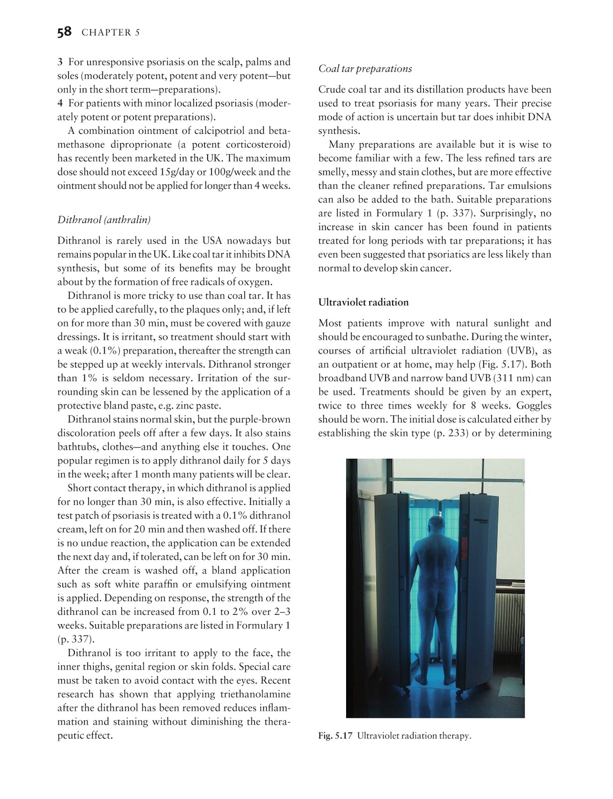
The median age of patients with MIDD is between 56 and 64 years old, and men make up two thirds of MIDD patients. Around 50% of patients with MIDD have multiple myeloma.
History
MIDD was first described in the medical literature by Kobernick et al. in 1957. They described it as "a non-amyloid kidney disease resembling diabetic glomerulosclerosis in non-diabetic myeloma patients." LCDD was further described by Randall et al. in 1976 which is why it is sometimes referred to as Randall-type MIDD. Preud'homme et al.
Further reading
"SEER Hematopoietic and Lymphoid Neoplasm Database/Monoclonal immunoglobulin deposition disease". SEER. Retrieved 29 May 2021.
Where are immunoglobulin deposits found in EBA?
Immunoelectron microscopy has shown immunoglobulin deposits in patients with EBA localized to the lamina densa zone of the basement membrane, below the lamina lucida. Standard transmission electron microscopy of lesional skin of patients with EBA shows that the anchoring fibrils are decreased or absent. These findings may be relevant in the development of the clinically evident skin fragility, as anchoring fibrils are postulated to play a role in epidermal–dermal adherence via the linkage of the hemidesmosome through the basement membrane. The lack of an inflammatory infiltrate in many patients with EBA suggests that autoantibodies may disrupt the interaction between anchoring fibrils and dermal matrix proteins.
Is MIDD a glomerular disease?
MIDD is not only a glomerular disease , and tubular lesions may be more conspicuous than the glomerular damage. Tubular lesions are characterized by the deposition of a refractile, eosinophilic, periodic acid–Schiff (PAS)–positive, ribbon-like material along the outer part of the tubular basement membrane. The deposits predominate around the distal tubules, the loops of Henle, and, in some instances, the collecting ducts, whose epithelium is flattened and atrophied. Typical myeloma casts are only occasionally seen in pure forms of MIDD. In advanced stages, a marked interstitial fibrosis including refractile deposits is frequently associated with tubular lesions.
Key Points
LCDD often features with symptomatic extrarenal involvement. LC cationic hypervariable regions possibly account for tissue deposition. Deep serum FLC response, achieved early in the disease course, predicts favorable renal and patient outcomes.
Abstract
Monoclonal immunoglobulin deposition disease (MIDD) is a rare complication of B-cell clonal disorders, defined by Congo red negative–deposits of monoclonal light chain (LCDD), heavy chain (HCDD), or both (LHCDD).
Introduction
Randall-type monoclonal immunoglobulin deposition disease (MIDD) is a rare complication of clonal B-cell disorders, defined by Congo red–negative nonorganized deposits of monoclonal immunoglobulins, usually light chains (LCDD), and less commonly, heavy chains (HCDD) or both (LHCDD), along the basement membranes of various organs.
Methods
Cases were extracted from the database from the French National Reference Center for MGRS.
Results
Two hundred fifty-five patients (median age 63.7 years, men 52%), diagnosed with LCDD (n = 212), HCDD (n = 23), or LHCDD (n = 20) between 1981 and 2015 in 55 centers in France, were included ( Table 1; Figure 1 ). The diagnosis was established on allograft biopsy in 9 patients.
Discussion
The present large series provides novel insights into the clinicopathological characteristics and prognosis of MIDD. First, it highlights that renal disease is heterogeneous, depending on the nature of deposited monoclonal immunoglobulin and underlying hematological disorder.
Authorship
Contribution: F.J. and C.C. collected data; F.J., C.C., V.J., C.S., and F.B. designed the study and wrote the manuscript; S.B. and C.S. performed the experiment from Figure 4; B.A., B.K., M.B., M.N., V.A., A.J., F.P., and J.P.F. actively corrected the manuscript and provided data; and D.N., V.G., J.M.G., and G.T. provided pathological data.
What is LCDD treatment?
Treatment of light-chain deposition disease (LCDD) is indicated for patients who present with systemic involvement, renal dysfunction, and associated presence of multiple myeloma. The goal of treatment in these patients is to suppress the production of light chains and damage to other organs. Appropriate medical management must be provided for organ dysfunction as needed, such as the use of angiotensin-converting enzyme (ACE) inhibitors or dialysis.
What is the function of Bortezomib in LCDD?
Bortezomib. In LCDD, monoclonal light chains interact with the receptors in mesangial cells and activate many pathways including the nuclear factor (NF)kB pathway. This results in increased cytokine production leading to cell proliferation and activation of genes responsible for collagen and tenascin production.
Is thalidomide an immunomodulatory drug?
Thalidomide is an immunomodulatory drug that has been extensively studied in amyloid light-chain (AL)–amyloidosis and multiple myeloma. The use in LCDD has been limited. A report in one young patient in whom conventional chemotherapy failed demonstrated that thalidomide with dexamethasone was able to provide a complete hematological response after 8 months. The patient had a sustained hematological response with improvement in renal function that lasted 31 months. [ 50]
Is plasma cell burden low in multiple myeloma?
Unlike in multiple myeloma, the plasma cell burden is quite low (< 5%) and the genetic abnormalities associated with adverse prognosis in multiple myeloma are absent. In patients with LCDD associated with multiple myeloma, the prognosis is quite poor and they should be treated per multiple myeloma guidelines. [ 38] .
What is monoclonal gammopathy?
Abnormal proliferation of plasma cells and some monoclonal B cells frequently cause the secretion of monoclonal immunoglobulins or immunoglobulin fragments into the serum, causing monoclonal gammopathy, which leads to various diseases including renal diseases. Therefore, monoclonal gammopathy is frequently associated with kidney diseases, including glomerular and tubulointerstitial diseases. Glomerular disease, with the deposition of monoclonal immunoglobulins or their components, includes monoclonal immunoglobulin deposition disease (MIDD), AL or AH amyloidosis, type I cryoglobulinemia, proliferative glomerulonephritis with monoclonal IgG deposits (PGNMID), immunotactoid glomerulopathy, and fibrillary glomerulonephritis. In addition, tubulointerstitial diseases with the deposition of monoclonal immunoglobulins or their components are constituted by light chain (myeloma) cast nephropathy, light chain associated Fanconi's syndrome (light chain proximal [crystal] tubulopathy), and crystal-storing histiocytosis. In the present review article, we demonstrate the clinicopathological characteristics of MIDD, which is one of the representative diseases of plasma cell dyscrasias, and discuss various renal diseases with the deposition of monoclonal immunoglobulins or their components in glomeruli and the tubulointerstitium. We recommend that these renal diseases are arranged as one disease category, "renal diseases with deposition of monoclonal immunoglobulins or their components", in order to simplify the understanding of complicated diseases in plasma cell dysplasia.
Is monoclonal gammopathy associated with kidney disease?
Therefore, monoclonal gammopathy is frequently associated with kidney diseases, including glomerular and tubulointerstitial diseases. Glomerular disease, with the deposition of monoclonal immunoglobulins or their components, includes monoclonal immunoglobulin deposition disease (MIDD), AL or AH amyloidosis, type I cryoglobulinemia, ...

Overview
Treatment
Early treatment is recommended in MIDD to prevent or reduce irreversible kidney damage. Treatment is directed at the underlying monoclonal gammopathy, and is intended to reduce the production of the monoclonal proteins and may include bortezomib-based treatment, an autologous stem cell transplant, and if the patient is considered eligible, an organ transplant. Bortezomib is frequently used as it is less harmful to the kidneys which may already have been d…
Symptoms and signs
When presenting to primary care physicians, patients with MIDD frequently have renal insufficiency, proteinuria and these symptoms are often accompanied by nephrotic syndrome, but patients may also present with acute renal failure. 83% of MIDD patients have renal insufficiency, and while almost all patients will have proteinuria, 40-50% will have proteinuria in the nephrotic range. Some patients may also have haematuria and hypertension but this is not found in all pati…
Pathogenesis
The underlying cause of MIDD is the production of monoclonal immunoglobulins. Monoclonal immunoglobulins are produced in diseases that feature abnormal proliferation of plasma cells. These diseases include monoclonal gammopathy of undetermined significance, smoldering multiple myeloma, multiple myeloma and Waldenström's macroglobulinemia. Almost 29% of monoclonal gammopathy patients may have MIDD. In MIDD, these abnormal immunoglobulins, …
Diagnosis
Techniques commonly used to aid the diagnosis of MIDD include serum protein electrophoresis, urine protein electrophroresis, serum or urine immunofixation, measurement of serum free light chains and renal biopsy. Immunofluorescence is then used on the biopsied sample to detect the protein deposits on the basement membrane. Increasingly, mass spectroscopy of the protein deposits may be used alongside or instead of immunohistochemical staining. This may be partic…
Epidemiology
The median age of patients with MIDD is between 56 and 64 years old, and men make up two thirds of MIDD patients. Around 50% of patients with MIDD have multiple myeloma.
MIDD is a rare disease, with incidence estimated at 1 person per million per year in Western countries.
History
MIDD was first described in the medical literature by Kobernick et al. in 1957. They described it as "a non-amyloid kidney disease resembling diabetic glomerulosclerosis in non-diabetic myeloma patients." LCDD was further described by Randall et al. in 1976 which is why it is sometimes referred to as Randall-type MIDD. Preud'homme et al. described LHCDD in 1980. Tubbs et al. described HCDD in 1992, which was grouped under the name of MIDD, first suggested by Buxba…
Further reading
• "SEER Hematopoietic and Lymphoid Neoplasm Database/Monoclonal immunoglobulin deposition disease". SEER. Retrieved 29 May 2021.