Full Answer
Is there any treatment for cornea dystrophy?
Treatment
- Medications and other therapies. Eye medication. ...
- Surgery. People who have surgery for advanced Fuchs' dystrophy can have much better vision and remain symptom-free for years afterward.
- Potential future treatments. A variety of new treatments are being investigated that could change how Fuchs' dystrophy is managed in the future.
Can corneal dystrophy be healed?
The progression of corneal dystrophy doesn’t have a reliable timeline; it can range from months to decades. Corneal dystrophy is an inherited condition, which means prevention is a mystery. The good news is that this condition is treatable. There are a number of promising medications, topical and oral, that may offset the effects of corneal dystrophy. There are even custom-designed contact lenses that improve the quality of vision while helping the eyes to heal.
When should you treat EBMD with PTK?
Successful treatment of EBMD is predicated upon optimizing conditions necessary for the formation of stable epithelial basement membrane adhesion complexes throughout the entire cornea, preferably before the development of vision-compromising morphological abnormalities in the visual axis.
What treatment options are there for corneal disease?
Treatment includes:
- Eyedrops/ointments
- Drying your swollen cornea with a hair dryer (at arm's length) two or three times a day
- Corneal transplant (full or partial)
How to find corneal dystrophy?
Most of the time your doctor will find a corneal dystrophy during a routine exam. A special tool called a slit lamp microscope let them see abnormal deposits on your cornea before you notice problems. If you have a family history of corneal dystrophy, be sure to mention it to your doctor.
What is the most common type of corneal dystrophy?
The most common type of this group is epithelial basement membrane dystrophy, also known as map-dot-fingerprint dystrophy.
What are the two layers of the cornea that are affected by corneal dystrophies?
Anterior or Superficial Corneal Dystrophies. These dystrophies affect the outer two layers of your cornea: the epithelium and the Bowman’s membrane.
What is the name of the disease that affects the front part of the eye?
By Dennis Newman. Medically Reviewed by Whitney Seltman, OD on June 17, 2020. Corneal dystrophies are a group of rare, genetic diseases that affect the cornea, the front part of your eye. There are more than 20 types, each with different symptoms. All cause a buildup of foreign material in one or more layers of your cornea.
How long does it take for a corneal dystrophia to get worse?
Keep in mind that corneal dystrophies get worse slowly. It often takes years, even de cades, before you’ll notice problems. Some people with corneal dystrophies have no symptoms.
How long does it take for dystrophies to start?
These dystrophies often start when you’re a child or teenager. Some may hurt your vision within a few years. With others, it may take decades before you’ll notice problems.
Why do doctors see dots on my cornea?
That’s because during an eye exam, your doctor may see dots, fingerprint shapes, or gray areas similar to a map on your cornea. Many people with this disease are symptom-free. These dystrophies usually affect the stroma, or center layer of your cornea. They may also progress into other layers.
What are the symptoms of corneal dystrophy?
What Is Corneal Dystrophy? 1 They tend to affect both eyes. 2 Their progression is gradual. 3 They affect only the eyes. 4 There is a hereditary connection. A family history of corneal dystrophy can suggest the development of the condition in a patient.
How to diagnose corneal dystrophy?
A routine eye examination can detect the development of corneal dystrophy. A full clinical examination is the best way to get an accurate diagnosis. This process will entail a patient history, especially determining if the patient has a family history of corneal dystrophy, and testing, like a slit lamp examination. With a slit lamp exam, a special microscope (a slit lamp) gives a doctor a high degree of magnification to study the buildup of particles and discover whether they are affecting corneal functions.
How old does a person with corneal dystrophy lose vision?
Without intervention, it may result in loss of vision by age 40. The sensitivity to light typical of corneal dystrophy is not universally felt by all people with this form of the condition, but eye pain is more common. Type 2 of this form of corneal dystrophy entails lesions developing on the stroma by age 20.
What is the purpose of eye testing for corneal dystrophy?
Regular eye testing can detect the development of corneal dystrophy and signal to a doctor whether treatment is required or if the condition is asymptomatic enough to not require intervention.
What is the term for a wide array of eye disorders that arise when particles build up on the cornea?
Corneal dystrophy is a term for a wide array of eye disorders that arise when particles build up on the cornea. Some forms of the condition are genetic. Many do not have any symptoms until later in life, when patients will experience pain, excessive tearing up, and gradual loss of vision. ( Learn More)
How many layers does the cornea have?
To better understand what corneal dystrophy is and how it works, it helps to look at the parts and functions of the cornea itself. The cornea has five layers. The epithelium is the outermost layer of the cornea, which acts as a protective shield. Bowman’s membrane, the second layer, is also used as a form of protection for the eye.
What happens when you lose your cornea?
When this happens, the cornea can lose its transparency, which in turn can cause blurred vision or a full loss of vision. While the different forms of corneal dystrophy can cause distinct symptoms, one common sign of the condition is what is known as recurrent corneal erosion.
How to know if you have a corneal dystrophy?
The only way to know for sure if you have a corneal dystrophy is to get a comprehensive dilated eye exam. Your eye doctor will use a microscope with a bright light attached (called a slit lamp) to check your eyes for signs of corneal dystrophies.
What causes corneal erosion?
Lattice dystrophy and map-dot-fingerprint dystrophy can both cause corneal erosion, when the outer layer of the cornea isn’t attached to the eye correctly and starts to erode (wear away). Treatments include eye drops, ointments, and special eye patches or contact lenses that stop your eyelid from rubbing against your cornea.
What is the difference between a normal cornea and a keratoconus?
It causes the middle and lower parts of the cornea to get thinner over time. While a normal cornea has a rounded shape, a cornea with keratoconus can bulge outward and become a cone shape. This different cornea shape can cause vision problems.
What is lattice dystrophy?
Lattice dystrophy usually begins in childhood. It causes material to build up on the cornea in a lattice (grid) pattern. As the material builds up, it can cause vision problems. Map-dot-fingerprint dystrophy (also called epithelial basement membrane dystrophy) is most common in adults ages 40 to 70. It causes a layer of the cornea ...
How to correct keratoconus?
Most people with keratoconus can correct their vision problems by wearing glasses, soft contact lenses, or special hard contact lenses that change the shape of the cornea. Your doctor may also recommend a procedure called corneal cross-linking to strengthen your cornea.
What to do if your eye is watery?
Watery eyes. Treatments include eye drops, ointments, and special eye patches or contact lenses that stop your eyelid from rubbing against your cornea. If you have severe corneal erosions or corneal scarring, you may need a surgical treatment, like laser eye surgery or a corneal transplant. Last updated: June 26, 2019.
How to tell if you have Fuchs' dystrophy?
Symptoms of Fuchs’ dystrophy include: 1 Blurry vision that’s worse in the morning and gets better later in the day 2 Glare and halos in your vision that make it hard to see things at night or in low light 3 Cloudy corneas 4 Sensitivity to light
What is corneal dystrophies?
Corneal dystrophies are a group of rare genetic eye disorders. With corneal dystrophies, abnormal material builds up in the cornea (the clear, front window of the eye). Most corneal dystrophies affect both eyes. They progress slowly and run in families.
How many types of corneal dystrophies are there?
There are more than 20 different types of corneal dystrophies. They are generally grouped into three categories: Anterior or superficial corneal dystrophies. These affect the outermost layers of the cornea: the epithelium and Bowman’s membrane. Stromal corneal dystrophies affect the stroma, which is the middle and thickest layer of the cornea.
Why is my cornea not clear?
Some people experience no symptoms. In others, the build-up of material in the cornea causes it to become opaque (not clear). This leads to blurred vision or vision loss. Many people also experience corneal erosion.
How many layers does the cornea have?
They progress slowly and run in families. The cornea has five layers: Epithelium: the outermost, protective layer of the cornea. Bowman’s membrane: this second protective layer is strong. Stroma: the thickest layer of the cornea. It is made up of water, collagen fibers and other connective tissue.
Which layer of the cornea is affected by stromal dystrophies?
Stromal corneal dystrophies affect the stroma, which is the middle and thickest layer of the cornea.
Which layer of the cornea is made up of cells that pump excess water out of the cornea?
Descemet’s membrane: a thin, strong inner layer that is also protective. Endothelium: the innermost layer made up of cells that pump excess water out of the cornea. Corneal dystrophies are caused by the build-up of foreign material in one or more of the five layers of the cornea.
Can a woman have corneal dystrophy?
Corneal dystrophies can appear at any age. Men and women are equally affected by most corneal dystrophies, except for Fuchs’ dystrophy. Fuchs’ affects women more frequently than men.
What is the treatment for corneal dystrophy?
Treatment includes eye drops and ointment, pain relievers, and medical and surgical management for severe cases.
How many types of corneal dystrophy are there?
There are over 20 types of corneal dystrophy, each affecting the corneal layers differently. Due to this, the symptoms and treatment for corneal dystrophy can vary. Treatment is very specific to the cause and severity of the disease.
What causes blurry vision in one eye?
This anterior corneal dystrophy causes scarring and clouding of Bowman’s membrane, resulting in blurry vision or double vision in one eye. It often results in painful, recurrent corneal erosions and severe visual impairment in early childhood. By 20 years of age, decreased vision and foreign body sensation are typical. It is also known as granular corneal dystrophy type III.
What is a group of over 20 corneal conditions that are slow-progressing, inherited disorders?
Corneal dystrophies are a group of over 20 corneal conditions that are slow-progressing, inherited disorders. They are non-inflammatory and result in a buildup of material in the cornea of both eyes. Typically, the dystrophy affects one layer of the cornea and progresses into the other layers.
Why does my cornea become cloudy?
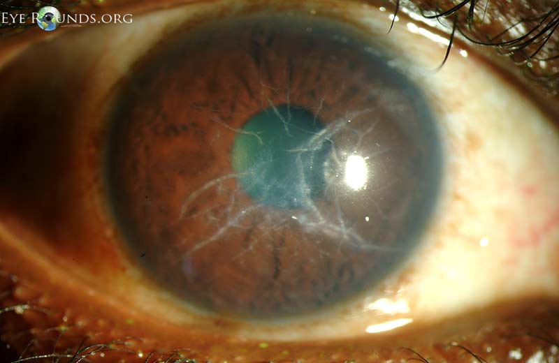
This dystrophy is due to abnormal overlapping protein fibers in the stroma (giving the appearance of a glass-like lattice). The protein fibers cause the cornea to become cloudy, decreasing vision .
What are the symptoms of Meesmann's corneal dystrophy?
Symptoms include slight irritation and a small amount of blur and decrease in visual acuity. Excessive tearing and light sensitivity are also common. Corneal clouding is not common in Meesmann corneal dystrophy.
How to treat lattice dystrophy?
The ocular symptoms of lattice dystrophy can be treated by drops and ointments. If symptoms become severe, a corneal transplant surgery may be necessary.
How to get rid of swollen cornea?
Dry your eyes with a hair dryer. Hold it at arm's length and direct warm — not hot — air across your face, especially in the morning when swelling is worse. This helps remove excess fluid in the cornea, which decreases swelling.
What test is used to measure the thickness of the cornea?
Corneal thickness. Your doctor might use a test called corneal pachymetry to measure the thickness of the cornea. Corneal tomography. Your doctor might obtain a special photograph of your cornea (tomography) to assess for early signs of swelling in your cornea. Corneal cell count.
How to diagnose Fuchs' dystrophy?
Your doctor will make the diagnosis of Fuchs' dystrophy by examining your eye with an optical microscope (slit lamp) to look for irregular bumps (guttae) on the inside surface of the cornea. He or she will then assess your cornea for swelling and stage your condition. Corneal thickness. Your doctor might use a test called corneal pachymetry ...
How to reduce glare in eyes?
In addition to following your doctor's instructions for care, you can try these techniques to help reduce glare or soothe your eyes. Apply over-the-counter (nonprescription) salt solution (5% sodium chloride) eyedrops or ointment. Dry your eyes with a hair dryer.
Is a corneal cell count required?
Sometimes your doctor might use a special instrument to record the number, shape and size of the cells that line the back of the cornea. However, this test is not required.
Can you have surgery for Fuchs's disease?
People who have surgery for advanced Fuchs' dystrophy can have much better vision and remain symptom-free for years afterward. Surgical options include:
Is Fuchs' dystrophy a nonsurgical disease?
After the genetic abnormality associated with most cases of Fuchs' dystrophy was discovered, there is a better understanding of how the disease might develop, and this offers the potential for nonsurgical therapies in the future. Various eyedrop treatments are being developed and may enter clinical trials in the future.
What is the best treatment for corneal dystrophy in dogs?
To treat corneal dystrophy, your vet may recommend a low-fat, high-fiber diet to combat cholesterol levels and various topical eye medications may also be used, but many dogs do not respond to treatment. In severe cases, surgery may be recommended but it is not without risks.
What to do if my dog has corneal dystrophy?
If you suspect your dog has developed corneal dystrophy, your veterinarian will perform a number of eye tests. They may also refer you to a veterinary ophthamologist for more advanced or specific testing as well to discuss a potential treatment plan. Eye pressure and tear production tests are often performed alongside checking for corneal ulcers and light reflexes. Visualization of the opacities in the corneas confirm diagnosis.
What is the difference between cataracts and corneal dystrophy?
Some people may confuse corneal dystrophy with cataracts, but these diseases affect different parts of the eye. Cataracts affect the lens in the eye while corneal dystrophy affects the cornea. There are three different types of corneal dystrophy and they are classified by the layer of the cornea that the disease occurs in.
How many types of corneal dystrophy are there?
There are three different types of corneal dystrophy and they are classified by the layer of the cornea that the disease occurs in.
What is the disease of the eyes that can affect dogs?
Adrienne Kruzer, BS, RVT, LVT, has worked with a variety of animals for over 15 years, including birds of prey, reptiles, and small mammals. Corneal dystrophy is a disease of the eyes that can affect dogs. Dogs of any age can develop it, but some breeds are at a higher risk than others. Corneal dystrophy can be a painful condition, ...
Why is the cornea white?
The cornea is the front, clear layer of the eye and its main job is to bend or refract light into the eye. With the development of corneal dystrophy, this clear layer becomes white and is no longer clear. This opacity is due to cholesterol or calcium deposits that build up in the cornea. Some people may confuse corneal dystrophy with cataracts, ...
Which layer of the cornea is affected by epithelial dystrophy?
Epithelial corneal dystrophy - The epithelium is the outermost or most superficial layer of the cornea and is affected in epithelial corneal dystrophy. Stromal corneal dystrophy - The stroma is the middle layer of the cornea and is affected in stromal corneal dystrophy. It is also known as macular corneal dystrophy.