Do breast calcifications mean that I have breast cancer?
While calcifications are usually harmless, they can be a sign that a woman is at risk for developing breast cancer and needs more testing. For instance, if the cluster of calcifications is tight or they are noted to present as lines of tiny calcifications, the radiologist may recommend additional mammogram images for further testing.
Should breast calcifications be removed?
If your mammogram shows microcalcifications that may suggest breast cancer, a biopsy is recommended. A biopsy means that a small area of the suspicious breast tissue is removed and examined under a microscope for cancer cells. The most common procedure after calcifications are found is a stereotactic breast core biopsy.
What if breast calcifications are cancerous?
Calcifications may be due to older age, past injury or an infection in the breast tissue. They are usually benign (not cancerous), but can be a sign of breast cancer. Your doctor will note if they have changed over time and follow-up tests may be needed to rule out cancer. How do you reduce calcification?
What causes calcifications in the breast?
Breast calcifications are often caused due to the natural aging process. As one gets old, the body goes through various changes and deterioration. The natural changes in the breast show up as breast calcifications. Over a period of time, blunt injury to the breast can lead to formation of calcifications in the breast.

How serious is calcification of the breast?
Although breast calcifications are usually noncancerous (benign), certain patterns of calcifications — such as tight clusters with irregular shapes and fine appearance — may indicate breast cancer or precancerous changes to breast tissue.
What percentage of breast calcifications are cancer?
Results. The screening sensitivity for calcifications was 45.5%, at a specificity of 99.5%. A total of 68.4% (n = 177) of cancer-related calcifications that could have been detected earlier were associated with invasive cancer when diagnosed.
What causes breast calcifications to increase?
A number of factors can cause calcification in a woman's breast, including normal aging, inflammation, and past trauma to the area. Calcium from your diet does not cause breast calcifications.
Should I worry about calcifications in breast?
Should I be worried? A: While calcifications could be a cause for concern and need further investigation, they're actually a common mammographic finding and are most often noncancerous (benign). However, additional imaging and testing is often necessary, as they could indicate cancer.
Do breast calcifications need to be removed?
They don't need to be removed and won't cause you any harm. If the calcifications look indeterminate (uncertain) or suspicious you will need further tests, as in many cases a mammogram won't give enough information.
How long does it take to recover from a stereotactic breast biopsy?
Watch for excessive bleeding, redness, skin changes, swelling or pain. Bleeding under the skin could present as a hard area (lump) that could take up to 6 weeks to resolve.
Should breast calcifications be biopsied?
Given your situation, though, your doctor should investigate any calcifications thoroughly. You may be more likely to have the area biopsied than a woman who is considered to be at average risk of breast cancer. Also, your doctor may recommend screening with breast MRI in addition to mammography.
What type of biopsy is done for breast calcifications?
Stereotactic breast biopsy is used when a small growth or an area of calcifications is seen on a mammogram, but cannot be seen using an ultrasound of the breast. The tissue samples are sent to a pathologist to be examined.
Should I have a mastectomy for DCIS?
In most cases, a woman with DCIS can choose between breast-conserving surgery (BCS) and simple mastectomy. But sometimes, if DCIS is throughout the breast, a mastectomy might be a better option. There are clinical studies being done to see if observation instead of surgery might be an option for some women.
What is the treatment for precancerous cells in the breast?
Surgery. For smaller DCIS tumors, you might get a lumpectomy, in which the abnormal cells and some breast tissue are removed. Some women decide to have a mastectomy, in which the breast is removed. After a mastectomy, you might choose to have breast reconstruction surgery.
Should I get a second opinion before breast biopsy?
Certainly a second opinion should be obtained before any definitive surgery, like a mastectomy, or a treatment with substantial side effects, such as radiation therapy or chemotherapy. People should not worry too much that a second opinion on their breast pathology will delay treatment.
How is DCIS treated?
Local treatment for DCIS usually involves breast-conserving therapy (BCT), which consists of lumpectomy (also called wide excision or partial mastectomy) followed in most cases by adjuvant radiation therapy (RT). Alternatively, mastectomy may be considered.
What is the procedure to remove calcified breast tissue?
A surgeon will perform the biopsy in an operating room under local or general anesthesia. Prior to the surgical procedure, a radiologist may use X-rays to identify the calcified breast tissue and will then mark the tissue to be removed -- with either a thin wire or with dye.
What is calcification in breast?
Breast calcifications are small calcium deposits that develop in a woman's breast tissue. They are very common and are usually benign (noncancerous). In some instances, certain types of breast calcifications may suggest early breast cancer. There are two types of breast calcifications: macrocalcifications and microcalcifications.
Can calcium cause calcification in breast?
A number of factors can cause calcification in a woman's breast, including normal aging, inflammation, and past trauma to the area. Calcium from your diet does not cause breast calcifications.
Is microcalcification a cancer?
Microcalcifications are small calcium deposits that look like white specks on a mammogram. Microcalcifications are usually not a result of cancer.
Can calcifications be seen on a mammogram?
Breast calcifications do not cause symptoms, as they are too small to be felt during a routine breast exam. Usually, breast calcifications are first noticed on a mammogram.
What are the different types of breast calcifications?
The two types of breast calcifications are microcalcifications and macrocalcifications.
How are breast calcifications diagnosed?
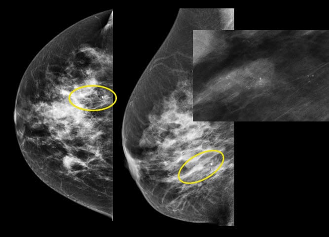
Calcifications may appear as bright white spots on mammograms. You can't feel them from the outside, so the only way to detect them may be through a mammogram.
When breast calcifications are a sign of cancer
Microcalcifications in a certain pattern may signal cancer, because when breast cells are growing and dividing, they make more calcium. So, if there’s an area of the breast where this growth is occurring, the calcium deposits would be grouped together.
What's next?
If you have microcalcifications, your doctor may order another mammogram, or a biopsy, or he or she may wait to order another mammogram after six months.
What does it mean when you see a breast calcification?
Although breast calcifications are usually noncancerous (benign), certain patterns of calcifications — such as tight clusters with irregular shapes and fine appearance — may indicate breast cancer or precancerous changes to breast tissue. On a mammogram, breast calcifications can appear as macrocalcifications or microcalcifications.
What is a calcification on a mammogram?
Breast calcifications are calcium deposits within breast tissue. They appear as white spots or flecks on a mammogram. Breast calcifications are common on mammograms, and they're especially prevalent after age 50.
Can breast calcifications be detected on a mammogram?
They're usually noncancerous, but certain patterns can be an early sign of cancer. If breast calcifications appear suspicious on your initial mammogram, you will be called back for additional magnification views to get a closer look at the calci fications.
What are the two types of breast calcifications?
The two main types of breast calcifications that can appear on a mammogram are macrocalcifications and microcalcifications . Macrocalcifications appear on the mammogram as a large round shape and are most often benign. You won’t need any additional testing or follow-up. Microcalcifications are small.
What causes calcification in breast?
Breast calcifications can form in several different ways. The most common is to form naturally as a part of the aging process. Calcification may also occur due to: 1 a noncancerous change in your breast, such as a fibroadenoma or breast cyst 2 infection 3 injury to your breast 4 surgery 5 breast implants 6 cancerous and noncancerous breast lesions
Why does calcification occur?
Calcification may also occur due to: a noncancerous change in your breast, such as a fibroadenoma or breast cyst. infection. injury to your breast. surgery. breast implants. cancerous and noncancerous breast lesions.
How often should you check for benign calcifications?
Calcifications that appear benign aren’t usually biopsied. But they should be monitored for any changes. Repeating mammograms every 6 to 12 months may be recommended to monitor benign calcifications. The radiologist will compare newer images to older images for any changes in the pattern or size of the calcifications.
Why do women get calcifications?
The most common is to form naturally as a part of the aging process. Calcification may also occur due to: a noncancerous change in your breast, such as a fibroadenoma or breast cyst.
Is breast calcification benign or noncancerous?
Most breast calcifications are noncancerous ( benign). Certain patterns of calcifications may be an indication of breast cancer. If calcifications are in tight clusters with irregular shapes, or if they grow in a line, that could indicate cancer.
Can a radiologist check for breast cancer?
If your mammogram shows breast calcifications, your radiologist may recommend other imaging tests or a biopsy. While calcifications can be benign, they can also be found in the breast in association with breast cancer. If your doctor has recommended that you get a biopsy or you wonder whether to have one, you can seek a second opinion ...