- Stage 0. Stage 0 means that the cancer is limited to the inside of the milk duct and is non-invasive. ...
- Stages I-III. Treatment for stages I to III breast cancer usually includes surgery and radiation therapy, often with chemo or other drug therapies either before (neoadjuvant) or after (adjuvant) surgery.
- Stage IV (metastatic breast cancer) Stage IV cancers have spread beyond the breast and nearby lymph nodes to other parts of the body. ...
- Recurrent breast cancer. Cancer is called recurrent when it comes back after primary treatment. ...
What treatment is the best for breast cancer?
- Oncolytics Biotech® Inc.
- Hologic, Inc.
- ImmunoGen Inc.
- BriaCell Therapeutics Corp.
Do breast calcifications ever go away?
Most cases of breast calcification do not need to be treated. On any future mammograms, the radiologist can compare the images to previous ones to determine if they have changed. If, however, one or more of the follow up tests indicate that the calcifications may be cancerous, your doctor will refer you to a doctor who specializes in cancer.
Can calcifications in the breast turn into cancer?
The majority of breast calcifications are not cancerous and they won’t turn into cancer. Instead, your doctor will try to figure out if the underlying cause is cancerous or not. If your breast calcifications are considered benign, your doctor may still recommend regular follow-up mammograms to keep an eye on possible changes.
Is the best treatment for breast cancer watchful waiting?
Very low-risk patients are those with this same criteria and also meet the following criteria:
- Stage T1C
- No more than two needles with cancer out of the standard 12-needle biopsy
- The cancer in any one needle can be no more than half of that needle.

How is breast calcification treated?
How are breast calcifications treated?Monitoring the tissue for any concerning changes.Removing the breast tissue or the entire breast.Chemotherapy and/or radiation.Targeted drug therapy.
What stage cancer are breast calcifications?
Are breast calcifications a sign of cancer? They're often benign, but calcifications can sometimes be an early sign of breast cancer. “The most common form of cancer we see with calcifications is ductal carcinoma in situ, which is considered stage 0 cancer,” Dryden says.
What happens if you have calcification in your breast?
Although breast calcifications are usually noncancerous (benign), certain patterns of calcifications — such as tight clusters with irregular shapes and fine appearance — may indicate breast cancer or precancerous changes to breast tissue.
Can you get rid of breast calcifications?
You may be recommended an operation to remove the area of calcification if it's not possible to get a biopsy of the area, or if the biopsy did not confirm a diagnosis. You may also need an operation if the biopsy results show an unusual change (called atypia), or the biopsy results show a sign of early cancer.
Should I be worried about breast calcification?
Should I be worried? A: While calcifications could be a cause for concern and need further investigation, they're actually a common mammographic finding and are most often noncancerous (benign). However, additional imaging and testing is often necessary, as they could indicate cancer.
How long does it take to recover from a stereotactic breast biopsy?
Watch for excessive bleeding, redness, skin changes, swelling or pain. Bleeding under the skin could present as a hard area (lump) that could take up to 6 weeks to resolve.
How do you treat calcification?
Treatment. People with painless joint or tendon calcification typically do not need treatment. No treatments can remove calcium deposits from the cartilage of the joints, so doctors tend to rely on glucocorticoid injections, oral colchicine, and NSAIDs to relieve any pain and underlying inflammation.
How often are breast calcifications cancerous?
Sometimes, breast calcifications are the only sign of breast cancer, according to a 2017 study in Breast Cancer Research and Treatment. The study notes that calcifications are the only sign of breast cancer in 12.7 to 41.2 percent of women who undergo further testing after their mammogram.
What causes breast calcifications to increase?
Sometimes calcifications indicate breast cancer, such as ductal carcinoma in situ (DCIS), but most calcifications result from noncancerous (benign) conditions. Possible causes of breast calcifications include: Breast cancer.
What type of biopsy is done for breast calcifications?
Stereotactic breast biopsy is used when a small growth or an area of calcifications is seen on a mammogram, but cannot be seen using an ultrasound of the breast. The tissue samples are sent to a pathologist to be examined.
Do breast calcifications need to be biopsied?
Given your situation, though, your doctor should investigate any calcifications thoroughly. You may be more likely to have the area biopsied than a woman who is considered to be at average risk of breast cancer. Also, your doctor may recommend screening with breast MRI in addition to mammography.
Can Apple cider vinegar get rid of calcium deposits?
Apple Cider Vinegar One of our stand-by treatments, apple cider vinegar is an effective option for treating calcium deposits as well. The vinegar dissolves the misplaced calcium and even restores the natural balance of nutrients in the body. Drink at least 1 tablespoon of ACV diluted in 8 ounces of water daily.
What is calcification in breast?
Breast calcifications are clusters of calcium that develop in the breast. Usually painless, they are found on routine mammograms. This condition is more common in women over age 50. Calcifications can be a sign that a woman is at risk for developing breast cancer.
What tests can be done to find calcifications in breast?
There are a number of tests that your healthcare provider can order to learn more about breast calcifications that have been found on a routine screening mammogram. These can include: Diagnostic mammogram: This is a more detailed mammogram than one that is done for routine screening.
What are the white spots on a mammogram?
Macrocalcifications : These appear as round and large bright white spots on a mammogram randomly scattered throughout the breast tissue. This is the most common type. They are typically not related to cancer and usually do not need follow up. Microcalcifications: These are smaller white spots on a mammogram.
Can calcifications show up on a mammogram?
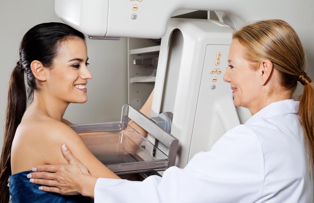
They are painless so women don’t know they have them unless they are detected by a mammogram. They are too small to feel, but can show up on a mammogram as small, bright, white spots. While calcifications are usually harmless, they can be a sign that a woman is at risk for developing breast cancer and needs more testing.
Can too much calcium cause calcification?
It is not known what causes calcifications to develop in breast tissue, but they are not caused by eating too much calcium or taking too many calcium supplements. They are seen on mammograms of about half of all women over age 50. However, they also are seen in about 10 percent of mammograms on younger women.
Is calcification cancerous?
If the calcifications are benign ( not cancerous), or probably benign, it is likely that the concerning calcifications are not cancer. Ultrasound: This is a procedure in which sound waves are used to create a picture of the breast tissue. This is noninvasive and painless.
Can a radiologist check for cancer on a mammogram?
On any future mammograms, the radiologist can compare the images to previous ones to determine if they have changed . If, however, one or more of the follow up tests indicate that the calcifications may be cancerous, your doctor will refer you to a doctor who specializes in cancer.
What is the treatment for stage IV breast cancer?
Treatment for stage IV breast cancer is usually a systemic (drug) therapy.
What is the difference between stage 2 and stage 3 breast cancer?
Stage II: These breast cancers are larger than stage I cancers and/or have spread to a few nearby lymph nodes. Stage III: These tumors are larger or are growing into nearby tissues (the skin over the breast or the muscle underneath), or they have spread to many nearby lymph nodes. Treatment of Breast Cancer Stages I-III.
What is stage 0 breast cancer?
Stage 0 means that the cancer is limited to the inside of the milk duct and is non-invasive. Treatment for this non-invasive breast tumor is often different from the treatment of invasive breast cancer. Ductal carcinoma in situ (DCIS) is a stage 0 breast tumor. Lobular carcinoma in situ (LCIS) used to be categorized as stage 0, ...
Is lobular carcinoma in situ a stage 0 tumor?
Ductal carcinoma in situ (DCIS) is a stage 0 breast tumor. Lobular carcinoma in situ (LCIS) used to be categorized as stage 0, but this has been changed because it is not cancer. Still, it does indicate a higher risk of breast cancer. See Lobular Carcinoma in Situ (LCIS) for more information.
What is the treatment for stage 1 breast cancer?
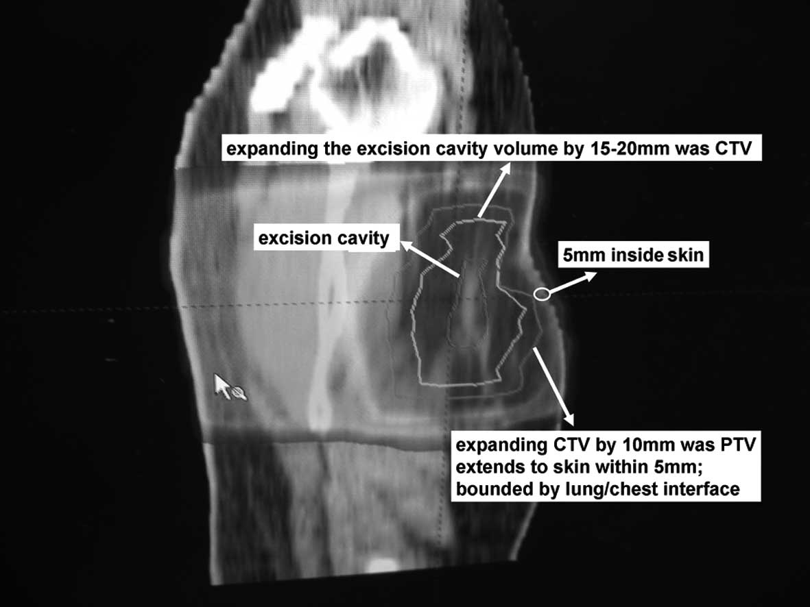
Local therapy (surgery and radiation therapy) Surgery is the main treatment for stage I breast cancer. These cancers can be treated with either breast-conserving surgery (BCS; sometimes called lumpectomy or partial mastectomy) or mastectomy.
What is the treatment for BCS?
Women who have BCS are treated with radiation therapy after surgery. Women who have a mastectomy are typically treated with radiation if the cancer is found in the lymph nodes.
What are the stages of breast cancer?
Most women with breast cancer in stages I to III will get some kind of drug therapy as part of their treatment. This may include: 1 Chemotherapy 2 Hormone therapy (tamoxifen, an aromatase inhibitor, or one followed by the other) 3 HER2 targeted drugs, such as trastuzumab (Herceptin) and pertuzumab (Perjeta) 4 Some combination of these
How big is a stage 3 breast tumor?
In stage III breast cancer, the tumor is large (more than 5 cm or about 2 inches across) or growing into nearby tissues (the skin over the breast or the muscle underneath), or the cancer has spread to many nearby lymph nodes.
Can stage 3 breast cancer spread to lymph nodes?
If you have inflammatory breast cancer: Stage III cancers also include some inflammatory breast cancers that have not spread beyond near by lymph nodes. Treatment of these cancers can be slightly different from the treatment of other stage III breast cancers.
Can you get radiation therapy before mastectomy?
If you were initially diagnosed with stage II breast cancer and were given treatment such as chemotherapy or hormone therapy before surgery, radiation therapy might be recommended if cancer is found in the lymph nodes at the time of the mastectomy.
Can you get a mastectomy with a large breast?
For women with fairly large breasts, BCS may be an option if the cancer hasn’t grown into nearby tissues. SLNB may be an option for some patients, but most will need an ALND.
What are the different types of breast calcifications?
The two types of breast calcifications are microcalcifications and macrocalcifications.
How are breast calcifications diagnosed?
Calcifications may appear as bright white spots on mammograms. You can't feel them from the outside, so the only way to detect them may be through a mammogram.
When breast calcifications are a sign of cancer
Microcalcifications in a certain pattern may signal cancer, because when breast cells are growing and dividing, they make more calcium. So, if there’s an area of the breast where this growth is occurring, the calcium deposits would be grouped together.
What's next?
If you have microcalcifications, your doctor may order another mammogram, or a biopsy, or he or she may wait to order another mammogram after six months.
What is calcification in breast?
Breast calcifications, or small calcium deposits in breast tissue, are signs of cellular turnover – essentially, dead cells – that can be visualized on a mammogram or observed in a breast biopsy. Calcifications are generally harmless and are often a result of aging breast tissue. On rare occasions, however, calcifications can be an early marker ...
What causes calcifications on mammograms?
There are a variety of causes for calcifications, including: Aging. A previous injury. Infection. Inflammation. Calcifications, unlike lumps, cannot be detected using touch.
What is the earliest form of breast cancer?
In some cases, calcifications on a mammogram represent the earliest form of breast cancer, which is called ductal carcinoma in situ (DCIS). In DCIS, the cancerous cells are in the breast’s milk ducts. DCIS is very treatable and highly curable – but in some cases, if left untreated, it has the potential to become invasive breast cancer.
What is a follow up mammogram?
The follow-up mammogram is used to take a closer look at the concerning calcifications to better determine if they are benign or in need of further testing. If deemed necessary, a biopsy will be recommended to check for underlying cancer. Most of the time, the biopsy will show that the calcification is not cancer.
What does calcification look like on a mammogram?
During a mammogram, calcifications appear as small white dots in the breast tissue. When they appear to be scattered and similar in appearance, they are usually benign (or harmless) and a biopsy or further testing is not needed.
Can breast cancer be caused by calcification?
As breast tissue ages and changes naturally, calcifications can be a normal byproduct of those changing cells. They cannot develop into cancer; rather, calcifications can be an indicator of some underlying process that involves the cancerous cells.
Is calcification on breast cancer harmless?
King, MD, FACS. Receiving the news that something abnormal has turned up on a routine mammogram can be frightening, but breast calcifications are usually harmless.
What is the survival rate of a woman with a crushed stone microcalcification?
Women with ‘ crushed stone ‘ microcalcifications, overall, tend to have a 15 year survival rate of 87% to 95%.
What are the factors that affect breast cancer staging?
Other factors traditionally associated with breast cancer staging and grading such as tumor size, nuclear features, and lymph node metastasis. Casting microcalcifications tend to be associated with tumors that have already reach a higher grade based on traditional measurements.
How high is DCIS cure rate?
DCIS has an extremely high cure rate, generally over 95%. Casting microcalcifications are perhaps the most serious indicators of the different textures frequently encountered, but their presence is not a significant prognostic indicator.
What does increased carbonate content in microcalcification mean?
Increased carbonate content in a microcalcification indicates that a cancer is growing in the viscinity.
How long does it take for a breast cancer patient to relapse?
Overall, the average relapse-free interval for patients with confirmed breast cancer associated with casting-type microcalcifications, is about 27 months.
Can casting microcalcifications cause lymph node metastasis?
Casting breast microcalcifications, when found in women who turn out to have multifocal DCIS, can often have higher incidence lymph node metastasis. Casting microcalcifications tend to be indicators of increased risk for systemic disease, and the presence of casting microcalcifications can influence adjuvant therapy decisions once the breast cancer is fully staged.
Is microcalcification a sign of breast cancer?
The presence of microcalcifications in an initial screening may or may not be indicative of acute or potential breast cancer. Research as to the predictive value of different microcalcification presentations is ongoing. However, there is reasonable evidence to suggest that of the three most common microcalcification textures, ...
