
Can a macular hole be repaired without surgery?
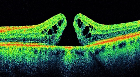
An untreated macular hole, left, and during treatment with the eye drops, right. Medicated drops may help close small macular holes over a two- to eight-week period, allowing some people to avoid surgery to fix the vision problem, a new study suggests.
How long is recovery after macular hole surgery?
The total recovery time is several months. Patients will be asked to maintain face down positioning after surgery, from one to seven days, depending on a variety of patient-specific factors. Patients are on post-operative eye drops for a few weeks. The gas bubble gradually resorbs over two to eight weeks.
Can a hole in the macula be repaired?
Vitrectomy surgery A macular hole can often be repaired using an operation called a vitrectomy. The operation is successful in closing the hole in around 9 out of 10 people who've had the hole for less than 6 months. If the hole has been present for a year or longer, the success rate will be lower.
What is the best treatment for macular holes?
Vitrectomy is the most common treatment for macular holes. In this surgical procedure, the vitreous gel is removed to stop it from pulling on the retina, and most commonly a gas bubble is placed in the eye to gently hold the edges of the macular hole closed until it heals.
Is a vitrectomy a serious operation?
Vitrectomy procedures are an effective surgery and severe complications are rare. According to the American Society of Retina Specialists, most surgeries have a 90 percent success rate.
How painful is a vitrectomy?
You might have some pain in your eye and your vision may be blurry for a few days after the surgery. You will need 2 to 4 weeks to recover before you can do your normal activities again. It may take longer for your vision to get back to normal.
How urgent is macular hole surgery?
Since macular hole surgery is never an emergency, it is sometimes worthwhile waiting a short time to see if a small macular hole will close on its own. When macular holes abort, or spontaneously close, the vision often returns all the way to normal over the course of about a year.
What is the success rate of macular hole surgery?
Anatomic success rates of macular hole surgery have been reported to be up to 89% without ILM peeling and up to 92% to 97% with peeling. This suggests that, despite ILM peeling, 3% to 8% of macular holes will remain persistently open.
How many days face down after macular hole surgery?
Thus, the results indicate that 3 days of strict face-down positioning are sufficient to achieve closure of even longstanding macular holes without removal of the ILM.
How long do I have to stay face down after vitrectomy?
Patients having vitreo-retinal surgery for a macular hole will need to posture face down for 14 days; for other conditions this is only necessary for 5 days.
What happens if you don't have surgery for macular hole?
Without prompt surgery or laser treatment, it can cause permanent vision loss. Macular pucker: Scar tissue on the macula “puckers” or wrinkles as it shrinks. If you have a macular pucker, your central vision may be distorted or blurry.
How long does vitrectomy surgery take?
A vitrectomy can take anywhere from one to several hours, depending on what condition you're treating. It may be just one in a series of procedures to repair a problem. You'll have the option to stay awake and use numbing drops or shots in your eye.
Is A Macular Hole The Same as ?
No. Macular holes and age-related macular degeneration are two separate and distinct eye conditions, although the symptoms for each are similar. Bo...
What Causes A Macular Hole?
Most of the eye’s interior is filled with vitreous, a gel-like substance that fills about 80 percent of the eye and helps it maintain a round shape...
What Are The Symptoms of A Macular Hole?
Macular holes often begin gradually. In the early stage of a macular hole, people may notice a slight distortion or blurriness in their straight-ah...
Are There Different Types of A Macular Hole?
Yes. There are three stages to a macular hole: 1. Foveal detachments (Stage I). Without treatment, about half of Stage I macular holes will progres...
How Do You Treat A Macular Hole?
Although some macular holes can seal themselves and require no treatment, surgery is necessary in many cases to help improve vision. In this surgic...
What Are The Risks of Surgery?
The most common risk following macular hole surgery is an increase in the rate of cataract development. In most patients, a cataract can progress r...
How Successful Is This Surgery?
Vision improvement varies from patient to patient. People that have had a macular hole for less than six months have a better chance of recovering...
What If I Cannot Remain in A Face-Down Position After The Surgery?
If you cannot remain in a face-down position for the required period after surgery, vision recovery may not be successful. People who are unable to...
How to tell if you have a macular hole?
In the early stage of a macular hole, people may notice a slight distortion or blurriness in their straight-ahead vision. Straight lines or objects can begin to look bent or wavy.
What causes macular holes in the eye?
Macular holes can also occur in other eye disorders, such as high myopia (nearsightedness), injury to the eye, retinal detachment , and, rarely, macular pucker.
What happens when the vitreous pulls away from the retina?
However, if the vitreous is firmly attached to the retina when it pulls away, it can tear the retina and create a macular hole. Also, once the vitreous has pulled away from the surface of the retina, ...
How does a stage 3 macular hole affect vision?
The size of the hole and its location on the retina determine how much it will affect a person’s vision. When a Stage III macular hole develops, most central and detailed vision can be lost.
What is the hole in the eye called?
A macular hole is a small break in the macula, located in the center of the eye’s light-sensitive tissue called the retina. The macula provides the sharp, central vision we need for reading, driving, and seeing fine detail. A macular hole can cause blurred and distorted central vision.
How many stages of macular holes are there?
Without treatment, about half of Stage I macular holes will progress. Partial-thickness holes (Stage II). Without treatment, about 70 percent of Stage II macular holes will progress. Full-thickness holes (Stage III). The size of the hole and its location on the retina determine how much it will affect a person’s vision.
What is the most common risk of macular hole surgery?
The most common risk following macular hole surgery is an increase in the rate of cataract development. In most patients, a cataract can progress rapidly, and often becomes severe enough to require removal.
What Causes a Macular Hole?
Age is the most common cause of macular hole. As you get older, the vitreous begins to shrink and pull away from the retina. Usually the vitreous pulls away with no problems. But sometimes the vitreous can stick to the retina. This causes the macula to stretch and a hole to form.
Macular Hole Diagnosis
Your ophthalmologist will put drops in your eye to dilate (widen) your pupil. This allows him or her to look through a special lens at the inside of your eye.
Macular Hole Treatment
Surgery called vitrectomy is the best way to treat a macular hole. Your ophthalmologist removes the vitreous that is pulling on your macula. Then he or she puts a gas bubble inside the eye. This bubble helps flatten the macular hole and hold it in place while your eye heals. The gas bubble slowly goes away on its own.
What is the best way to treat a stage 2 macular hole?
A fairly new technology called optical coherence tomography can be used to monitor you closely for possible progression into stage 2 macular hole. A Stage 2 or greater macular hole is typically treated by surgery performed by a retinal specialist.
What is a macular hole?
It sounds frightening, but a macular hole is actually a hole in the macula of your eye. 1 The macula is a highly specialized area of the central retina that gives us the ability to see fine detail. We use our macula and central vision to view detailed objects when reading or driving. Usually occurring in people over the age of 60, ...
How many stages of macular hole?
Types of Macular Holes. Macular holes are classified based on size and progression. There are four stages of a macular hole: Stage 1 (macular cyst): 1 A new macular hole sometimes appears as a yellow macular cyst. Up to 50% of macular cysts spontaneously go away and the macula returns to normal. Stage 2 (early macular hole): 1 The cyst begins ...
How much chance of developing a macular hole in one eye?
If you develop a macular hole in one eye, you have about a 30% chance of it developing one in the other eye. If you have a macular hole in one eye and the other eye has a posterior vitreous detachment, your chance of developing another macular hole begins to decrease. If you notice any change in your central vision, see your eye doctor right away.
What is a stage 3 hole?
Stage 3 (full thickness macular hole): 1 A stage 3 hole is defined by its great size. People with stage 3 holes often develop significant vision problems. Stage 3 macular holes are also defined by a rim of elevated tissue. Stage 4: A stage 4 macular hole is similar to a stage 3 but the patient also has a posterior vitreous detachment.
Can macular holes be treated?
If a macular hole is not caused by trauma or occurring along with other eye diseases, the hole can be treated with some reasonable chance of success. Larger and older macular holes have a decreased chance of successful treatment. Very early macular holes are monitored by an optometrist or an ophthalmologist.
Who monitors macular holes?
Very early macular holes are monitored by an optometrist or an ophthalmologist. Your eye doctor may use an Amsler Grid to check your central vision. 2 Your eye doctor may also dilate your eyes and take digital retinal photographs.
What is a macular hole?
Macular hole occurs when some or all of the neurons in the center of the macula are pulled out of position. Macular holes are different from age-related macular degeneration (AMD), a disease in which deposits called drusen form under the retina and the light-sensitive cells (photoreceptors) of the macula slowly break down (dry AMD), ...
Why do macular holes form?
They are caused by age-dependent contraction of the jelly in the center of the eye, called the vitreous. The vitreous is the substance that fills in the region from the lens to the back of the eye. As the vitreous shrinks, it pulls on the retina.
What is the name of the hole in the retina that causes blurring of the central vision?
A macular hole is basically what it sounds like; a hole in the central part of the retina, which is called the macula. This hole causes blurring or distortion of the central vision. Learn about the similarities and differences between macular hole and macular degeneration.
Why do you need outpatient surgery?
Others require outpatient surgery to prevent further vision loss, and, in some cases, improve vision . The surgery involves removing the vitreous jelly to stop it from pulling on the retina, and then replacing it with gas that holds the retinal in place as long as the patient is in a face-down position.
Can the vitreous separate from the retina?
In most people, the vitreous separates cleanly from the retina, but in some patients, it is tightly stuck to the retina and tears a hole. Then, fibers on the surface of the retina can pull on the hole to enlarge it.
How to treat macular holes?
A vitrectomy is the most typical treatment for macular holes. In this surgery, a retinal expert gets rid of the vitreous gel to stop it from pulling on the retina. Then the expert inserts a mixture of air and gas into the area once occupied by the vitreous.
Where are macular holes located?
The macula, where holes in some cases establish, is an extremely small spot in the center of the back of the eye (retina).
Why does my retina slosh?
The clear vitreous diminishes and becomes more liquid with aging, triggering it to slosh around. Because the vitreous is connected to the retina with small hairs of cells, it can pull on the retina as it shrinks. Often, this shrinkage can tear off a little piece of the retina, causing a hole. If this missing out on piece ...
Why do I have a hole in my macula?
Another direct reason for macular holes due to vitreous shrinking is when the hairs remain connected to the retina and break away from the vitreous .
What is the macula made of?
The macula likewise has plenty of light-sensitive cells called cones. The entire rest of the retina is made up of photosensitive cells called rods that see black and white shading, shape and movement (such as for night vision and side vision). Because macular holes frequently relate to aging processes, they are most likely to develop ...
How long does a macular bubble stay in the eye?
While the bubble is doing its job, you need to lie face down so that the bubble remains in the right place in the eye, in some cases for as long as 2 to 3 weeks!
Do women have more macular holes than men?
Likewise, women have a somewhat higher risk for macular holes than men. When a macular hole establishes, many people see an unexpected decline in vision in one eye. Macular holes, tears and cysts are not the same as another age-related eye disease called macular degeneration, which likewise occurs more frequently among those over age 60. ...
How to treat macular holes?
Traditionally, treatment for macular holes has involved a vitrectomy, an invasive eye surgery in which the transparent gel from the middle of the eye is removed ; the eye is then filled with a gas bubble. It's a fairly straightforward operation, but it still carries risk.
Who is the ophthalmologist who treats macular holes?
Ophthalmologist and retinal surgeon Dimitra Skondra, MD, Ph.D., knows that developing any new treatment can be fraught with false hopes and unexpected setbacks. She is cautiously optimistic, however, that a new method she and her colleagues are developing to treat macular holes with eye drops means some people can avoid having invasive surgery ...
Symptoms
Causes
- The back cavity of the eye is filled with a gel-like substance called vitreous. In certain places, the vitreous adheres to the retina by tiny fibers. As we age, vitreous begins to liquefy and collapse on itself. When this begins to happen, the vitreous may pull away from the retina. This is called posterior vitreous detachment. Most older adults never notice this process (which is normal) bu…
Types of Macular Holes
- Macular holes are classified based on size and progression. The four stages of a macular hole are: 1. Stage 1 (macular cyst):1A new macular hole may appear as a yellow macular cyst. Up to 50% of macular cysts spontaneously go away and the macula returns to normal. 2. Stage 2 (early macular hole):1The cyst begins to take on an oval, crescent or horseshoe shape. Vision begins t…
Treatment
- If a macular hole is not caused by trauma and doesn't occur along with other eye diseases, the hole can be treated with a reasonable chance of success. Larger and older macular holes have a decreased chance of successful treatment. Very early macular holes are monitored by an optometrist or an ophthalmologist. Your eye doctor may use an Amsler grid...
A Word from Verywell
- If you develop a macular hole in one eye, you have about a 30% chance of it developing one in the other eye. If you have a macular hole in one eye and the other eye has a posterior vitreous detachment, your chance of developing another macular hole begins to decrease. If you notice any change in your central vision, see your eye doctorright away. Early detection and treatment o…