Mounting evidence indicates that the presence of measurable (“minimal”) residual disease (MRD), defined as posttherapy persistence of leukemic cells at levels below morphologic detection, is a strong, independent prognostic marker of increased risk of relapse and shorter survival in patients with acute myeloid leukemia (AML) and can be used to refine risk-stratification and treatment response assessment.
Full Answer
What is the normal range for MRD levels in leukemia?
Jun 04, 2021 · Subject terms: Cancer genomics, Acute myeloid leukaemia, Translational research, Acute myeloid leukaemia. Introduction. Disease relapse remains the major cause of treatment failure in acute myeloid leukemia (AML) treated with allogeneic hematopoietic stem cell transplantation (allo-HSCT)1. Measurable residual disease (MRD) detection is highly valuable …
What are the MRD end points for acute myeloid leukemia (APL)?
Mar 22, 2018 · However, it should be noted that MRD tests with MRD quantified below <0.1% may still be consistent with residual leukemia, and several studies have shown prognostic significance of MRD levels below 0.1%. 12,29-32 Thus, cutoff levels below 0.1% (eg, <0.01%) may define patients with particularly good outcome.
How should MRD markers be evaluated for AML cells?
Jun 12, 2018 · Use 500 000 to 1 000 000 white blood cells, use the best aberrancy available and relate it to CD45 + white blood cells. To define “MRD-negative” and “MRD-positive” patient groups, a cutoff of 0.1% is recommended. If true MRD <0.1% is found, report this as “MRD-positive <0.1%, may be consistent with residual leukemia.”.
What is the percentage of MRD measurement Following cutoff MRD level?
May 07, 2020 · MLL monitoring. Liu et al found that any MLL expression (>0.0%) after allo-HCT was highly predictive for relapse in multivariate analysis (>90% patients relapsed, hazard ratio [HR], 18.643). 34 Most MRD positivity emerged at 3 to 5 months posttransplant, and the median time to relapse after MLL detection was 109 days.

What is normal MRD?
What is positive MRD?
Is there standard level of MRD detection?
Is MRD negative remission?
What does MRD in leukemia really mean?
How do you test for MRD?
- Flow cytometry. Flow cytometry uses a sample of bone marrow cells. ...
- PCR. PCR looks for atypical genetic characteristics in specific segments of DNA. ...
- NGS. NGS testing can quickly examine stretches of DNA or RNA to look for atypical genetic characteristics.
What is considered MRD negative?
How much does a MRD blood test cost?
Which is the most sensitive method of minimal residual disease testing in chronic myelogenous leukemia?
What is partial remission?
Is remission the same as cure?
Cure means that there are no traces of your cancer after treatment and the cancer will never come back. Remission means that the signs and symptoms of your cancer are reduced. Remission can be partial or complete.Jun 17, 2019
Is residual disease curable?
What is MRD in leukemia?
Mounting evidence indicates that the presence of measurable (“minimal”) residual disease (MRD), defined as posttherapy persistence of leukemic cells at levels below morphologic detection, is a strong, independent prognostic marker of increased risk of relapse and shorter survival in patients with acute myeloid leukemia (AML) and can be used to refine risk-stratification and treatment response assessment. Because of the association between MRD and relapse risk, it has been postulated that testing for MRD posttreatment may help guide postremission treatment strategies by identifying high-risk patients who might benefit from preemptive treatment. This strategy, which remains to be formally tested, may be particularly attractive with availability of agents that could be used to specifically eradicate MRD. This review examines current methods of MRD detection, challenges to adopting MRD testing in routine clinical practice, and recent recommendations for MRD testing in AML issued by the European LeukemiaNet MRD Working Party. Inclusion of MRD as an end point in future randomized clinical trials will provide the data needed to move toward standardizing MRD assays and may provide a more accurate assessment of therapeutic efficacy than current morphologic measures.
What is RT-qPCR used for?
RT-qPCR is used to amplify leukemia-associated genetic abnormalities. Optimized RT-qPCR assays are more sensitive than MFC, with a detection range of 10 −4 to 10 −6. 24,43 Additionally, quantitative assays that measure number of leukemic transcripts can be informative of whether transcript levels are rising or falling and can potentially inform further therapy, although benefits of MRD-directed therapy in AML are not yet firmly established. 44 Viable targets for molecular MRD monitoring include leukemic fusion genes such as promyelocytic leukemia gene retinoic acid receptor-α ( PML-RARA ), core-binding factor subunit β myosin heavy chain 11 ( CBFB-MYH11 ), and runt-related transcription factor 1 ( RUNX1)/RUNX1 translocated to 1 ( RUNX1T1 ), and mutant NPM1. 24 Wilms’ tumor gene ( WT1) should not be used as an MRD marker because of poor sensitivity and specificity unless no other MRD markers are available. 24 The ELN MRD Working Party recommends against use of Fms-like tyrosine kinase internal tandem duplication ( FLT3 -ITD), FLT3 -TKD, NRAS, KRAS, IDH1, IDH2, MLL -PTD, and expression levels of EVI1 as single markers of MRD because they are prone to frequent losses or gains; however, these mutations may have prognostic significance if accompanied by other MRD markers. 24 Given available molecular targets, RT-qPCR assessment of MRD is thought to be applicable to only ∼50% of all AML cases and less than ∼35% in older patients ( Figure 1 ), whereas MFC can detect MRD in ∼90% of patients when a comprehensive antibody panel is used. 19,28,44,46,47,50 Limitations of RT-qPCR–based MRD assays are their dependence on specific mutations, requiring individual reference standard curves based on target serial dilutions. 51 Digital PCR, a high-throughput technology that generates absolute quantification, can clonally amplify target nucleic acids and does not require a reference standard curve, has greater assay sensitivity than RT-qPCR. 52 For example, digital PCR can detect a variety of NPM1 mutation subtypes without the need for multiple plasmid standards. 53,54
What is next generation sequencing?
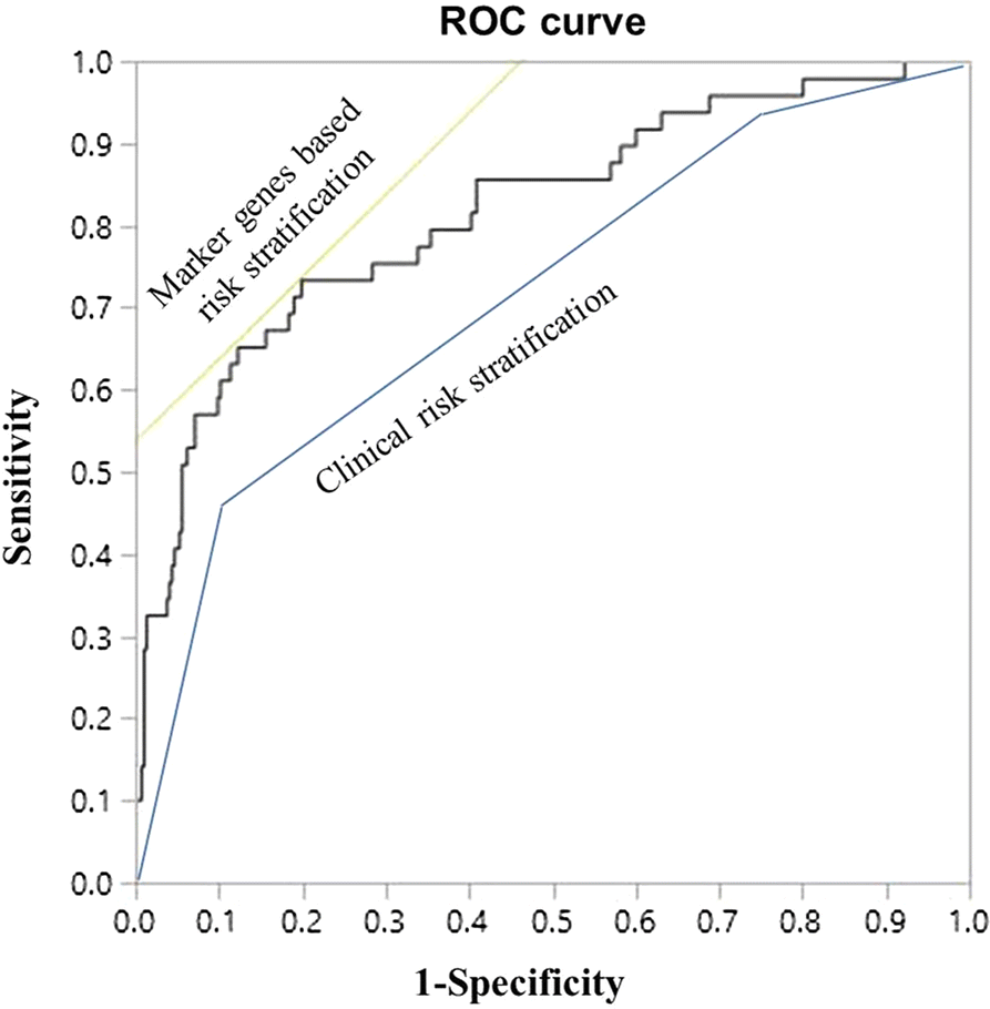
Next-generation DNA sequencing technologies, which allow parallel and repeated sequencing of millions of small DNA fragments, can be used to evaluate a few genes or an entire genome. 55 The ability of NGS to assay large numbers of mutated genes could help trace the evolution of malignant clones, which cannot be done with RT-qPCR. 15 Studies have demonstrated the feasibility of NGS to monitor mutations for which targeted therapies are available, such as FLT3 - ITD 56 and IDH1/2, 57 and mutations with prognostic relevance, such as CEBPA and NPM1 in patients with AML. 10 A recent study compared a targeted 28-gene NGS panel for detection of common AML mutations (with variant allele frequency [VAF] ≥5%) and a 10-color MFC assay of different-from-normal MRD in patients with AML before allo-HSCT. 58 Results of the 2 assays were concordant in 71% of patients. For patients in CR or CR with incomplete hematologic recovery (CRi), MRD measured by NGS was much greater than the estimated percentage of aberrant blasts detected by MFC, suggesting that residual mutations persisted in non-blast compartments during remission. Patients found to be MRD-positive with both assays had the highest risk of relapse compared with patients who were negative by both assays and with patients who had discordant assay results. 58 Similarly, in 340 patients with AML in CR or CR with CRi, there was a 69.1% concordance of MRD detection in the bone marrow using a 54-gene NGS evaluation and an MFC assay; however, persistent mutations were detected by NGS only in 64 patients. 23 Four-year relapse rate was highest among patients with MRD detected by both methods (73.3%), followed by those with MRD only on NGS (52.3%), those with MRD only on MFC (49.8%), and those who were MRD-negative on both assays (26.7%). 23
What is WT1 in leukemia?
WT1 is a nearly universal leukemia antigen that can be measured in PB but is also overexpressed in normal regenerating BM cells. Patients who do not clear their pretransplant high BM- WT1 transcripts (>250 copies) at 3 months post-allo-HCT or who show a continuous increase of PB- WT1 transcripts are at risk for relapse. 57,58 Patients with sustained low WT1 levels after allo-HCT (BM <100, PB <50 copies) had excellent outcomes. 58-60 We have recently commenced PB- WT1 testing using a commercially available European Leukemia Net–certified RT-qPCR kit as part of developing release criteria for administration of WT1- specific T cells within a forthcoming multicenter study. 61,62
Is FLT3-ITD mut subclonal?
FLT3-ITD mut is a poor MRD marker, as this mutation is typically subclonal. 35 A recently described NGS-based FLT3-ITD mut MRD assay is currently being evaluated in its ability to predict relapse after allo-HCT in the gilteritinib randomized trial mentioned above. 36
What is chimerism analysis?
Chimerism analysis detects host-derived genetic material that cannot be equated with persistence of leukemia cells. Low-level host DNA (<1%) can be found for a longer period after transplant (eg, due to recipient stromal cells), 38 bringing into question the clinical utility of the highly sensitive variant-allele–specific RT-qPCR–based or droplet digital PCR (ddPCR) techniques (sensitivity 0.1% to 0.01%) as compared with the standard STR-based PCR (sensitivity 1% to 5% according to microsatellite marker). 39-42 Fluctuating low-level mixed chimerism in PB at the early posttransplant phase may be due to viral infections and expansion of residual, recipient-derived, virus-specific T cells. 41 Chimerism patterns, kinetics, and clinical interpretation are strongly related to the transplant practice (eg, MAC vs reduced-intensity conditioning [RIC], T-cell depletion vs none) and to the underlying disease (eg, malignant vs nonmalignant). 43-45 Lineage-specific chimerism analysis may increase its specificity in predicting relapse. 46,47 Mixed chimerism in T cells is common after T-cell–depleted or RIC transplants and is weakly associated with later disease relapse. 45 A prospective study showed that the decrease of CD34 + -specific donor chimerism to <80% had 100% sensitivity and 86% accuracy in predicting relapse. This contrasts with the 14% sensitivity and 46% accuracy of conventional chimerism testing. 48 As a general rule, the speed and extent of the decrease of donor chimerism predicts relapse with a higher specificity than a static approach considering chimerism levels only at individual time points. 41 We routinely monitor chimerism by STR-PCR in PB every 2 weeks within the first 3 months and at least monthly thereafter. We additionally check monthly chimerism in ficoll-isolated mononuclear and polymorphonuclear fractions and in T cells isolated by magnetic beads. 39 BM-chimerism analysis is routinely performed on days +30, +100, +270, and +365. A continuous decrease of donor chimerism in the non–T-cell compartment or drop of donor chimerism to <80% in the magnetic-bead–isolated CD34 + subsets raises suspicion for impending relapse. Our decision making for a preemptive intervention encompasses other MRD markers and clinical parameters (eg, pretransplant risk assessment, new-onset cytopenia).
What is MRD in myeloma?
What Is Minimal Residual Disease (MRD) in Myeloma? Minimum residual disease (MRD) is a measurement of whether myeloma cells remain in the body after treatment. MRD is tested by analyzing samples of blood or bone marrow.
What is MRD used for?
MRD is being used within clinical trials for new myeloma treatments.5 Participants in clinical trials are tested during studies to find out whether one treatment produces more people with negative MRD test results (no remaining myeloma cells) over another treatment.5 The treatment that provides the most MRD-negative results may be more effective.5. ...
How to understand leukemia?
Understanding Blood Counts in Leukemia 1 Complete blood count (CBC) is a common blood test often performed for people living with leukemia. 2 If a CBC shows high or low numbers of any type of blood cell, this can help doctors better understand how your leukemia and any treatments for leukemia are affecting your body. 3 Anxiety about blood tests and waiting for results is normal, but members of MyLeukemiaTeam offer each other support.
What is the test for leukemia?
When you give a blood sample, it may be tested in the laboratory in many different ways. Common blood tests for leukemia include complete blood count (CBC), genetic analysis of cancer cells, and minimal residual disease (MRD) — a measurement of how many leukemia cells remain in the body after treatment.
What type of leukemia causes a large number of immature white cells in the blood?
Different types of leukemia can be indicated by different blood test results. Acute lymphocytic leukemia (ALL) may cause a large number of immature white cells (lymphoblasts) in the blood, as well as low numbers of red blood cells and platelets. Acute myeloid leukemia (AML) may cause pancytopenia.
What is CBC test?
Article written by. Annie Keller. Complete blood count (CBC) is a common blood test often performed for people living with leukemia. If a CBC shows high or low numbers of any type of blood cell, this can help doctors better understand how your leukemia and any treatments for leukemia are affecting your body.
What does a complete blood count show?
A complete blood count shows the current number of cells in your blood and what types of cells they are. Blood is made of three main types of cells: red blood cells, white blood cells, and platelets.
What are the three main types of blood cells?
Blood is made of three main types of cells: red blood cells, white blood cells, and platelets. Red cells (RBCs) are also referred to as erythrocytes. Their primary function is to carry oxygen from the heart and lungs to different parts of the body. White cells (WBCs) are also known as leukocytes.
What is the function of a red cell?
Red cells (RBCs) are also referred to as erythrocytes. Their primary function is to carry oxygen from the heart and lungs to different parts of the body. White cells (WBCs) are also known as leukocytes. They work as a first line of defense in the immune system, fighting bacteria and viruses that may enter the blood.
Can leukemia return?
Leukemia may return, or relapse, after apparently disappearing. Chemotherapy and targeted drugs are the primary leukemia treatments. Radiation is sometimes used, and stem cell therapy is an option for advanced leukemia that doesn't respond to treatment.
How long does chemo last for AML?
For kids with AML, she says, those who remain in remission after six months of intensive chemo don't require further treatment.
What are the different types of leukemia?
These are the most common types of leukemia: 1 Acute lymphocytic leukemia. ALL develops from lymphocytes. The leukemia cells quickly spread to the blood and sometimes to lymph nodes and bodily organs including the spleen, liver, brain and spinal cord. ALL is the most common type of leukemia in children, teens and adults under 40. 2 Acute myeloid leukemia. AML involves overproduction of a type of myeloid cells. Most AML cases occur in older adults, according to the American Society of Clinical Oncology. Although AML can be diagnosed at any age, it's uncommon in people younger than 45, with an average age of diagnosis of 68. 3 Chronic lymphocytic leukemia and chronic myeloid leukemia. Chronic forms of leukemia, CLL and CML arise later in life and gradually grow over the years. Chronic leukemia is more common in men.
How to tell if you have leukemia?
Someone with leukemia may have the following symptoms: 1 Pale appearance. 2 Weakness, fatigue or shortness of breath while playing or during activity. 3 Severe or frequent infections. 4 Recurring fever or chills. 5 Easy bruising. 6 Frequent nosebleeds. 7 Bleeding that continues for a long time, even with minor injuries or cuts. 8 Tiny red or purplish skin spots, called petechiae. 9 Bone or joint pain. 10 Swollen lymph nodes in the neck or elsewhere. 11 Reduced appetite and unintended weight loss. 12 Abdominal pain.
How many people will be diagnosed with leukemia in 2020?
An estimated 60,500 Americans will be diagnosed with leukemia in 2020, according to the Surveillance, Epidemiology and End Results program of the National Cancer Institute. Currently, the five-year relative survival rate after being diagnosed with leukemia is about 64%, according to SEER. "Relative survival" compares survival ...
Where does leukemia spread?
The leukemia cells quickly spread to the blood and sometimes to lymph nodes and bodily organs including the spleen, liver, brain and spinal cord. ALL is the most common type of leukemia in children, teens and adults under 40. Acute myeloid leukemia.
How long does it take to cure leukemia?
Cure is possible with leukemia. For ALL, Gruber says, cure is typically defined as five years of remission after diagnosis. For AML, she says, cure is typically defined as retaining remission for three years after diagnosis. Helping kids stay as healthy as possible throughout their treatment is the first step.
