In a minority of cases, surgical intervention is the only option, whereby a resection and anastomosis of the affected segment of bowel is necessary to limit bleeding. Bowel resection in patients with angiodysplasia is associated with a relatively high mortality, thus should only be considered if absolutely necessary.
What is the definitive treatment for angiodysplasia?
Surgical resection is the definitive treatment for angiodysplasia. Partial or complete gastrectomy for the management of gastric angiodysplasia has been reported to be followed by bleeding in as many as 50% of patients. Is Angiodysplasia cancerous?
What is angiodysplasia?
This results in the loss of pre-capillary sphincter competency and in turn causes the formation of small arterio-venous communications characterized by a small tuft of dilated vessels. The main features of angiodysplasia are rectal bleeding and anaemia. This typically presents in one of three ways:
What is the prognosis of angiodysplasia?
Bleeding from angiodysplasia is usually chronic and recurrent, but massive hemorrhage occurs in about 10% to 15% of affected patients. Angiodysplasias are acquired lesions associated with aging.
How is small bowel angiodysplasia treated in cirrhosis?
Treatment with continuous octreotide LAR 20mg once a month, reduces transfusion requirements in persons with diffuse small bowel angiodysplasia. The use of short-acting SMS analogues is recognized in acute variceal hemorrhage secondary to portal hypertension in cirrhosis.
What is Angiodysplastic lesion?
Angiodysplasia is an important vascular lesion of the gut and a source of significant morbidity from bleeding. This lesion is probably responsible for approximately 6.0% of cases of lower gastrointestinal (GI) bleeding and 1.2-8.0% of cases of hemorrhage from the upper GI tract.
What is angioectasias?
Angioectasia are an acquired vascular malformation associated with advanced age. Pathogenesis of colonic angioectasia formation is multifactorial and commonly attributed to mild chronic venous obstruction and to chronic mucosal hypoxemia resulting in increased vascular endothelial growth factor (VEGF) expression 3 .
What is angiodysplasia associated with?
Angiodysplasia has been reported to be associated with aortic stenosis. Heyde first reported this association in 1958, describing Heyde syndrome as the combination of calcific aortic stenosis and GI bleeding due to angiodysplasia of the colon.
What causes angiodysplasia of colon?
Causes. Angiodysplasia of the colon is mostly related to the aging and breakdown of the blood vessels. It is more common in older adults. It is almost always seen on the right side of the colon.
What is the treatment for angiodysplasia?
Surgical resection is the definitive treatment for angiodysplasia. Partial or complete gastrectomy for the management of gastric angiodysplasia has been reported to be followed by bleeding in as many as 50% of patients. Rebleeding was attributed to other angiodysplastic lesions.
What is Angioectasias in the distal rectum?
Angioectasia is characterized by focal accumulation of dilated vessels in the mucosa and submucosa of the intestinal wall[1]. This condition can occur anywhere in the gastrointestinal (GI) tract, and most commonly occurs in the colon[2,3]; however, 15% of cases are thought to be located in the small bowel[4].
Can angiodysplasia heal itself?
Treatment options for angiodysplasia Sometimes, bleeding caused by angiodysplasia stops on its own without medical intervention. But you may require treatment to control bleeding and reverse anemia. Treatment depends on the severity of the condition and whether you have anemia.
Is angiodysplasia an AVM?
Angiodysplasias (also known as arteriovenous malformations, or AVMs) account for less than 10% of all cases of hematochezia, but may be the most common cause of lower GI bleeding in patients older than 65. Colonic AVMs are found in less than 1% of the population and are usually asymptomatic.
What causes bleeding blood vessels in the stomach?
Angiodysplasia is when you have abnormal or enlarged blood vessels in your GI tract. These blood vessels can become fragile and bleed. Benign tumors and cancer. Benign tumors link and cancer link in the esophagus, stomach, colon, or rectum may cause bleeding when they weaken the lining of the GI tract.
Is angiodysplasia seen on colonoscopy?
Angiodysplasia is usually diagnosed as an incidental finding during colonoscopy for colorectal cancer screening exams or when evaluating the patient for acute or chronic blood loss related anemia. The initial diagnostic modality depends on the characteristics of bleeding and suspicion for the location of the source.
What is acquired angiodysplasia?
Acquired angiodysplasia – begins as reduced submucosal venous drainage in the colon due to chronic and intermittent contraction of the colon, giving rise to dilated and tortuous veins. This results in the loss of pre-capillary sphincter competency and in turn causes the formation of small arterio-venous communications characterized by a small tuft of dilated vessels.
What imaging is used for small bowel angiodysplasia?
Figure 2 – Capsule endoscopy is the preferred imaging choice for small bowel angiodysplasia.
What is angiography for GI?
Angiography can involve either radionuclide scanning, CT scanning, or MRI scanning to image the GI tract vascular supply after the injection of a radio-opaque contrast agent into the vessels. 10% of patients with angiodysplasia will present with a major GI bleed, for which patients should be managed accordingly.
What is the most common vascular abnormality of the gastrointestinal tract?
Angiodysplasia is the most common vascular abnormality of the gastrointestinal tract, responsible for approximately 6% of lower GI bleeding cases and up to 8% of upper GI bleeds. It is caused by the formation of arteriovenous malformations between previously healthy blood vessels, most commonly in the caecum and ascending colon.
When is mesenteric angiography indicated?
Mesenteric angiography may also be indicated in GI bleeding from any other location when en doscopic therapy has failed (to treat or localise) or when endoscopy is not a suitable option (for example in patients who are not fit for endoscopy). The sensitivity of angiography ranges from 58-86% and increases depending on the bleeding rate of the lesion.
What is the first line of treatment for a bleeding site?
In persistent or severe cases, when bleeding sites are identified, there are two main methods of treatment; endoscopic therapies and radiographic therapies (mesenteric angiography): Endoscopy – usually the first line of management, with most widely used technique being argon plasma coagulation.
What blood tests are ordered for GI bleeding?
Blood tests will be usually ordered as part of the routine assessment for any patient presenting with GI bleeding, including a FBC*, U&Es, LFTs, and clotting. Depending on the clinical picture, a Group and Save or Crossmatch may be warranted if there is a potential need for transfusion.
What is the treatment for angiodysplasia?
Actively bleeding angiodysplasia- In most patient’s management should follow a similar path as in managing upper and lower GI bleeding for other reasons, which is hemodynamic resuscitation, frequent complete blood count monitoring, and blood transfusion if needed. Treatment decisions should be dependent on hemodynamic stability and whether the patient is actively bleeding or not.
What is angiodysplasia in the GI tract?
Angiodysplasia is an abnormal, tortuous, dilated small blood vessel in the mucosal and sub mucosal layers of the GI tract. It is the most common vascular abnormality in the GI tract. Although usually readily seen by colonoscopy and angiography, they are often difficult to diagnose in pathologic specimens. This activity reviews the evaluation, histopathology, and treatment of angiodysplasia. It explains the role of the interprofessional team in managing patients with this condition.
What causes GI bleeding in end stage renal failure?
End-stage renal disease - Peptic ulcer disease remains the most common cause of GI bleeding in end-stage renal disease patients. Still, angiodysplasia is another sizeable cause of both upper and lower GI bleeding in these patients and accounts for almost 20% to 30%, respectively. According to one report, one-half of patients with recurrent bleeding in chronic renal failure were because of angiodysplasia. [14][15]
What is the most common vascular abnormality in the GI tract?
In the general population, the most common vascular abnormality in the GI tract is angiodysplasia, which mainly occurs in patients over 60 years of age. [8][9]The prevalence of angiodysplasia increases with age. It may have no symptoms or present with GI bleeding.[10] There are reports of non-variceal upper GI bleeding caused by angiodysplasia in approximately 5% to 10% of patients.[11] Small bowel angiodysplasia is the most common cause of obscure GI bleeding (OGIB) in patients older than 50 years old. In contrast, small bowel tumors are the common cause in patients under 50 years old with OGIB[3][12]. The most common site of angiodysplasia in the GI tract is the colon.[13] Angiodysplasia has been reported to be associated more with some conditions in literature such as end-stage renal disease (ESRD), Von Willebrand disease, left ventricular assist device (LVAD), and aortic stenosis (Heyde syndrome).
Can angiodysplasia be detected on a colonoscopy?
Although usually readily apparent on colonoscopy and angiography, angiodysplasia is often extremely difficult to detect on the gross examination of a resected specimen without the use of specific injection techniques. As a result of vascular injection studies of resected colons, the determination was that angiodysplasia develops as a result of intermittent partial obstruction of small veins that drain the colonic mucosa and submucosa as they course through the muscularis propria. Over time, obstruction of the penetrating veins of the muscularis propria leads to dilation and tortuosity of the submucosal veins and, consequently, the venules and capillaries that drain them.
Can angiodysplasia be seen on mucosal surface?
In specimens examined after formalin fixation, the lesions are usually not visible on the mucosal surface. If a vascular lesion is detected, the histologic examination typically reveals a discrete cluster of dilated, tortuous veins and venules within the submucosa, some associated with dilated capillaries in the overlying mucosa as well. [28]
Can angiodysplasia cause a GI bleed?
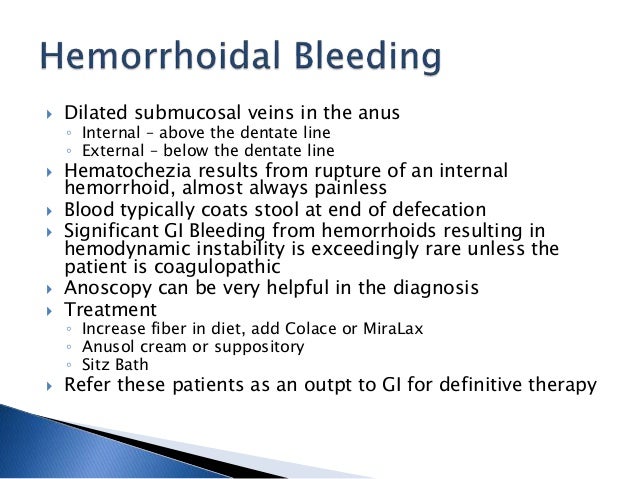
Patients with angiodysplasia may be asymptomatic or present with mild to moderate occult lower GI bleeding without abdominal pain. History should include all the same elements as it would in the evaluation of upper and lower GI bleeding. Patients may present with occult blood in stool and iron deficiency anemia. Physical examination in a hemodynamic patient may show symptoms and signs of anemia. A systolic ejection murmur during the cardiac examination may raise suspicion for underlying aortic stenosis. Angiodysplasia lesions may be found incidentally in endoscopy for other reasons. Orthostasis or hypotension may rarely occur in acute and heavy bleeding. Although bleeding stops spontaneously in most patients, it may recur. Angiodysplasia-related GI bleeding can happen anywhere in the GI tract but more frequently seen in the cecum, rectosigmoid area, the rest of the colon, small intestine, and stomach in descending order. [29]
What is the definitive treatment for angiodysplasia?
Surgical resection is the definitive treatment for angiodysplasia.
How long does colonic angiodysplasia last after laser obliteration?
Patients with colonic angiodysplasia generally have a 60% chance of remaining free of bleeding at 24 months after laser obliteration.
What is hemodynamically stabilized?
Initially, hemodynamically stabilize all patients with active bleeding with intravenous fluids and packed red blood cells as needed. In addition, correct coagulopathies. Admit the patient with colonic angiodysplasia to the intensive care unit (ICU) if the patient is hemodynamically unstable.
What is right hemicolectomy?
Right hemicolectomy for angiodysplasia is a second-line therapy after endoscopic ablation, if repeated endoscopic coagulation has failed, if endoscopic therapies are not available , and for life-threatening hemorrhage.
Is angiodysplasia of colonic origin managed by endoscopic obliteration?
Angiodysplasia of colonic origin has been managed by endoscopic obliteration. Heater probe and multipolar electrocoagulation probe have been more successful than monopolar electrocoagulation. Rebleeding rates for monopolar electrocoagulation are approximately 50%, with the transfusion requirement resembling that of patients receiving no therapy.
Why is angiography important?
Angiography plays a more important role in the preoperative localization of small bowel lesions immediately before surgical resection, because intraoperative palpation, endoscopy, and visual inspection through multiple enterotomies are of little value with angiodysplasia. Injection of dyes, such as methylene blue, indigo carmine, and fluorescein, ...
Can angiodysplasia be controlled with angiography?
Angiodysplasia that presents with acute hemorrhage can be controlled effectively with angiography, although it is seldom needed. Angiography is appropriate in severely ill patients who are not candidates for surgical intervention. In these patients, transcatheter embolization of selected mesenteric arteries has been quite effective. However, the rate of complications is sufficiently high and must be balanced against the risk of surgical resection.
How to diagnose angiodysplasia?
Angiodysplasias are often difficult to diagnose in pathologic specimens. In resection specimens examined in the fresh state, one may see only a small focus of enhanced vascular markings and erythema, but even these subtle signs may be absent. In specimens examined after formalin fixation, the lesions are usually not visible on the mucosal surface. Few laboratories are equipped to perform injection studies, which require processing of fresh specimens. With fixed resection specimens, slicing the bowel wall with a sharp blade at the site of suspected mucosal abnormalities helps reveal the lesion. If a vascular lesion is detected, histologic examination usually reveals a discrete cluster of dilated, tortuous veins and venules within the submucosa ( Fig. 10-13A ), some associated with dilated capillaries in the overlying mucosa as well (see Fig. 10-13B ).
What is angiodysplasia in elderly?
Angiodysplasia is a common cause of chronic, low-grade or acute, massive lower gastrointestinal bleeding in elderly patients .41,42 These lesions are acquired vascular ectasias, possibly caused by chronic, low-grade colonic obstruction. 41 Angiodysplasias are composed of clusters of dilated, tortuous, thin-walled veins, venules, and capillaries localized in the colonic mucosa and submucosa ( Fig. 64-10 ). The mucosal layer overlying the vascular tuft may be thin or ulcerated. These lesions are single or multiple and small (usually < 5 mm) and are usually found in the cecum or ascending colon. 41
What is the second most common cause of lower GI bleeding in elderly patients?
Angiodysplasia (angiectasia) is the second most common cause of lower GI bleeding in elderly patients.45 Angiodysplasia is characterized by the presence of a cluster of abnormally dilated blood vessels in the submucosa and mucosa of the lower GI tract.
What is the treatment for AVMs?
If found during an acute bleed, AVMs can be treated using cautery methods or argon plasma coagulation. Patients who experience recurrent bleeding from AVMs should be evaluated for angiodysplasia of the upper GI tract.
Where is vascular ectasia found?
Angiodysplasia or vascular ectasia is a thin-walled, dilated, punctate red vascular structure in the mucosa or submucosa of the bowel; it typically involves adjacent venules, capillaries, and arterioles. Angiodysplasia is found in the colon, especially the right colon, in up to 1% of persons and is found also in the stomach and small bowel ...
What percentage of patients with GI bleeding have angiodysplasia?
Angiodysplasias of the stomach have been found to be the cause of blood loss in 4% to 7% of patients with GI bleeding.18,25 Angiodysplasias in the stomach or duodenum are found incidentally in approximately 50% of cases. 26
Where are angiodysplasia veins located?
Angiodysplasia are small clusters of dilated and tortuous veins, which appear in the mucosa of the colon and in the small intestine.
Summary
Second most common cause of lower gastrointestinal bleeding in patients >60 years of age.
Definition
A degenerative vascular malformation of the gastrointestinal tract characterised by fragile and leaky blood vessels. Subsequently, gastrointestinal bleeding and anaemia occur. Lesions, single or multiple, are located most commonly in the caecum and ascending colon.
What is angioectasia in medical terms?
Angioectasias were defined pathologically as dilated submucosal veins with overlying ectasia of mucosal venules and capillaries. The term angiodysplasia was used interchangeably with angioectasia although the equivalence of these terms has been debated.
Where does angioectasia occur?
This condition can occur anywhere in the gastrointestinal (GI) tract, and most commonly occurs in the colon [2,3]; however, 15% of cases are thought to be located in the small bowel [4].
What is the most common vascular anomaly in the GI tract?
Introduction. Gastrointestinal (GI) angioectasias represent dilated, ectatic, thin-walled vessels in the mucosa or submucosa and are the most common vascular anomalies in the GI tract. Angioectasias are most frequently located in the colon and less frequently found in the upper GI tract or in the small bowel.
Is angiodysplasia related to cancer?
One of these is Osler-Weber-Rendu syndrome. The condition is not related to cancer. It is also different than diverticulosis, which is a more common cause of intestinal bleeding in older adults.
What is the definitive treatment for angiodysplasia?
Furthermore, what is the treatment for Angiodysplasia? Surgical resection is the definitive treatment for angiodysplasia. Partial or complete gastrectomy for the management of gastric angiodysplasia has been reported to be followed by bleeding in as many as 50% of patients.
Where does angioectasia occur?
This condition can occur anywhere in the gastrointestinal (GI) tract, and most commonly occurs in the colon [2,3]; however, 15% of cases are thought to be located in the small bowel [4].
Pathophysiology
Clinical Features
- The main features of angiodysplasia are rectal bleeding and anaemia. This typically presents in one of three ways: 1. Asymptomatic– only diagnosed incidentally during colonoscopy (around 10% cases) 2. Painless occult PR bleeding(majority of case) 3. Acute haemorrhage(10-15% of cases) As the AV lesions can occur throughout the GI tract, the degree o...
Differential Diagnoses
- The major differential diagnoses for painless GI bleeding include oesophageal varices (which may present with acute lower GI bleeding if large enough volume), GI malignancies, diverticular disease, or coagulopathies.
Investigation
- Laboratory Tests
Blood tests will be usually ordered as part of the routine assessment for any patient presenting with GI bleeding, including a FBC*, U&Es, LFTs, and clotting. Depending on the clinical picture, a Group and Save or Crossmatchmay be warranted if there is a potential need for transfusion. *Ap… - Imaging
In patients presenting with symptoms of GI bleeding, it is essential to exclude any malignancy. Patients with occult angiodysplasia will likely receive an upper GI endoscopy (if medically fit) and/or colonoscopydepending on the suspected site of bleeding. Small bowel bleeds are trickie…
Management
- 10% of patients with angiodysplasia will present with a major GI bleed, for which patients should be managed accordingly. Risk factors associated with poor outcomeinclude advancing age, liver disease, patient presenting in hypovolaemic shock, and current inpatients (who have almost 4 times associated mortality compared to an outpatient at presentation). Patients admitted with a…
Complications
- The complications of angiodysplasia are mainly related to the treatment, asre-bleeding post-therapyis relatively common. Endoscopic techniques have a very small risk of bowel perforation, whereas mesenteric angiography carries risks of haematoma formation, arterial dissection, thrombosis and bowel ischaemia.