
Top10homeremedies.com
Feb 11, 2020 · Lobar pneumonia references a form of pneumonia that affects a specific lobe or lobes of the lung. This is a bacterial pneumonia and is most commonly community acquired. Antibiotics are almost always necessary to clear this type of pneumonia. The antibiotic is chosen based on the causative organism identified or suspected.
Trueremedies.com
Sep 01, 2021 · Conclusion. Lobar pneumonia is a distinct clinico-pathological entity caused by S. pneumoniae, demonstrated by PCR testing and/or cytological examinations.Bacteriologic studies frequently give falsenegative results. Lobar pneumonia is characterized by three main histopathological patterns (congestion or microbeous edema, and red and gray hepatization) …
Allremedies.com
The mainstay of treatment of lobar pneumonia is an antibiotic therapy, which is augmented by supportive treatment including oxygen administration, fluid resuscitation, and pulmonary toileting. Patient Information
What are the four stages of lobar pneumonia?
Multilobar pneumonia (MLP) may have poorer outcomes and is a constituent of some prognostic indices. ... Our aim was to systematically-review and meta-analyse the impact of multi-lobar involvement in pneumonia. ... Odds-ratios (OR) for the association between MLP and mortality, unfavourable outcomes, and poor treatment response were pooled ...
What antibiotic is best for pneumonia?
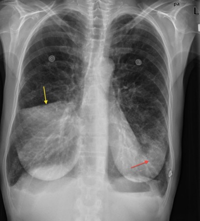
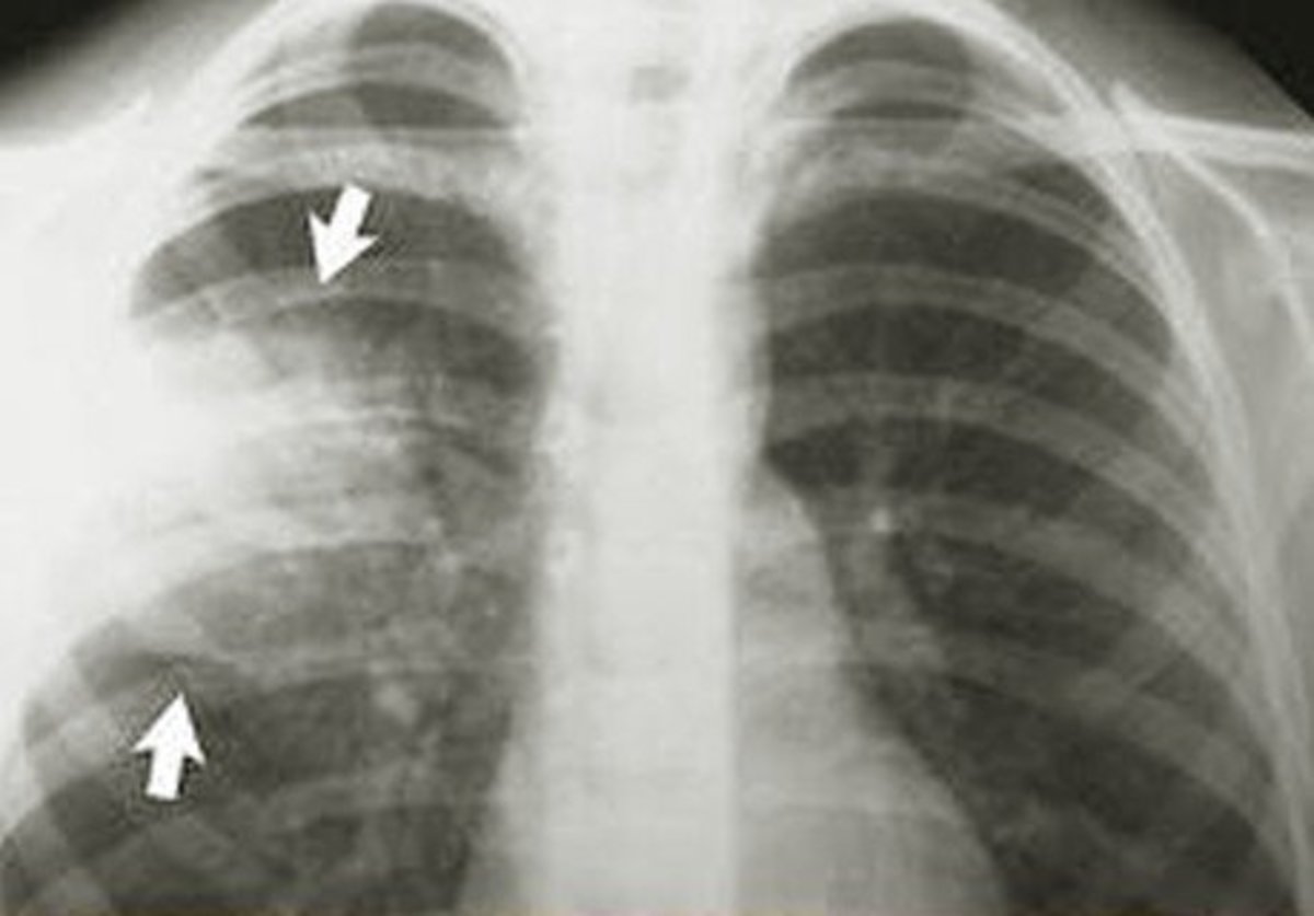
On imaging it presents an opaque pulmonary consolidation which is unusually round, and can resemble a lung mass. However it quickly resolves with antibiotics. Diagnosis The most common organisms which cause lobar pneumonia are Streptococcus pneumoniae, also called pneumococcus, Haemophilus influenzae and Moraxella catarrhalis.
What to do if you get pneumonia?
Jun 13, 2020 · Treatment. Treatment for pneumonia involves curing the infection and preventing complications. People who have community-acquired pneumonia usually can be treated at home with medication. Although most symptoms ease in a few days or weeks, the feeling of tiredness can persist for a month or more.
How do you cure pneumonia?
No report of Lobar pneumonia is found for people with Aphonia. ... or treatment provided by a qualified healthcare provider. All information is observation-only. Our phase IV clinical studies alone cannot establish cause-effect relationship. Different individuals may respond to medication in different ways.
What is the best antibiotic to lobar pneumonia?
Macrolides. The best initial antibiotic choice is thought to be a macrolide. Macrolides provide the best coverage for the most likely organisms in community-acquired bacterial pneumonia (CAP). Macrolides have effective coverage for gram-positive, Legionella, and Mycoplasma organisms.
Is lobar pneumonia treatable?
Treatment for pneumonia involves curing the infection and preventing complications. People who have community-acquired pneumonia usually can be treated at home with medication. Although most symptoms ease in a few days or weeks, the feeling of tiredness can persist for a month or more.13 Jun 2020
Is lobar pneumonia serious?
It is a serious infection in which the air sacs fill with pus and other liquid. Lobar pneumonia affects one or more sections (lobes) of the lungs. Bronchial pneumonia (also known as bronchopneumonia) affects patches throughout both lungs.
What is the most common treatment for pneumonia?
Mild pneumonia can usually be treated at home with rest, antibiotics (if it's likely be caused by a bacterial infection) and by drinking plenty of fluids. More severe cases may need hospital treatment.
How do you get lobar pneumonia?
Bacteria. The most common cause of bacterial pneumonia in the U.S. is Streptococcus pneumoniae. This type of pneumonia can occur on its own or after you've had a cold or the flu. It may affect one part (lobe) of the lung, a condition called lobar pneumonia.13 Jun 2020
Is lobar pneumonia painful?
Characteristic Clinical Features Patients may have fever, cough, and purulent sputum. Pleuritic chest pain is relatively common, and hemoptysis may occur. Signs and symptoms of pneumonia may be milder or even absent in the elderly.
How long does it take to recover from lobar pneumonia?
It may take time to recover from pneumonia. Some people feel better and are able to return to their normal routines within a week. For other people, it can take a month or more. Most people continue to feel tired for about a month.
What are the 4 stages of lobar pneumonia?
Stage 1: Congestion. Stage 2: Red hepatization. Stage 3: Grey hepatization. Stage 4: Resolution.21 Jun 2021
What part of the lung does lobar pneumonia affect?
Lobar pneumonia is a form of pneumonia characterized by inflammatory exudate within the intra-alveolar space resulting in consolidation that affects a large and continuous area of the lobe of a lung. It is one of three anatomic classifications of pneumonia (the other being bronchopneumonia and atypical pneumonia).
What is lobar pneumonia?
The term lobar pneumonia refers to the infection of one lobe of a lung, whereas bronchopneumonia refers to a more widespread infection. From: The Respiratory System (Second Edition), 2010.
What are the 3 stages of pneumonia?
Stages of PneumoniaStage 1: Congestion. During the congestion phase, the lungs become very heavy and congested due to infectious fluid that has accumulated in the air sacs. ... Stage 2: Red hepatization. ... Stage 3: Gray hepatization. ... Stage 4: Resolution.
What are the danger signs of pneumonia?
The signs and symptoms of pneumonia may include:Cough, which may produce greenish, yellow or even bloody mucus.Fever, sweating and shaking chills.Shortness of breath.Rapid, shallow breathing.Sharp or stabbing chest pain that gets worse when you breathe deeply or cough.Loss of appetite, low energy, and fatigue.More items...•30 Jul 2021
What causes a lobar pneumonia?
Examples of bacterial causes of lobar pneumonia include Haemophilus influenzae, Klebsiella spp, Staphylococcus spp, Legionella spp, and Streptococcus pneumoniae. These agents may follow a hematogenous spread pattern or infect the lobes by aspiration.
What is the workup for pneumonia?
Workup for pneumonia is essential to determine the level of severity of the disease and the treatment plan to be adopted. There are various diagnostic tools employed in the workup for pneumonia. These tools include the PSI/PORT, CURB 65 and the APACHE systems. PSI/PORT is a joint acronym for pneumonia severity index/patient outcome research team score, CURB-65 is also an acronym for confusion, uremia, respiratory rate, blood pressure, and age >65. These indices are measured and used to categorize the patients into groups of varying disease severity and treatment plans. APACHE stands for Acute Physiology And Chronic Health Evaluation, which takes into consideration several laboratory indices for matching disease severity with the appropriate treatment plans.
What is the most common cause of death in the United States?
Acute lower respiratory tract infection s are the most frequent infective causes of death in the United States. This group of infections is also associated with a greater morbidity than any other disease worldwide. However, there are statistical variations in the prevalence of the disease and the causative pathogens between countries and geographic regions, thereby limiting data on worldwide epidemiological studies of the disease.
What is the best way to diagnose pneumonia?
Direct means of obtaining diagnostic material in patients with pneumonia include percutaneous lung aspiration, transbronchial lung biopsy, video-assisted thoracoscopy, and open lung biopsy. These procedures are usually reserved for cases of severe pneumonia in impaired hosts and in pediatric populations, in whom sputum is not routinely available.
Why is pneumonia important?
Acute pneumonia is an important cause of morbidity and mortality worldwide. While the majority of cases of acute community acquired pneumonia (CAP) are due to bacteria, especially Streptococcal pneumoniae and less commonly the atypical organisms, respiratory tract viruses also contribute to a substantial proportion.
Why is pneumonia so common in tropical countries?
The higher incidence of pneumonia in tropical countries is primarily due to immunocompromise due to HIV infection but is also due to increased carriage of pneumococci by children and adults, large family size, crowding in small houses, exposure to domestic and tobacco smoke and the impaired immunity due to poor diet and parasitic diseases. Individuals with increased susceptibility to pneumonia include those with reduced splenic function (sickle cell disease, post-splenectomy), pregnant women, patients with diabetes mellitus and those with excess alcohol intake. Bacterial pneumonia may be preceded by a viral infection such as influenza that damages mucosal defence mechanisms.
What is AFOP in respiratory failure?
AFOP is a histopathologic pattern first reported in patients with acute respiratory failure, with predominantly basal and bilateral areas of consolidation at imaging. It is characterized by abundant fibrin deposition within the alveolar air spaces, with hyperplasia of type II pneumocytes, associated organizing-type pneumonia, and absence of hyaline membranes (which constitute the hallmark of diffuse alveolar damage). AFOP currently is considered to be a histopathologic variant of OP that may be associated with more rapid progression of disease. As with OP, AFOP may be encountered in the context of various underlying conditions, including infection.
What are the sequelae of pneumonia?
Acute pneumonia can lead to parenchymal damage and long-term sequelae. The most common sequelae of acute pneumonia are bronchiectasis and Swyer-James syndrome. Bronchiectasis is enlargement of the diameter of the bronchi that is related to damage to the bronchial walls. It is best demonstrated by high-resolution CT, where the diagnostic finding is that the bronchus in question is larger in diameter than the adjacent pulmonary artery ( Fig. 3-30 ). Swyer-James syndrome is characterized by unilateral lung hyperlucency that is thought to be secondary to a virus-induced necrotizing bronchiolitis that leads to an obliterative bronchiolitis (see Fig. 3-30 ). Radiography shows a hyperlucent and enlarged lung with a static lung volume. The pulmonary vessels are less prominent than on the normal side.
What is low power view of Klebsiella pneumonia?
Low-power view of Klebsiella pneumonia shows extensive congestion and fibrinous exudate admixed with inflammatory reaction. These features, although not specific for Klebsiella pneumonia, should prompt the use of cultures and special stains.
What is Brown Hopps stain?
The Brown-Hopps histochemical stain is useful in the diagnosis of Klebsiella pneumonia, as it may demonstrate the presence of bacterial forms, which may not be readily identifiable in the conventional H&E. Note the presence of numerous organisms compatible with Klebsiella .
What is lobular pneumonia?
Lobular pneumonia is a form of pneumonia characterized by inflammatory exudate within the intra-alveolar space resulting in consolidation that affects a large and continuous area of the lobe of a lung.
What is the most common organism that causes lobar pneumonia?
The most common organisms which cause lobar pneumonia are Streptococcus pneumoniae, also called pneumococcus, Haemophilus influenzae and Moraxella catarrhalis. Mycobacterium tuberculosis, the tubercle bacillus, may also cause lobar pneumonia if pulmonary tuberculosis is not treated promptly. Other organisms that cause lobar pneumonia are Legionella pneumophila and Klebsiella pneumoniae.
What is the term for the accumulation of cellular debris in the lungs?
This leads to the accumulation of cellular debris within the lungs. This leads to consolidation or solidification, which is a term that is used for macroscopic or radiologic appearance of the lungs affected by pneumonia. Bacterial pneumonia is mainly classified into lobar and diffuse
How to get rid of pneumonia?
Get plenty of rest. Don't go back to school or work until after your temperature returns to normal and you stop coughing up mucus. Even when you start to feel better, be careful not to overdo it. Because pneumonia can recur, it's better not to jump back into your routine until you are fully recovered.
What is the best medicine for pneumonia?
It may take time to identify the type of bacteria causing your pneumonia and to choose the best antibiotic to treat it. If your symptoms don't improve, your doctor may recommend a different antibiotic. Cough medicine.
How long does it take for a person to feel tired after pneumonia?
Although most symptoms ease in a few days or weeks, the feeling of tiredness can persist for a month or more. Specific treatments depend on the type and severity of your pneumonia, your age and your overall health. The options include: Antibiotics. These medicines are used to treat bacterial pneumonia.
What tests are done to determine if you have pneumonia?
If pneumonia is suspected, your doctor may recommend the following tests: Blood tests . Blood tests are used to confirm an infection and to try to identify the type of organism causing the infection. However, precise identification isn't always possible. Chest X-ray.
What to do if pneumonia isn't clearing?
If your pneumonia isn't clearing as quickly as expected, your doctor may recommend a chest CT scan to obtain a more detailed image of your lungs. Pleural fluid culture. A fluid sample is taken by putting a needle between your ribs from the pleural area and analyzed to help determine the type of infection.
What is a sputum sample?
A sample of fluid from your lungs (sputum) is taken after a deep cough and analyzed to help pinpoint the cause of the infection. Your doctor might order additional tests if you're older than age 65, are in the hospital, or have serious symptoms or health conditions. These may include: CT scan.
How fast can you breathe in a minute?
Your breathing is rapid (30 breaths or more a minute) You need breathing assistance. Your temperature is below normal. Your heart rate is below 50 or above 100. You may be admitted to the intensive care unit if you need to be placed on a breathing machine (ventilator) or if your symptoms are severe.
Diagnosis
- Direct means of obtaining diagnostic material in patients with pneumonia include percutaneous lung aspiration, transbronchial lung biopsy, video-assisted thoracoscopy, and open lung biopsy. These procedures are usually reserved for cases of severe pneumonia in impaired hosts and in pediatric populations, in whom sputum is not routinely available. Open lung biopsy remains the …
Contraindications
- Biopsy procedures are rarely indicated in the previously well patient with acute pneumonia. The indications and usefulness of these invasive procedures remain controversial. Blind lung aspiration has provided a diagnostic yield of 30% to 82% in adults and children with diffuse lung infiltrates, although false-negative rates of up to 18% have been reported.98,123,124 Computed …
Research
- Acute pneumonia has long been considered a risk factor for NIV failure.3 A trial evaluating NIV use in heterogeneous respiratory failure showed very poor outcome in the group of patients with pneumonia, with all such patients requiring intubation.48 Another study evaluated NIV use in patients with hypoxemic respiratory failure and identified community-acquired pneumonia as a s…
Signs and symptoms
- The symptoms and signs of lobar pneumonia may be confusing. In early pneumonia, the diagnosis may have to be made in a patient with symptoms, fever and shallow tachypnoea in the absence of any auscultatory signs. The patient will often point to the place where pain occurs when asked to cough. When pleurisy is diaphragmatic, the patient may present with suspected a…
Management
- The aetiological cause of pneumonia cannot usually be determined at the bedside but a clinical assessment of severity is more important as it can be used to guide management. In particular, young patients with uncomplicated lobar pneumonia can be managed at home with oral therapy. Patients with indicators of severity (age, co-existing disease, multi-lobar disease, shock, hypoxia…
Epidemiology
- Mycoplasma pneumoniae, Chlamydia pneumoniae and Legionella pneumophila also cause pneumonia in adults but are rare in Africa. In South-east Asia and northern Australia, melioidosis should be considered as a possible cause of both acute and unresolving pneumonia, especially in the debilitated or immunocompromised. Appropriate media are needed to culture the organism …
Cause
- Corynebacterium diphtheriae produces a gray pseudomembrane in the pharynx and larynx, which may lead to respiratory obstruction and sudden death. Sudden death from acute epiglottitis occurs from respiratory obstruction caused by swelling of the epiglottic folds, uvula, and vocal cords. The most common cause of acute epiglottitis in developing countries is H. influenzae (Ty…
Medical uses
- Among atypical pneumonias, mycoplasma infections respond best to a tetracycline or erythromycin; legionella infections respond to macrolides, alone or combined with rifampicin.
Treatment
- Bronchopneumonia is most frequently associated with pneumococci or haemophilus, so that amoxicillin, tetracycline or trimethoprim should be effective. More rarely, bronchopneumonia is caused by Staph. aureus, and flucloxacillin, fusidic acid or clindamycin is urgently required for treatment. Acute exacerbations of chronic bronchitis are almost invariably associated with eithe…
Classification
- AFOP is a distinct histologic type of organizing pneumonia. Another is granulomatous organizing pneumonia. It is characterized by radiologic findings of pulmonary nodules or masses and on histology by the presence of nonnecrotizing granulomas and organizing pneumonia.
Other animals
- Fibrinous lobar pneumonia, purulent peritonitis, and mesenteric lymphadenopathy are the primary lesions reported in callitrichids infected with K. pneumoniae (Gozalo and Montoya, 1991, 1992). Enteritis and hepatomegaly were reported in a colony of common marmosets by Campos et al. (1981). Purulent meningitis, consolidative pneumonia, intestinal hemorrhages, peritonitis, and ai…
Toxicity
- Fatal multisystemic infection of African green monkeys resulted in abscess formation in the abdomen, liver, lungs, skin, and CNS. Often, abdominal masses were present entrapping and adhering to loops of bowel and were histologically characterized by chronic pyogranulomatous inflammation and fibrous adhesions (Twenhafel et al., 2008).
Prognosis
- In the initial AFOP study, patients fell into one of two clinical patterns of disease. Slightly over half of patients presented with a fulminant illness progressing rapidly to death, with the remaining patients experiencing an indolent course with eventual recovery. No clinical or histologic features correlated with the eventual outcome, and an optimal therapy was not elucidated. Because the o…