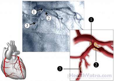
What is Angiography Treatment & Thier Cost
- Angiography Treatment Cost. Practical techniques for multivessel coronary supply route ailment (CAD) with stable angina and protected ventricular capacity are treatment (MT), percutaneous coronary mediation (PCI), and coronary conduit sidestep ...
- Coronary Angiography Treatment. ...
- Restorative Issues to Consider Before Having an Angiogram
Is an angiogram a dangerous procedure?
An angiogram is a procedure to make the arteries visible for the doctor to see blood flow through the arteries. Angiograms are used to diagnose and determine treatment options for Peripheral Arterial Disease (PAD). PAD is a disease in which plaque builds up in the arteries that carry blood to your head, organs, and extremities.
What is an angiogram and how is it performed?
What is Angiography Treatment & Thier Cost. An angiogram is an X-beam strategy that can be both demonstrative and restorative. It is viewed as the best quality level for assessing blockages in the blood vessel framework. An angiogram identifies blockages utilizing X-beams taken during the infusion of a complexity operator (iodine color).
What are the different ways to do an angiogram?
Dec 22, 2021 · Doctors can do an angiogram on different parts of the body, such as: the heart, during the diagnosis or treatment of some aspects of heart disease the brain, to help diagnose a stroke or the risk of a stroke the chest or lungs, for example, to detect bleeding the kidneys, to look for high pressure ...
What are the risks of having an angiogram?
An angiogram is a procedure that uses X-ray contrast to look at the blood vessels (arteries or veins) in your body. Cleveland Clinic is a non-profit academic medical center. Advertising on our site helps support our mission. We do not endorse non-Cleveland Clinic products or services. Policy Why do we do an angiogram?

How serious is an angiogram?
Angiography is generally a safe procedure, but minor side effects are common and there's a small risk of serious complications. You'll only have the procedure if the benefits outweigh any potential risk.
What is the procedure for a angiogram?
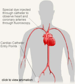
In a coronary angiogram, a catheter is inserted into an artery in the groin, arm or neck and threaded through the blood vessels to the heart. A coronary angiogram can show blocked or narrowed blood vessels in the heart. A coronary angiogram is a procedure that uses X-ray imaging to see your heart's blood vessels.Dec 14, 2021
Why would a doctor order an angiogram?
Why do we do an angiogram? When blood vessels are blocked, damaged or abnormal in any way, chest pain, heart attack, stroke, or other problems may occur. Angiography helps your physician determine the source of the problem and the extent of damage to the blood vessel segments that are being examined.Jul 16, 2019
What happens after an angiogram?
After an angiogram, your groin or arm may have a bruise and feel sore for a day or two. You can do light activities around the house but nothing strenuous for several days. Your doctor may give you specific instructions on when you can do your normal activities again, such as driving and going back to work.
Is an angiogram painful?
Will an angiogram hurt? Neither test should hurt. For the conventional angiogram you'll have some local anaesthetic injected in your wrist through a tiny needle, and once it's numb a small incision will be made, in order to insert the catheter.
Which is better CT scan or angiogram?
CT angiography of the heart is a useful way of detecting blocked coronary arteries. CT angiography may also cost less than catheter angiography. No radiation remains in a patient's body after a CT exam. The x-rays used for CT scanning should have no immediate side effects.
Can angiogram clear blockage?
Long-term outlook after a coronary angiogram Narrowed coronary arteries may possibly be treated during the angiogram by a technique known as angioplasty. A special catheter is threaded through the blood vessels and into the coronary arteries to remove the blockage.
What is the recovery time from an angiogram?
Most people feel fine a day or so after having the procedure. You may feel a bit tired, and the wound site is likely to be tender for up to a week. Any bruising may last for up to 2 weeks.
What happens if they find a blockage during an angiogram?
If your doctor finds a blockage during your coronary angiogram, he or she may decide to perform angioplasty and stenting immediately after the angiogram while your heart is still catheterized. Your doctor will give you instructions to help you prepare.Oct 8, 2021
Should I be worried about having an angiogram?
Angiograms are generally safe, complications occur less than 1% of the time. However, there are risks with any test. Bleeding, infection, and irregular heartbeat can occur. More serious complications, such as heart attack, stroke, and death can occur, but they are uncommon.Dec 2, 2014
Are you awake during an angiogram?
During the angiogram, you are awake, but are given medications to help you relax. A thin tube (catheter) is placed in the femoral artery (groin area) through a small nick in the skin about the size of the tip of a pencil. The catheter is guided to the area to be studied.Jul 25, 2012
Is angiogram same as angiography?
Angiography, angiogram, or arteriograms are terms that describe a procedure used to identify narrowing or blockages in the arteries in the body. The procedure is the same regardless of what area of the body is being viewed.
For What Reason Would I Have a Coronary Angiogram?
You’d generally have an angiogram since you have indications of coronary illness (CHD, for example, chest torment, and regularly because different tests, similar to an electrocardiogram ( ECG ), have proposed you may have CHD. CHD is brought about by the development of fat stores in the coronary courses.
Angiography Treatment Cost
Practical techniques for multivessel coronary supply route ailment (CAD) with stable angina and protected ventricular capacity are treatment (MT), percutaneous coronary mediation (PCI), and coronary conduit sidestep unite (CABG).
Coronary Angiography Treatment
This can help analyze heart conditions, plan future medications, and do specific methodology. The heart has four chambers: the two little rooms at the top are called atria, and the two bigger chambers at the base are called ventricles.
Why do doctors use angiograms?
Doctors use angiograms to examine blood vessels. Angiogram results can help doctors diagnose and treat blood vessel problems and cardiovascular diseases. During the procedure, a doctor gently guides a catheter through an artery until it reaches the area of the body under investigation. Once the catheter reaches the correct location in the body, ...
What is the difference between angiogram and angioplasty?
Angiogram vs. angioplasty. During an angioplasty, a doctor inserts an inflatable balloon or mesh splint into a blocked or narrow artery. When it is in the right place, the doctor will inflate or expand the balloon or splint, improving the blood flow in that artery. Doctors often perform angioplasties during angiograms.
What are the conditions that angiograms can diagnose?
They use angiogram results to diagnose the following conditions: aneurysms, or bulges that develop in weakened artery walls. atherosclerosis, which occurs when plaques and fa tty material collect on the inner walls of the arteries. pulmonary embolisms, or blood clots.
What does an abnormal angiogram mean?
An abnormal angiogram result may indicate that a person has one or more blocked arteries. In these cases, the doctor may choose to treat the blockage during the angiogram.
Where do they put contrast dye in angiogram?
To perform a traditional angiogram, a doctor inserts a long, narrow tube called a catheter into an artery located in the arm, upper thigh, or groin. They will inject contrast dye into the catheter and take X-rays of the blood vessels. The contrast dye makes blood vessels more visible on X-ray images.
What is the blood vessel abnormality on an angiogram?
The term “angiogram” refers to a number of diagnostic tests that doctors can use to identify blocked or narrow blood vessels. Angiograms also help doctors diagnose a range of cardiovascular diseases, including coronary atherosclerosis, vascular stenosis, and aneurysms.
How long does it take for a doctor to remove a catheter?
After taking the X-ray images, the doctor will remove the catheter and apply steady pressure on the area for about 15 minutes. This ensures that there is no internal bleeding.
What is an angiogram?
An angiogram is a procedure that uses X-ray contrast to look at the blood vessels (arteries or veins) in your body. Cleveland Clinic is a non-profit academic medical center. Advertising on our site helps support our mission. We do not endorse non-Cleveland Clinic products or services. Policy.
Why do you need an angiography?
On occasion, it may be necessary for you to spend the night in the hospital. Angiography allows your physician to view how blood circulates within vessels at specific locations in the body. This diagnostic test is used to locate the specific source of an abnormality in the neck, kidneys, legs, or other sites.
How long do you have to wait to take glucophage before a blood test?
Please notify the radiology nurse upon arrival that you are diabetic. Do NOT take Glucophage® (metformin hydrochloride) for 48 hours before the test or 48 hours after the test to reduce the risk of kidney complications.
How long do you have to be monitored for a radiology exam?
You will be monitored for 4 to 6 hours. At that time, the radiology nurse will discuss at-home instructions with you. You will be provided with a written form of these instructions. Please follow these at home.
What to drink after a syringe procedure?
Drink only clear liquids for breakfast the day of your procedure. Clear liquids include clear broth, tea, black coffee and ginger ale. NOTE: People who are scheduled to have general anesthesia during the procedure may have nothing to eat or drink after midnight.
How long before a blood test can you take dipyridamole?
Do NOT take dipyridamole (Persantine®) or warfarin (Coumadin®) within 72 hours before the test, and 24 hours after the test. These medications are often referred to as blood-thinning pills. Do not take Plavix® for 5 (five) days prior to the procedure.
Why is angiography important?
Angiography is a common medical procedure used to visualize blood flow within the body. It may be important to diagnose various medical conditions. It also presents an opportunity to intervene and treat blockages and other abnormalities, especially those that affect the heart and brain. Discover the reasons it is performed, techniques, ...
What is cerebral angiography used for?
Cerebral angiography may be used to treat narrowing that contributes to transient ischemic attacks or stroke risk. In the hours following a stroke, it may be possible to extract a clot and reverse symptoms like weakness, numbness, loss of speech, or vision changes.
Why is microangiography used?
Microangiography may be used to image the smaller blood vessels supplying other organs, particularly to address localized bleeding. It may also be useful in detecting and treating cancerous tumors since rapidly growing tumors are highly vascular. Depriving the tumor of its blood supply may be an effective adjunctive therapy.
Why do you need an angiogram?
Often an angiogram is performed with both a diagnostic portion, to better visualize the nature of the problem, and a treatment portion, in which an intervention immediately corrects the underlying problem. Unlike other tests, it is often unnecessary to gather information to review and be used at a later date.
How long after angiography should you not smoke?
If there is a serious problem, it may be necessary to contact the healthcare provider and get emergency medical assistance. For 24 hours following angiography, the patient should not drink, smoke, or perform tasks that require coordination (such as operating vehicles or heavy machinery).
What is abnormal blood flow?
Abnormal blood flow affects the organs supplied by the vessels, and may increase the risk for chest pain ( angina ), heart attack , stroke, and other disorders. Besides the obvious diagnostic use, angiography may also be used to deliver treatment.
How long before angiography should I drink?
To prepare for angiography, it is important to avoid eating in the eight hours leading up to the procedure. Drinking clear liquids until two hours before the procedure will help keep blood vessels patent, flexible, and more easily accessible.
What is the most common angiogram?
The most common angiograms include pulmonary angiogram (of the chest), coronary angiogram (of the heart), cerebral angiogram (of the brain), carotid angiogram (of the neck and the head), peripheral angiogram (of the arms and legs) and aortogram (of the aorta). An angiogram is used to detect aneurysms (bulge within the blood vessels).
What is the best test for heart failure?
An angiogram or angiography is usually recommended by the doctor only if other less invasive testing techniques have been exhausted or haven’t been able to come up with concrete results. Certain less invasive alternatives include a heart stress test, ECG (Electrocardiography) and echocardiography.
Why do you use fluorescent dye in angiograms?
The fluorescent dye is used for an angiogram which is injected into the blood as it helps in highlighting the blood vessels which are present at the back of the eye thus it becomes easier to take a photograph of them.
Why do doctors use dye on X-rays?
Dye helps in getting a clear X-ray thus enable your doctor to have a clear view of blood vessels from the lungs. In this radionuclide is injected in the arm vein to see the progress of the blood cells and the heart is traced through the scanner. This RNA procedure helps the doctor check the functioning of the heart.
What is an angiography test?
What is an angiography? An angiography, also known as an angiogram, is an X-Ray test that makes use of a dye along with a camera in order to take clear pictures of the circulation of blood inside a vein or an artery. This procedure can be performed for the veins or the arteries of the chest, back, arms, head, belly and the legs.
What is an angiogram?
An angiogram is used to detect aneurysms (bulge within the blood vessels). Any blockage or narrowing of the blood vessels that affect proper blood flow can also be detected by this procedure. The possibility of a coronary artery disorder being present, as well as its condition, can be determined by this technique.
Where is contrast dye injected into a catheter?
In this, a catheter is inserted into the veins artery of the leg, and then it is passed into the blood vessel. After this contrast dye is injected into the catheter to get a proper image of the blood vessels. Magnetic resonance angiography:
Why do you need an angiogram for chest pain?
Conventional angiograms are more often used for patients who show a high risk for heart disease and have one or more of these symptoms: Unexplained pain in the chest, jaw, arm, or neck; other tests are often performed to rule out injury, temporomandibular joint disorder (TMJ), Lyme disease, etc. Increased chest pain.
What is an angiogram?
An angiogram is a radiological procedure. There are actually two types of angiograms. Both use advanced x-ray technology to deliver images of arteries to a monitor. Computed tomography angiography (CTA) is noninvasive, meaning that there are no incisions made on the body.
Why do you have to be strapped on a bed for an x-ray?
The patient may be strapped on a bed if the procedure requires them to be tilted for x-ray machines to get a better view. Patients might be given nitroglycerin to dilate or widen, the arteries, or a beta blocker to slow the heart rate. A coronary angiogram requires more preparation because it is invasive.
Is angiogram painful?
Some patients find angiograms to be uncomfortable, but few describe either procedure as actually painful. For most patients, the hardest part is remaining still throughout the procedure. The dye may initially deliver a burning feeling as it makes its way into the arm and to the chest.
What is the most common type of cardiac catheterization procedure?
Cardiac catheterization procedures can both diagnose and treat heart and blood vessel conditions. A coronary angiogram, which can help diagnose heart conditions, is the most common type of cardiac catheterization procedure. During a coronary angiogram, a type of dye that's visible by an X-ray machine is injected into the blood vessels of your heart.
What is the procedure called to check for blocked blood vessels?
Close. Coronary angiogram. Coronary angiogram. To complete a coronary angiogram, a catheter is inserted in an artery in your groin or arm and threaded through your blood vessels to your heart. Your doctor uses the angiogram to check for blocked or narrowed blood vessels in your heart. A coronary angiogram is a procedure ...
What does an angiogram show?
An angiogram can show doctors what's wrong with your blood vessels. It can: Show how many of your coronary arteries are blocked or narrowed by fatty plaques (atherosclerosis) Pinpoint where blockages are located in your blood vessels. Show how much blood flow is blocked through your blood vessels.
Why do you need an angiogram?
Your doctor may recommend that you have a coronary angiogram if you have: Symptoms of coronary artery disease, such as chest pain (angina) Pain in your chest, jaw, neck or arm that can't be explained by other tests. New or increasing chest pain (unstable angina)
What to do before angiogram?
Before your angiogram procedure starts, your health care team will review your medical history, including allergies and medications you take. The team may perform a physical exam and check your vital signs — blood pressure and pulse. You'll also empty your bladder and change into a hospital gown.
What are the signs of infection after a catheter is inserted?
You have signs of infection, such as redness, drainage or a fever. There's a change in temperature or color of the leg or arm that was used for the procedure. Weakness or numbness in the leg or arm where the catheter was inserted. You develop chest pain or shortness of breath.
Where is the catheter placed in the heart?
In a cardiac catheterization procedure, doctors insert a catheter in an artery in your wrist (radial artery) or in your groin (femoral artery).The catheter is then threaded through your blood vessels to your heart.
