
What is intraosseous lipoma?
Intraosseous lipoma is a very rare lesion that constitutes no more than 0.1% of all bone tumors. 1)It may undergo varying degrees of degenerative changes and manifests with areas of fat necrosis, cystic changes, and calcification.
What are the signs and symptoms of expansile intraosseous lipomas?
Although there are typically no signs of aggressive behavior, expansile intraosseous lipomas may outgrow the cortical border. This can be used to differentiate them from older bone infarcts, which may also develop cystic degeneration over time.
What is calcaneal intraosseous lipoma (CIP)?
Images 6a and 7a courtesy of Tudor Hughes, M.D. Calcaneal intraosseous lipoma is an uncommon benign bone tumor which, as it slowly expands and remodels adjacent osseous structures, acquires a typical appearance that should be readily diagnostic on plain radiographs and MRI.
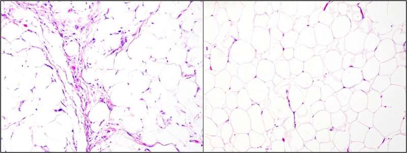
What are the lesions of lipomas composed of?
The lesions are composed of mature adult fat and may contain combinations of solid, liquefied and/or necrotic fat.
What causes intraosseous lipoma?
Intraoseous lipoma has an unknown aetiology. Three theories have been considered: a traumatic origin and later fat degeneration, infections, or osseous fat infarction with metaplasia and third, at present moment most studies think that intraosseous lipoma is a primary tumour of marrow fat.
Can lipoma grow inside a bone?
Lipoma can be located in the intraosseous region or adjacent to bone and referred to as intraosseous, parosteal, or periosteal lipoma respectively. Those that are in such sites may contain osseous and/or chondral components [5, 6]. Less than 1% of lipomas were ossified in one study of 635 cases [2, 4].
What is a bone lipoma?
Lipoma is a rare benign intraosseous neoplasm, constituted by adipose cells that can also arise on the surface of bone. It corresponds to less than 0.1 % of primary bone neoplasms, and 15 % of them are surface tumors. Main incidence is in the fifth decade of life.
Is lipoma invasive?
Lipomas are benign, slow-growing tumors that are usually located in the subcutaneous tissues. Surgical excision is the mainstay of treatment for lipomas [1], though they frequently require an incision equal to the diameter of the tumor.
Can a lipoma turn cancerous?
Cancerous tumours of the fat cells are called liposarcomas. They are a type of soft tissue sarcoma. It is very rare for lipomas to turn into a cancerous sarcoma. It is still important to tell your doctor if your lipoma changes in any way or if you get any new lumps.
What kind of doctor removes lipomas?
Lipoma removal is usually carried out by a Dermatologist but may also be carried out by other with training in skin surgery procedures.
What is an intraosseous lesion?
Introduction. The intraosseous lipoma is the most common lipogenous lesion of bone. Intraosseous lipoma is found most frequently in the intertrochanteric region of the proximal femur (34%), with the calcaneal intraosseous lipoma being the next most prevalent, found in 8-15% of cases.
Do intramuscular lipomas need to be removed?
Basically, an intramuscular lipoma in the extrathoracic muscle layer, together with its intramuscular fatty tissue, must be removed.
What does an ultrasound of a lipoma look like?
Traditionally it has been taught that the sonographic appearance of a simple lipoma is consistent with a hyperechoic mass with no posterior acoustic enhancement (4). These soft-tissue masses are lower in reflectivity than muscle but more reflective than adjacent subcutaneous fat.
At what size should a lipoma be removed?
All lipomas in the upper extremities measuring larger than 5 cm in a single dimension should be surgically removed due to malignant potential.
Is lipoma surgery painful?
With local anesthesia, you may still feel pressure or pushing, but you should not feel any pain. If your lipoma is large or deep, you may be given general anesthesia. General anesthesia will keep you asleep and free from pain during surgery.
What is the best treatment for lipoma?
Most lipomas don't need treatment. If a lipoma is bothering you, your provider can remove it surgically. Lipoma removal procedures are safe and effective, and you can usually go home the same day. As an alternative to lipoma surgery, your provider may recommend liposuction to remove the lipoma.
What is the most common bone tumor in bone 6?
Intraosseous lipoma. Intraosseous lipomas are rare benign lesions that account for about 0.1-2.5% of all bone tumors . It is, however, the most common lipogenic tumor in bone 6.
Where can lipomas be found?
Although intraosseous lipomas can be found essentially anywhere within the skeleton, the lower limb accounts for the majority of cases.
What is stage 3 of sclerotic bone?
In addition to the signal characteristics of stage 2 lesions, stage 3 lesions may contain fluid-equivalent cavities and signal-void bony septa and are surrounded by thickened, signal-void rims of sclerotic bone.
Can intraosseous lipomas be treated?
Intraosseous lipomas that do not affect bone stability may be treated conservatively. Cases with imminent fractures are treated by curettage and bone grafting. There are no recurrences after surgical therapy. However, malignant transformations have been sporadically described 1.
Can intraosseous lipomas outgrow the cortical border?
Although there are typically no signs of aggressive behavior, exp ansile intraosseous lipomas may outgrow the cortical border. This can be used to differentiate them from older bone infarcts, which may also develop cystic degeneration over time.
Is T2 granulation tissue in lipomas?
Moreover, in intraosseous lipomas, T2-hyperintense granulation tissue of central necrosis is surrounded by viable fat tissue. Conversely, in bone infarcts, fat tissue in the center is surrounded by granulation tissue at the periphery.
Can CT attenuation be narrowed down to fat-containing lesions of the bone?
By CT attenuation or MRI signal characteristics, the differential diagnosis can be narrowed down to fat-containing lesions of the bone. The fatty components of intraosseous lipomas may display varying degrees of involution and necrosis. Based on these features, Milgram and co-workers 2 proposed three categories:
What is intraosseous lipoma?
Intraosseous lipomas composed solely of fat (Milgram stage 1 lesions) are radiolucent, well-circumscribed lesions that frequently are associated with mild, focal, expansile remodeling. 1 On MRI, the lesion is geographic, rounded, without cortical interruption, with a peripheral rim of high signal intensity on T1- weighted images (1a,1b). Homogeneous fat suppression should occur on fat-suppressed T2-weighted or inversion recovery-weighted images (1c,1d). In Milgram stage 2 or 3 lesions, the ossifications and calcifications may produce a distinctive radiographic appearance (5a).
Where is intraosseous lipoma most common?
Intraosseous lipoma is found most frequently in the intertrochanteric region of the proximal femur (34%), with the calcaneal intraosseous lipoma being the next most prevalent, found in 8-15% of cases. 1,4 The high incidence of proximal femoral and calcaneal sites is considered to be a function of the relative paucity of trabecular bone in both of these locations, a characteristic that is also responsible for the “pseudolesion” appearance seen on radiographs at these sites. The sparse trabecular bone at the anteroinferior calcaneus has led to the supposition that intraosseous lipomas represent an “overshoot” phenomenon that develops during the transition of hematopoietic to fatty marrow, and therefore that intraosseous lipomas in these sites might more correctly be considered hamartomas rather than neoplasms. 2,3 Many of these lesions are found incidentally in the calcaneus on routine radiographs, however a significant percentage of these lesions present due to pain, which is reported up to 66% of the time. 4 The symptoms may result from remodeling of bone due to expansion, or due to intralesional ischemia, noted to be a common pathogenetic consequence of a long-standing calcaneal intraosseous lipoma.
Is calcaneal intraosseous lipoma a benign tumor?
Calcaneal intraosseous lipoma is an uncommon benign bone tumor which, as it slowly expands and remodels adjacent osseous structures, acquires a typical appearance that should be readily diagnostic on plain radiographs and MRI. Calcaneal intraosseous lipomas may present with bone pain up to 66% of the time from chronic expansion, or with acute symptoms related to pathologic (insufficiency) fracture. 1,3 Although there exists a differential diagnosis, including bone infarct, unicameral bone cyst, aneursymal bone cyst, chondromyxoid fibroma, osteoblastoma and giant cell tumor, the presence of patently obvious fat signal intensity on T1-weighted MR images should allow a near 100% sensitivity and specificity in the diagnosis of calcaneal intraosseous lipoma. Bone infarcts can occasionally be a source of diagnostic confusion, as infarcts contain fat and often have a low signal intensity rim. However, it should be emphasized that calcaneal intraosseous lipoma is an expansile lesion that remodels cancellous and cortical bone, which is never a feature of bone infarction. 3
How rare are bone lipomas?
Bone lipomas are rare tumours, that constitute up to 0.1% of bone tumours [1,2]. With an ever increasing use of cross-sectional imaging in the diagnostics of the musculoskeletal system, and especially of MRI, the detection rate of those lesions has increased [3].
Is intraosseous lipoma rare?
Intraosseous lipoma is a very rare lesion, which constitutes not more than 0.1% of bone tumors. The introduction of cross-sectional imaging, especially MRI, seems to have increased the detection rate of these lesions. Case Report: The authors presented 6 cases of intraosseous lipomas in bones of the lower extremities.
Is lipoma the last phase of involution?
It is also believed that lipomas may constitute the last phase of involution of other focal lesions of bones, a bone infarct in particular. Stage III intraosseous lipoma described by Milgram may be difficult to distinguish from a bone infarct in histopathological examination [4].
Is soft tissue lipoma more common in women?
Soft tissue lipomas are much more common in women (8:1). However, epidemiology of intraosseous lipomas, with a slightly higher incidence in men, does not support the relationship between these two types of tumors [3]. It seems that the nature of intraosseous lipomas is yet to be unequivocally defined.
Epidemiology
Diagnosis
Clinical Presentation
Pathology
Radiographic Features
Treatment and Prognosis
- Intraosseous lipomas that do not affect bone stability may be treated conservatively. Symptomatic cases or those with imminent fractures are treated by curettage 8 and bone grafting. There are no recurrences after surgical therapy 8. However, malignant transformations have been sporadically described 1.
Differential Diagnosis
Introduction

Pathology and Staging
Imaging Considerations
Discussion
Conclusion
References