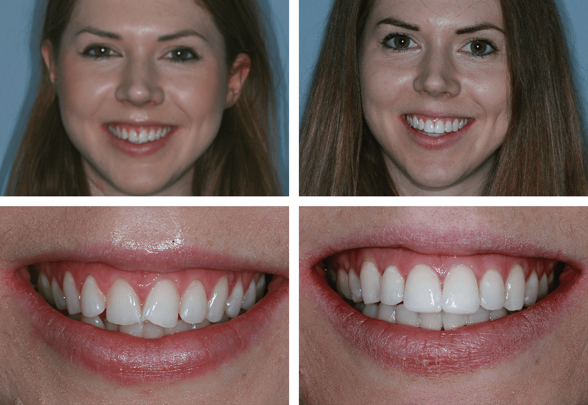
How should documentation for orthodontic treatment be selected?
Nov 12, 2013 · Orthodontic records are required for an orthodontic diagnosis and treatment plan , . Although records are mainly used for these purposes, monitoring facial growth and development with or without orthodontic treatment also plays an important role in research and clinical audit . Traditionally, dental casts, intra- and extra-oral photographs, different radiographic images, and …
Do different diagnostic records contribute to orthodontic treatment planning?
Carlos Barrero, in Diagnosis and Treatment Planning in Dentistry (Third Edition), 2017. Patient considerations. A fundamental determinant in orthodontic treatment planning is the patient’s own perceived need for that treatment. For most patients, the willingness to accept orthodontic treatment is motivated by a desire to improve appearance; a direct correlation can be made …
How do Orthodontists approach diagnosis?
Nov 12, 2013 · Background Traditionally, dental models, facial and intra-oral photographs and a set of two-dimensional radiographs are used for orthodontic diagnosis and treatment planning. As evidence is lacking, the discussion is ongoing which specific records are needed for the process of making an orthodontic treatment plan. Objective To estimate the contribution and …
What is treatment planning in orthodontics?
Orthodontic treatment planning is usually based on detailed subjective information obtained from the patient and objective diagnostic records (clinical examination, photograph evaluation, cast...

What is planning orthodontic treatment?
Planning orthodontic treatment should be part of a multidisciplinary approach where the protocol is jointly developed to produce the best outcomes and reduce the burden of care on the patients.
Can orthodontics be used for malocclusion?
Dental Problems. If the orthodontic problem in the adolescent is strictly dental, conventional orthodontic treatment can be used to manage the malocclusion. Identification and management of dental orthodontic problems have already been discussed and basically do not change with the age of the patient.
What is the purpose of a lower second premolar extraction?
Lower second premolar extractions provide greater mesial movement of the lower first molars for the correction of class II to class I molar relationships where space requirements in the lower anterior segment are small. This approach is common in treatment of class II division 1 dental malocclusion.
What is class II malocclusion?
In certain forms of class II malocclusion, where the lower arch is well aligned, protrusion may be corrected by extraction of first premolars in the upper arch only . The postorthodontic occlusion would have a normal overjet and overbite, with maxillary second premolars and molars in a full cusp class II relationship with the mandibular arch. Under these conditions the mesiobuccal cusp of the maxillary first molar articulates in the embrasure between the mandibular first molar and second premolar. The distobuccal cusp of the maxillary molar articulates with the mandibular first molar mesiobuccal groove ( Figure 16-5 ). Earlier it was suspected that finishing in class II molar relationship can lead to temporomandibular joint disorder and compromised occlusal stability. However, studies have found that occlusal stability of class II malocclusion finished in class II molar relationship was at par with those finished in class I molar relationship ( Jason et al. 2010 ).
What is a brachyfacial patient with skeletal pattern and Class II malocclusion?
Brachyfacial patient with skeletal pattern and Class II malocclusion, presenting with a deep overbite, buccal crossbite of the upper first molars, marked proclination of the upper and lower incisors, increased overjet and accentuated curve of Spee.
What is an orthodontic anchor?
The term orthodontic anchorage was first introduced by Edward Angle and can be explained as resistance to unwanted movement . The goal is to maximize desired tooth movements and minimize the unwanted ones. 1 As orthodontic treatment advanced in complexity and in frequency, more recent techniques, using temporary skeletal anchorage, were developed to help correct more severe discrepancies. These techniques allowed the orthodontist to move teeth against a rigid fixation, allowing for more focused movements of teeth. This type of rigid fixation allowed for greater interaction between the orthodontist and the oral and maxillofacial surgeon and vastly enhanced the treatment planning for the orthodontist. 2
What is the biotype of gingiva?
There are significant intrinsic biologic variations between humans with respect to the morphologic characteristics of the gingiva; this is known as the gingival biotype.52,73,138 The evaluation of the individual's gingival biotype is important to orthodontic treatment planning, because thick and thin gingival biotypes are frequently associated with varied osseous patterns. These two tissue types are likely to respond very differently to similar orthodontic forces by demonstrating different patterns of osseous remodeling. With an understanding of the nature of the individual's biologic risk factors (e.g., gingiva, alveolar bone, breathing pattern, degree of malocclusion), appropriate periodontal, orthodontic, and surgical preventative procedures and precautionary measures may be instituted to provide a more favorable tissue environment to minimize alveolar bone loss and gingival recession. Historically, Ochsenbein and Maynard discussed the importance of thick versus thin gingiva with regard to restorative treatment planning.150 In addition, in a group of patients reviewed by Olsson and colleagues, a thick periodontal biotype (85% of population) was found to be more prevalent than a thin periodontal biotype (15% of population). 152 Thick gingival tissue is dense in appearance, with a fairly large zone (length) of attachment. The gingival topography is relatively flat, which is suggestive of a full underlying bony architecture. Thin gingival tissue tends to be delicate and almost translucent in appearance. The tissue is friable, with a minimal zone of attachment; this is suggestive of minimal bone over the labial roots of the teeth. The diametrically opposite thin and thick gingival biotypes will respond differently when subjected to inflammation, mechanical trauma, orthodontic forces, or surgical insults. Results from at least some clinical studies indicate that, as long as a tooth is being orthodontically moved with light forces and within or into (and not out of) the alveolar process, then the risk of harmful side effects on the marginal (gingival) soft tissues is minimal. 212-215 In current clinical practice, pretreatment gingival augmentation (i.e., free gingival grafts, lateral pedicled flaps, and autogenous and allogenic connective tissue grafts) to improve the presenting thin keratinized gingiva—in combination with light orthodontic force—generally achieves objectives and limits recession and alveolar bone loss.
What is used for orthodontic treatment?
Traditionally, dental models, facial and intra-oral photographs and a set of two-dimensional radiographs are used for orthodontic diagnosis and treatment planning. As evidence is lacking, the discussion is ongoing which specific records are needed for the process of making an orthodontic treatment plan.
Do you need a cephalogram for orthodontics?
Cephalograms are not routinely needed for orthodontic treatment planning in Class II malocclusions, digital models can be used to replace plaster casts, and cone-beam computed tomography radiographs can be indicated for impacted canines. Based on the findings of this review, the minimum record set required for orthodontic diagnosis and treatment planning could not be defined.
What is the first step of the screening process?
In the first step of the screening process, two observers (RR, HB) independently screened the retrieved records on the basis of title and abstract according to the eligibility criteria. After reviewing the title and abstract, articles were classified as included, excluded, or unclear. Any disagreements were resolved by discussion and consensus.
What is the role of an orthodontist in a dental case?
The responsibility of the orthodontist is to diagnose and treatment plan the case, decompensate the dentition , and provide the surgeon a stable intra-arch dental framework. The surgeon must then place the skeletal components into the most pleasing and functional position possible to achieve the pretreatment goals established for the patient. Clear communication is essential as the goals of presurgical orthodontic treatment generally are opposite that of the routine orthodontic regimen used to camouflage the skeletal discrepancies in patients who choose not to pursue surgery. Successful occlusal relationships can be achieved with orthodontic treatment alone in minor skeletal discrepancies and at times even in more significant deficiencies; however, it is frequently at the expense of a pleasing facial appearance or a precarious position of a tooth that can risk its vitality relative to its position within the cortical and cancellous bone alveolus. These same patients who have had orthodontic treatment without consideration for combined surgical-orthodontic management may seek plastic surgical procedures later in life to address this residual skeletal deformity. Skeletal correction, once the occlusion is achieved, is compromised and frequently requires the use of prosthetic implants and soft tissue procedures to camouflage the deformity: the results of which are less than ideal.
How does computerized system help orthodontists?
A computerized system is built to help orthodontist in planning the treatment procedures of craniofacial deformities. CT scans (head) for 10 patients were processed and their three-dimensional reconstructions were developed. For each patient a profile representing the present state is obtained. Another predicted profile for the patient drawn and the orthodontic formulates his plan depending on the distance measured between the two profiles. The system provides a new method for segmentation and visualization of the teeth crowns and roots. The three axes of each tooth were computed to enable the translation and rotation.
What is the science of malocclusion?
Malocclusion is a manifestation of genetic and environmental interaction on the development of the orofacial region. It is important to consider genetic factors to understand the cause of existing problems, which has influence the outcome of treatment. The review aims to provide information to the dental practitioner and orthodontist on basics of genetics and common disorders with gene impairments. These genetic factors in turn have an impact on outcome of orthodontic treatment.
What is LCR in dentistry?
Background Lateral Cephalometric Radiographs (LCR) are a common decision-making aid in orthodontic treatment planning and are routinely used in clinical practice. The aim of this present study was to test the null hypothesis that LCR evaluation does not alter specific components of orthodontic treatment planning in Class II patients. Materials and Methods Records of 75 patients, who had been treated at the Department of Orthodontics, Centre of Dental Medicine, University of Zurich comprised the study material. Inclusion criteria were: (1) adolescents between the age of 12-15, (2) permanent dentition with Class II buccal segment relationship (3) absence of craniofacial and dento-alveolar malformations. Fifteen orthodontists from the dental faculties of Istanbul University, Istanbul and Ege University, Izmir filled out Likert-type linear scale questionnaires without knowing that they would repeat the same procedure with and without LCRs at two different time points. Equivalence and clinical relevance were assessed using (%95 CI) Wilcoxon signed rank tests. Results Extraction decision did not differ between groups (p=0.68). Preference of functional appliance use (p=0.006) and inter-maxillary fixed functional appliance (p=0.043) was different among groups. Conclusion LCR evaluation has minor influence on treatment planning procedure of Class II patients. It might be beneficial to consider its prescription not in a routine manner but as a supplementary tool considering possible reduction of radiation exposure.
What is RPE in dental?
Rapid palatal expansion (RPE) has been used primarily to treat dental crossbites or for space gaining to prevent extractions with little or no attempt made to coordinate or normalize the transverse skeletal pattern. Traditionally, maxillary orthopedics has been performed using the dental units only as anchorage (e.g., Hyrax or Haas appliances). Dental anchorage not only has created limited skeletal orthopedic change, but also can cause significant adverse periodontal outcomes and unstable side effects. There is a clear correlation between buccal tooth movement and gingival recession and bone dehiscences. These adverse periodontal responses with RPE indicate the importance of early treatment. The beneficial periodontal effects of transverse skeletal correction have been a primary focus of our research for the past 35 to 40 years. We have emphasized the importance of correcting transverse skeletal discrepancy to: 1) prevent periodontal problems; 2) achieve greater dental and skeletal stability; 3) improve dentofacial esthetics by eliminating or improving buccal corridors; and 4) improve airway resistance. When it may be critical to save the natural dentition, we do not want to introduce adverse dental/skeletal changes for adolescent patients and/or patients with advanced periodontal disease. New advances in skeletal anchorage should permit orthopedic change without adverse dental changes by applying force directly to the maxillary bone; an innovative technique to maximize the skeletal maxillary changes in the transverse dimension is explained in this chapter. Furthermore, diagnosis of the transverse dimension—the use of cone-beam computed tomography (CBCT) for 3D evaluation of skeletal changes, the benefits of the skeletal transverse changes of the whole maxillofacial complex and its periodontal response, the changes in airway and non surgical RPE with bone-anchored appliances utilizing temporary anchorage devices (TADs)—is described and discussed.
What is skeletal class II malocclusion?
Abstract Background: Treating a skeletal Class II malocclusion in non- growing patient has always been a controversial subject in orthodontics, whether to go for a surgical line of treatment or a conservative approach (camouflage). Treatment choices available are particularly difficult for young patients because of the uncertainty regarding future growth of mandible. Surgical treatment has generally been considered necessary for elderly patients with severe skeletal Class II problems. Temporary anchorage devices (TADs) have expanded the capabilities of clinicians, and allowed the correction of borderline orthognathic surgical cases to be treated with an orthodontics-only approach. This had greatly reduced psychological, economical burden on the patient and helped clinicians to achieve good functional and esthetic results as compared to surgical intervention. This case report discusses the management of one such skeletal Class II malocclusion using temporary anchorage devices (TADs).
What is a midline diastema?
Maxillary midline diastemas are a common esthetic problem that dentists must treat. Many innovative therapies have been used, varying from restorative procedures to surgery (frenectomies) and orthodontics. The importance of the presence of a maxillary midline diastema resides in its position and the concern it causes to patients. This specific diastema has been attributed to genetic and environmental factors, even though it is often a normal feature of growth, especially in primary and mixed dentition. The need for treatment is mainly attributed to esthetic and psychological reasons, rather than functional ones. Although it is often the case, treatment plans should not be selected empirically but rather should be based on adequate scientific documentation. This paper reviews different treatment techniques to manage the situation and presents three cases to illustrate a range of restorative, prosthetic and orthodontic options. Keywords: Midline diastema; dental spacing; etiology; treatment; relapse. DOI: 10.3329/bjms.v9i4.6691Bangladesh Journal of Medical Science Vol.09 No.4 July 2010 pp.234-237
Continuing Education (CE)
The continuing education article below is available to subscribers of Orthodontic Practice US. In order to earn continuing education credits, you must be a Free or Paid subscriber and complete a short quiz about the content of the article. Our Free CE is limited to only 2 free credit hours per year.
Educational aims and objectives
This clinical article aims to examine how correct diagnosis and treatment planning continue to be the foundation of orthodontic therapy.
Expected outcomes
Orthodontic Practice US subscribers can answer the CE questions by take the quiz to earn 2 hours of CE from reading this article. Correctly answering the questions will demonstrate the reader can:
Introduction
Artists paint a canvas from what they know, what they feel, and what they see. To a remarkable degree, many orthodontists approach diagnosis in the same way. While it may be permissible for artists to create paintings from what they know or feel, orthodontists would do well to evaluate objectively only what they see.
Patient data collection
The first step in forming an accurate diagnosis in any of the healing arts begins with a collection of information from patients regarding their concerns.
Systems analyses
All of the early cephalometric analyses and treatment planning regimens relied on osseous tissues for correctly aligning the teeth. Tweed, 5,6 Steiner, 7,8 Ricketts, 9 and Williams10 preferred to place the mandibular incisors in a predetermined position to achieve treatment goals.
The tri-dimensional diagnosis and treatment plan
The three-dimensional diagnosis and treatment plan is nothing more than an attempt to evaluate and use the data collected from the examination and the various analyses in the horizontal, vertical, and transverse dimensions.
What is treatment planning?
Treatment planning To outline a plan for organizing orthodontic treatments. The objective of treatment planning is to design the strategy that a wise and prudent clinician, using his or her best judgment, would employ to address the problems while maximizing benefit to the patient and minimizing cost and risk.
What is an ideal goal?
Ideal goal Ideal goal is usually defined from the standpoint of – - aesthetic - function and health of the oral tissues, - stability of the treatment result. Always try to achieve an ideal goal. Ideal goal should not be rejected just because of the lack of skill by the dentist.
