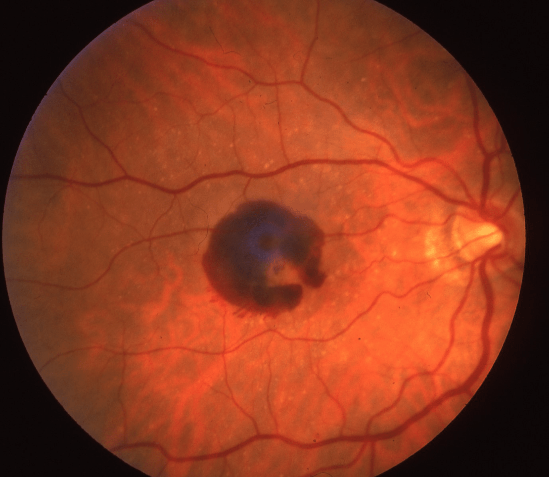
What causes macular hemorrhage?
Submacular hemorrhage frequently results from a choroidal neovascular membrane secondary to age-related macular degeneration. Other conditions associated with CNVM, including myopia, trauma, ocular histoplasmosis and angioid streaks, can also lead to submacular hemorrhage.Jan 6, 2014
How serious is a retinal bleed?
When retinal hemorrhages occur, symptoms range from the undetectable to severe vision problems. Vision problems are often temporary, but in some instances, they can be permanent. If you are experiencing vision problems of any kind, it is important that you seek treatment from professional, experienced eye doctors.Apr 13, 2017
How do you get retinal hemorrhage?
A retinal hemorrhage occurs when blood vessels in the retina begin to bleed. A hemorrhage, or bleeding, happens when these tiny blood vessels are damaged by injury or disease. Retinal hemorrhage can be caused by diabetes, high blood pressure, head injuries and even sudden changes in air pressure.
What does it mean when your retina is bleeding?
Vitreous hemorrhage is a condition where bleeding into the vitreous occurs and clouds your eye's view. Bleeding usually comes from the blood vessels that feed the retina at the back of the eye. Changes caused from complications of diabetes are the most common cause of vitreous hemorrhage in adults.
How long does a retinal hemorrhage last?
Retinal Nerve Fiber Layer (RNFL) Hemorrhages Flame shaped hemorrhages: These hemorrhages are diffuse, found in the posterior pole, and last approximately 6 to 12 weeks.Feb 17, 2022
How is retinal hemorrhage treated?
Treatment. Retinal hemorrhages, especially mild ones not associated with chronic disease, will normally reabsorb without treatment. Laser surgery is a treatment option which uses a laser beam to seal off damaged blood vessels in the retina.
Can you recover from retinal hemorrhage?
You may not need treatment, because a retinal hemorrhage often heals by itself. If your bleeding is caused by a medical condition, your healthcare provider will treat that illness. You may need any of the following: Steroid medicine may be given if you have macular degeneration.
Can stress cause retinal hemorrhage?
As we know, stress often has a negative impact on a person's physical well-being and can even be hard on the eyes. If you frequently experience stress you might wonder, can stress cause retinal detachment? The simple answer is no, stress cannot cause retinal detachment.
Can blood thinners cause retinal hemorrhage?
Anticoagulant and antiplatelet drugs may increase the risk for retinal or subretinal bleeding by 50% in people who have a combination of neovascular age-related macular degeneration (nAMD) and hypertension, researchers say.Feb 17, 2016
Is bleeding in the back of the eye serious?
Eye bleeding deeper or at the back of the eye may sometimes cause redness. Bleeding in the eye can happen for several reasons. Most of the time, you will not have blood leaking from your eye. Depending on the location in the eye, bleeding can be harmless or it may lead to complications if left untreated.
What is subretinal hemorrhage?
Subretinal hemorrhages are frequently focal, dense, round, deep, and often centered on the fovea, associated with the occurrence or the extension of a lacquer crack. There is no associated serous retinal detachment. On FA, the hemorrhages may totally obscure the crack, but ICG angiography may help detect the rupture in the Bruch's membrane ...
What causes sudden loss of vision?
Sudden visual loss may occur as a result of macular hemorrhage at the site of the fovea when Bruch's membrane decompensates (Fuchs' fleck). Gradual visual loss and metamorphopsia may result from breaks in Bruch's membrane.
How effective is vitrectomy?
Vitrectomy surgery has been effective in reducing vision loss when traction retinal detachment threatens the center of the macula or vitreous hemorrhage fails to clear. The use of laser delivered from within the eye at the time of surgery and other advanced surgical techniques make vitrectomy a valuable procedure to prevent vision loss and restore vision.
Where are atrophic changes found?
Atrophic changes are found in the sclera, around the disc, in the choroid, in Bruch's membrane and the retina in the central area, and in the peripheral parts of the retina. The atrophic thinning of the sclera is confined to its posterior half, where it may be very attenuated. Atrophic choroidal changes are also mainly in the central area of the fundus, with gradual disappearance of the small vessels. Lacunae appear, making irregular areas of atrophy around the disc and at the macula. The fundus changes appear to be primary in nature and are not just due to the mechanical effects of stretching. Clinically, the retinal changes are secondary to changes in the choroid. Genetic factors play a prominent part, possibly by means of primary changes in the RPE.
What is SCORE study?
The SCORE study is a multicenter, randomized, phase III trial to compare the effectiveness and safety of standard care versus triamcinolone acetonide injection for the treatment of macular edema associated with CRVO and BRVO. Standard care is defined as observation of macular edema in CRVO and for BRVO immediate observation of eye with dense macular hemorrhage and then grid photocoagulation when clearing of hemorrhage permits or immediate grid photocoagulation in eyes without dense macular hemorrhage.
What is the perfusion status of a CRVO?
The CVOS classified the perfusion status of a CRVO as perfused, nonperfused, or indeterminate based on fluorescein angiographic characteristics. Angiographic assessment of perfusion status in CRVO is based on the photographic protocol from the CVOS which used a conventional wide-angle fundus camera with sweeps of the midperiphery 30 seconds after intravenous injection of sodium fluorescein.
Is visual acuity normal?
Visual acuity. Visual acuity may be normal for a long time. Sudden loss may occur when there is hemorrhaging at the macula. Visual loss may also be gradual, because of pigmentary changes and lacquer cracks in Bruch's membrane. Ultimately there is severe visual loss as a result of extensive chorioretinal atrophy.
What causes retinal hemorrhage?
Retinal hemorrhage can have many causes, including: 1 Medical conditions, such as hypertension, diabetes, lupus, anemia, infections and leukemia. 2 Eye diseases, such as macular degeneration, which is also known as age-related macular degeneration (AMD or ARMD). The macula is the small area at the center of the retina that is responsible for our sharpest vision. 3 Head injuries caused by accidents. 4 Shaken baby syndrome or other forms of child abuse. 5 A sudden change in air pressure during activities such as mountain climbing or scuba diving. The change in air pressure may decrease the amount of oxygen available to the retina. 6 Some medications, such as blood thinners. 7 On very rare occasions, straining due to constipation, severe coughing or vomiting.
Why is it important to protect your retina?
It's essential to protect your retina so you can enjoy a lifetime of good eyesight. Many problems with the retina can be detected by your eye doctor before you notice symptoms.
What is the most common form of macular degeneration?
This eye disease causes a loss of central vision that occurs in two forms: “dry” (atrophic) and “wet” (exudative). The “ Dry ” form of macular degeneration is the most common one.
Can AMD cause vision loss in elderly?
It is referred to as Age-related Macular Degeneration. It is the leading cause of severe vision loss in elders above the age of 60. If someone in your family has or had this disease you may be at a higher risk for developing it. Consult with your eye doctor about such risk.
What causes a zigzag line in the retina?
The “wet” form of macular degeneration is caused by the growth of abnormal blood vessels from the choroid underneath the macula. This is called Choroidal Neovascularization. These blood vessels leak blood and fluid into the retina, leading to distorted vision that makes straight lines look zigzag and wavy, as well as blind spots and loss of central vision. These abnormal blood vessels and the bleeding from them eventually form a scar, leading to permanent loss of central vision.
Can AMD cause blurry vision?
In its early stages, AMD may not have signs and may be unrecognized. Gradually it progresses or affects both eyes. The first symptom of macular degeneration is usually Blurred Vision with a dim, blurry spot in the centre of your eyesight. This spot may get bigger or darker with time.
How does Visudyne work?
This is a two-step treatment. It involves a light-sensitive drug (Visudyne), which is used to damage abnormal blood vessels. Your Eye Doctor will inject the drug into the bloodstream to be absorbed by the abnormal blood vessels in the eye. He/she will then shine a cold laser into the eye to activate the drug. And damaging the abnormal blood vessels.
Does low nutrition cause AMD?
Researchers have linked nutrients such as lutein and zeaxanthin, vitamin C, vitamin E and zinc to reducing the risk of certain eye diseases , including AMD.
What is the condition of the macula?
Age-related Macular Degeneration (AMD) is a disease of the retina. This eye disease happens when there are changes to the macula, which is a small portion of the retina that is located on the inside back layer of the eye. It occurs when a part of the retina called the macula is damaged. With AMD one loses central vision. The person cannot see fine details, whether looking at something close or far. But the peripheral (side) vision will stay normal. For instance, imagine you are looking at a fan. With AMD, you might see the wings of the fan but not the central part.
Can AMD cause blurry vision?
In its early stages, AMD may not have signs and may be unrecognized until it progresses or affects both eyes. The first symptom of macular degeneration is usually Blurred Vision with a dim, blurry spot in the center of your vision. This spot may get bigger or darker with time.
What is the yellow pigment in the macula called?
The “dry” form of macular degeneration is identified by the presence of yellow deposits, called drusen, in the macula. A few small drusen may not cause any changes in vision. However, as they grow in numbers and size, they may lead to a dimming or distortion of vision. Then people start noticing when they read.
What causes a zigzag line in the retina?
The “wet” form of macular degeneration is caused by the growth of abnormal blood vessels from the choroid underneath the macula. This is called Choroidal Neovascularization. These blood vessels leak blood and fluid into the retina, leading to distorted vision. That makes straight lines look zigzag and wavy, as well as blind spots and loss of central vision. These abnormal blood vessels and bleeding from them eventually form a scar, leading to permanent loss of central vision.