What causes angiomatoid fibrous histiocytoma? Histiocytomas develop when histiocytes (normal immune cells found in various parts of your body) grow rapidly and produce more histiocytes. AFH has been linked to genetics, inherited conditions and radiation treatment, but the exact cause is unknown.
Full Answer
Does aneurysmal fibrous histiocytoma recur?
Aneurysmal fibrous histiocytoma (AFH) is a type of fibrohistiocytic tumour. We present a case of a patient who presented with a skin papule on the thigh region. Histopathological examination following total excision of the lesion revealed an AFH. No sign of recurrence was present 6 months after surgery.
What causes Angiomatoid fibrous histiocytoma?
(Etiology) The exact cause and mechanism of Angiomatoid Fibrous Histiocytoma formation is unknown. It is considered that the origin may be muscular-related, or connected to certain types of cells present in the lymph nodes, which synthesize collagen
What is the prognosis of fibrous histiocytoma?
Prognosis is excellent when lesions are well-defined and located just below the skin surface and are completely removed by surgical procedures; this reduces their recurrence risk too. The probability of Angiomatoid Fibrous Histiocytoma recurrence is moderate at 11%, after surgical removal of the tumor.
What are the signs and symptoms of fibrous histiocytoma?
Note: Benign fibrous histiocytoma rarely occurs in your mouth. But when it does, you may experience dysphasia (difficulty swallowing), dyspnea (shortness of breath) or difficulty speaking. Fever. Weight loss. Anemia.

What is treatment for benign fibrous histiocytoma?
The treatment of choice for BFH is wide resection of the tumor, which results in an excellent prognosis and low recurrence rate. In agreement with cases reported in the literature, our case confirms that wide excision is adequate to prevent the recurrence of the tumor.
What causes fibrous histiocytoma?
What causes angiomatoid fibrous histiocytoma? Histiocytomas develop when histiocytes (normal immune cells found in various parts of your body) grow rapidly and produce more histiocytes. AFH has been linked to genetics, inherited conditions and radiation treatment, but the exact cause is unknown.
What is aneurysmal fibrous histiocytoma?
Aneurysmal fibrous histiocytoma (AFH; also referred to as aneurysmal benign fibrous histiocytoma) is a rare variant of cutaneous cellular fibrous histiocytoma. It accounts for 0.3% of soft tissue sarcomas/borderline tumors, and is classified by the WHO under tumors of uncertain differentiation [1, 2].
Is malignant fibrous histiocytoma curable?
Malignant fibrous histiocytoma is a potentially curable disease. The most important part of the treatment is complete surgical removal, usually followed by adjuvant radiation therapy.
How do you treat histiocytoma in dogs?
Treatment for Histiocytomas in Dogs In most cases, histiocytomas in dogs require no treatment, especially if your dog does not experience discomfort. Since we expect histiocytomas to disappear in less than 3 months, growths that last longer are surgically removed and tested to confirm the tumor type.
Do histiocytomas go away?
The histiocytoma is a benign skin growth that usually goes away by itself within a couple of months.
What is cellular fibrous histiocytoma?
Abstract. Cellular fibrous histiocytoma, a variant of fibrous histiocytoma, is a designation used for lesions showing increased cellularity with a fascicular growth pattern and frequent extension into the subcutis.
What is aneurysmal Dermatofibroma?
Aneurysmal dermatofibroma (ADF, also referred to as aneurysmal fibrous histiocytoma) is a rare soft-tissue tumor with a local recurrence rate as high as 20% after traditional surgical excision (TSE).
Do Dermatofibromas recur?
Background: Cellular dermatofibromas, a variant of dermatofibroma, are reported to recur at rates of 26% to 50%.
What is a malignant fibrous histiocytoma?
Listen to pronunciation. (muh-LIG-nunt FY-brus HIS-tee-oh-sy-TOH-muh) A type of cancer that usually forms in the soft tissue, but it may also form in bone. It can occur anywhere in the body, but it usually occurs in the legs (especially the thighs), arms, or back of the abdomen.
Is a Histiocytoma cancerous?
A malignant fibrous histiocytoma is a type of cancer that's most often found in soft tissue such as muscles and tendons. In very rare cases it starts in bones. When this happens, it's most often in the leg bones. Cancer cells start to take over and destroy the bone.
Is Stage 4 a sarcoma terminal?
A sarcoma is considered stage IV when it has spread to distant parts of the body. Stage IV sarcomas are rarely curable. But some patients may be cured if the main (primary) tumor and all of the areas of cancer spread (metastases) can be removed by surgery. The best success rate is when it has spread only to the lungs.
What are the possible Complications of Angiomatoid Fibrous Histiocytoma?
Deep-seated tumors ( those buried in the body tissues), may create problems for adjoining tissues and organs, hindering their normal function
How is Angiomatoid Fibrous Histiocytoma Diagnosed?
The diagnostic tests may vary based on location of the tumor. Angiomatoid Fibrous Histiocytoma is diagnosed by:
What are the symptoms of AFH?
The signs and symptoms of AFH include: Most tumors are asymptomatic, with no indication of pain or any external sensation. The well-defined benign nodules (usually containing multiple cysts) grow at a slow rate.
Why does fibrocytoma form?
They are occasionally thought to occur in response to an injury/trauma
What is the surgical treatment for AFH?
Surgical treatment: Wide surgical excision of AFH tumor with removal of the entire lesion is to be performed. If the tumor is not fully removed, then it might recur
Where does AFH occur?
AFH usually occurs in the limbs (at the knee and elbow joints). The head, neck, and trunk are other regions where it is noticed from time to time. There have been rare instances of AFHs being found in the lungs, abdominal cavities, and female genitalia.
What is a histopathological study?
Histopathological studies conducted on a biopsy specimen. X-ray studies (of the affected region, in non-skin tumors) MRI scan (of the affected region, in non-skin tumors) Many clinical conditions may have similar signs and symptoms.
What are the findings of AFH?
Consistent with the MR appearance in the 6 cases reported in the literature, our case also demonstrated (a) multiple internal cystic areas, (b) an enhancing fibrous pseudocapsule which was markedly hypointense on T1- and T2WI, and (c) foci of susceptibility artifacts representing hemosiderin. Conversely, inconsistent findings in the 6 previously reported cases included pattern of enhancement, if present or reported, and the presence, or absence of fluid-fluid levels. A summary and comparison of the findings in the cases are presented in Table 1. Taken together, these findings can be found with metastasis, hemangioma, hematoma, malignant fibrous histiocytoma, myxoid chondrosarcoma, leiomyosarcoma with necrosis, malignant ossifying fibromyxoid tumor, and various other sarcomas [14,15,16,17]. MR has traditionally been used for staging and followup, however a recent report highlights the potential utility of positron emission tomography in staging [18].
What is AFH diagnosis?
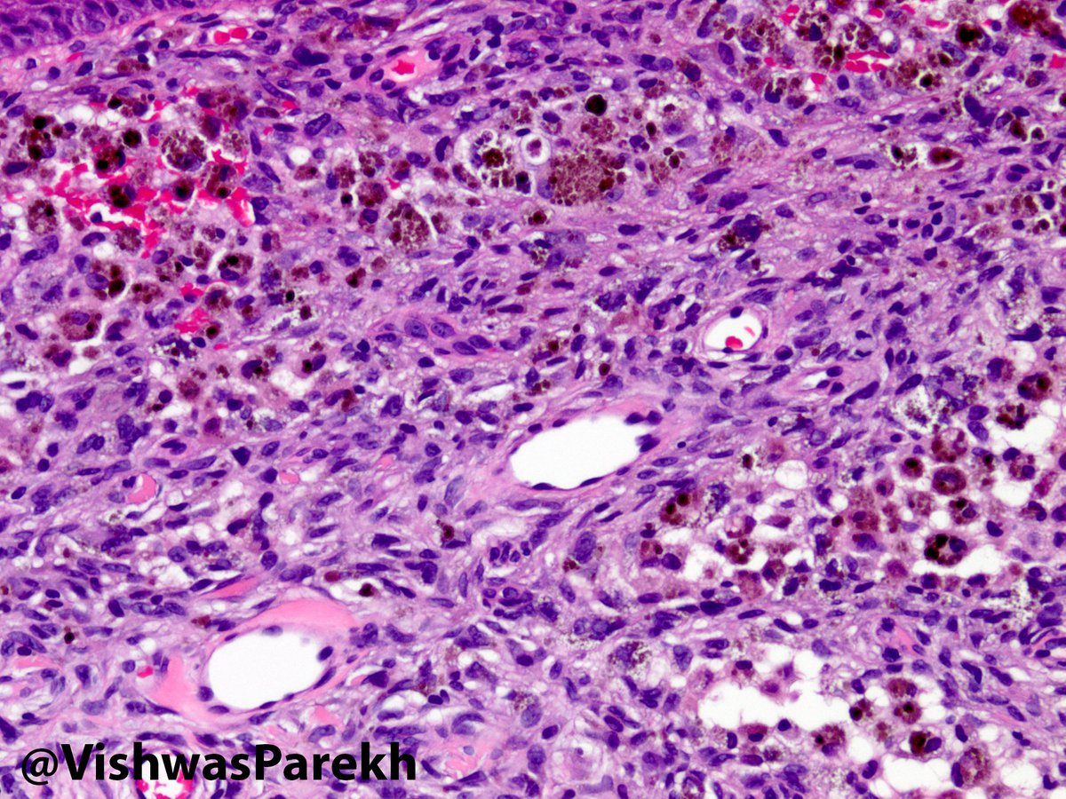
The diagnosis of AFH is made based on histopathology and immunohistology. Macroscopically, AFH is generally firm and circumscribed. The characteristic microscopic appearance includes distributions of ovoid to spindle cells with bland, vesicular nuclei, lymphoplasmocytic infiltrate with intervening blood-filled cystic spaces, and a fibrous pseudocapsule [22,23]. Immunohistochemistry variably demonstrates positivity for desmin, CD68 and CD 99 [24]. Lastly, cytogenetic analysis has recently added to the diagnosis of AFH, with the EWSR1-CREB1 fusion gene present in the majority of AFH [4].
Is angiomatoid malignant fibrous histiocytoma a histogenesis?
When Enzinger initially described “angiomatoid malignant fibrous histiocytoma” in 1979 [1], the histogenesis was controversial. Today, the precise line of differentiation remains unknown, but this entity is no longer termed “malignant” due to its benign microscopic appearance and favorable prognosis [2]. Additionally, the 2002 World Health Organization (WHO) classification removed it from the malignant fibrous histiocytoma subtype of sarcoma (now synonymous with undifferentiated pleomorphic sarcoma) and placed it under the category of tumors of uncertain differentiation as angiomatoid fibrous histiocytoma [3].
Is Angiomatoid fibrous histiocytoma a hematoma?
Angiomatoid fibrous histiocytoma (AF H) is a rare soft tissue tumor most commonly occurring in children, adolescents, and young adults. Clinically and radiographically the lesion is easily confused with a hematoma, soft tissue hemangioma, or malignant fibrous histiocytoma. While the lesion is rare, due to the potential for local recurrence and metastasis, it is imperative to consider this lesion in the differential diagnosis of a soft tissue mass in a child or adolescent. Here, we present the clinical, radiologic, and pathologic findings of a case of AFH.
Is AFH a preoperative diagnosis?
Making a pre-operative diagnosis of AFH is challenging with no distinct clinical or imaging findings to lead to diagnosis. Nonetheless, soft tissue malignancies have been stratified according to age and location to suggest one diagnosis more than another. For example, 80 % of of rare malignant soft tissue masses in a 6 – 15 year old patient in the hand and wrist, upper extremity, axilla and shoulder, lower extremity, hip, groin and buttocks, or trunk are either most likely or second most likely to be AFH according to a study of 39,179 soft tissue lesions over a 10 year period [12,13].
Is fibrous histiocytoma a tumor?
Angiomatoid fibrous histiocytoma is a rare soft tissue tumor with intermediate malignant potential. While nonspecific, a mass with MR features including cystic areas, an enhancing fibrous pseudocapsule, and internal foci blood products in the extremity of a child or adolescent should prompt the consideration of AFH in the differential. Wide surgical excision with clear margins and post-excisional monitoring is warranted.
How big is a dermatofibroma?
Giant dermatofibromas have been described, ranging in size from 35 to 300mm. Other histologic variants of dermatofibroma include lipidized, hemosiderotic, keloidal, granular cell, palisading, atrophic, clear cell, myxoid, lichenoid, balloon cell, and signet-ring cell.
Can dermatofibroma be a male or female?
DF may occur in any individual of either sex with no racial predilection. Women are much more likely to develop these lesions than men, with a 1:4 male:female ratio of dermatofibroma. DFs more commonly develop in young adults but can appear at any age.
Is dermatofibroma a benign disease?
There is little value in imaging studies or serologic tests with solitary lesions of DF. However, the presence of multiple dermatofibromas (defined as at least 15 ), while most commonly a benign finding, can be indicative of a state of immune suppression and has been linked with HIV infection, myasthenia gravis, systemic lupus erythematosus, and diabetes. In these instances, further serologic testing may be indicated.
How is a dermatofibroma diagnosed?
Dermatofibroma is usually easy to diagnose clinically, supported by dermoscopy. The most common dermoscopic pattern is a central white area surrounded by a faint pigment network. However different patterns may be seen in skin of colour.
What are the complications of dermatofibroma?
Because dermatofibromas are often raised lesions, they may be traumatised, for example by a razor.
What is a dermatofibroma?
A dermatofibroma is a common benign fibrous nodule usually found on the skin of the lower legs.
What are the clinical features of dermatofibroma?
A dermatofibroma usually presents as a solitary firm papule or nodule on a limb.
Can dermatofibromas be seen in men?
Dermatofibromas are mostly seen in adults. People of every ethnicity can develop dermatofibromas. Ordinary dermatofibromas are more common in women than in men, although some histologic variants are more commonly identified in males.
Can dermatofibroma be removed surgically?
Usually, only reassurance is needed. If it is nuisance or causing concern, the lesion can be removed surgically. Recurrence is common as the lesion often extends beyond the clinical margin.
Abstract and Figures
Aneurysmal fibrous histiocytoma (AFH) is a rare variant of cutaneous fibrous histiocytoma, with low malignant potential and infrequent metastatic progression. We present the case of a 19-year-old female with a large AFH of the neck metastatic to soft tissue and treated with radiation therapy and molecularly targeted therapy.
References (11)
ResearchGate has not been able to resolve any citations for this publication.
