Medication
Treatment can range from prescription medications to home remedies to more extensive treatment for complications that arise. Treatment also depends on several factors, such as the person’s age, how they tolerate medications, and the severity of their injury. There is also the risk for serious complications if a hyphema is not treated.
Procedures
- eye drops (steroid drops to limit swelling and/or dilating drops to assist with pain).
- patch over the impacted eye.
- bed rest.
- minimal eye motion (this suggests no reading).
- head elevated a minimum of 40 degrees when sleeping (to assist body take in blood).
- check eye pressure daily.
Self-care
Understanding Hyphema: Symptoms, Treatment & More
- Possible Complications of a Hyphema. When a hyphema is present, there are certain complications that a person can experience. ...
- Getting a Diagnosis. A complete medical history is needed to make a diagnosis. ...
- Treatment Options. There are different treatment options that can be used to treat a hyphema. ...
- Hyphema Prevention. ...
Nutrition
- Eat a healthy diet that includes lots of fruits and vegetables.
- Get regular exercise.
- Stay hydrated.
- Limit caffeine consumption.
How is hyphema treated?
How to treat a hyphema?
What are the possible complications of hyphema?
What causes high eye pressure and how to reduce it?

What is the treatment for hyphema?
Medical treatment for an isolated hyphema typically is topical. Topical corticosteroids (systemic for severe cases) may reduce associated inflammation, although the effect on the risk for rebleeding is debatable. Topical cycloplegic agents are also useful for patients with significant ciliary spasm or photophobia.
What medications is contraindicated in hyphema?
The antiplatelet effect of aspirin tends to increase the incidence of rebleeding in patients with traumatic hyphema and should be strictly avoided. Nonsteroidal anti-inflammatory drugs (NSAIDs) with analgesic activity, such as mefenamic acid (Ponstel) or naproxen (Aleve), share this deleterious antiplatelet effect.
Why is atropine given in hyphema?
Secondly, topical atropine (1%) is indicated in hyphemas occupying more than 50% of the anterior chamber to reduce the incidence of posterior synechiae formation and avoid pupillary block.
What doctors can take care of hyphema?
Hyphema is usually caused by a blunt injury to the face or eye. Because this is a serious injury, you will need to see an eye specialist (ophthalmologist) right away.
Which disorder is a common complication of a hyphema?
The 2 major acute complications of hyphemas are acute intraocular hypertension and re-bleeding. [15] Acute intraocular hypertension is most likely encountered in the emergency department.
What are the complications of hyphema?
Complications of traumatic hyphema include increased intraocular pressure, peripheral anterior synechiae, optic atrophy, corneal bloodstaining, secondary hemorrhage, and accommodative impairment.
What is atropine sulfate used for?
Atropine sulfate eye drops is used to dilate the pupil before eye exams. It is also used to treat an eye condition called amblyopia (lazy eyes) and other eye conditions (eg, cycloplegia).
How do you put atropine in your eye?
Tilt your head back, look upward, and pull down the lower eyelid to make a pouch. Hold the dropper directly over your eye and place one drop into the pouch. Look downward and gently close your eyes for 1-2 minutes. Place one finger at the corner of your eye (near the nose) and apply gentle pressure for 2 to 3 minutes.
Can you use injectable atropine in the eye?
Atropine sulfate is used in the eye to dilate the pupil. It may also be used to control pain in the eye due to corneal and uveal disease and in treating secondary glaucoma.
What is the first aid in case of hyphema?
Clean the wound with mild soap and water. Rinse for several minutes under running water. Apply antibiotic ointment to prevent infection. Cover the wound with gauze or a bandage.
How is hyphema diagnosed?
How is a hyphema diagnosed?comprehensive eye exam to test your ability to see.eye pressure check.examination of inside of the eye with a special microscope called a slit lamp.a CT scan might be ordered to check for fracture of the orbit (socket) if there was trauma to the eye.
Is hyphema a medical emergency?
It can interfere with vision and cause a dangerous increase in eye pressure, in which case a hyphema is considered a medical emergency that requires urgent medical attention to protect overall eye health and minimize the risk of permanent vision loss.
What Causes Hyphema?
In many cases, a person develops a hyphema because of an injury or blow to the eye. This injury causes a tear of the iris or pupil of the eye and allows blood to accumulate.
Signs & Symptoms of Hyphema
Hyphema can cause a range of symptoms and signs. If you have a hyphema, you may experience the following symptoms and signs, including:
Can Hyphema be a Sign of Something Serious?
Yes. Hyphemas may be a sign of something more serious. It is important to seek medical care if you believe you have a hyphema.
When is Hyphema an Emergency?
A hyphema can become an emergency when a person does not receive proper treatment, and vision loss worsens. The hyphema can become more severe when a person does not receive medical care and elevate intraocular pressure.
How to Manage Hyphema Symptoms
If you have a hyphema, there are steps that you can take to lessen symptoms. You can follow the recommendations listed below:
How is Hyphema Diagnosed?
If you believe that you have a hyphema, visit your local eye clinic and speak to the ophthalmologist about any symptoms.
What Treatment Options are Available for Hyphema?
Your treatment options will vary according to the cause of the hyphema and severity grading.
How to treat hyphema?
There are different treatment options that can be used to treat a hyphema. Protect the affected eye by wearing a special shield over it. Help the eye drain by raising the head of the bed. Rest and avoid physical activity for the specified amount of time.
What is needed to make a diagnosis of hyphema?
A complete medical history is needed to make a diagnosis. It is important to determine if the person recently experienced any issues that could cause bleeding in the eye or trauma that affected the eye. The doctor will physically examine the eye to look for a hyphema or any signs of trauma.
What causes hyphema in the eye?
A hyphema occurs when blood collects in the eye, which can lead to a blockage of vision. ( Learn More) Trauma is the usual cause of this condition.
How long does it take for hyphema to heal?
When hyphema is mild, it may heal without the need for medical intervention. In mild cases, the healing time is usually about one week. If the person has swelling in the eye, the doctor might prescribe eye drops for this. They work to reduce discomfort and pain. These should be taken exactly as directed.
What is the grade of hyphema?
There are different severity grades associated with a hyphema. Grade 0 : A microscope is necessary to see the red blood cells, but the blood pooling is not visible. Grade 1: The pooled blood fills less than a third of the chamber. Grade 2 The pooled blood fills up to half of the chamber.
Can hyphema be treated?
There is also the risk for serious complications if a hyphema is not treated. ( Learn More) There are several tests that the doctor might perform to make an accurate diagnosis. If trauma was the cause, it is also important to perform additional testing to look for other potential issues, such as a concussion.
Is a broken blood vessel a hyphema?
It is important to note that a broken blood vessel is a separate issue referred to as subconjunctival hemorrhage. This hemorrhage is usually harmless and not painful. However, a hyphema usually causes pain. Without prompt and proper treatment, a hyphema can lead to permanent vision issues.
What is the best treatment for isolated hyphema?
Medical treatment for an isolated hyphema typically is topical. Topical corticosteroids (systemic for severe cases) may reduce associated inflammation, although the effect on the risk for rebleeding is debatable. Topical cycloplegic agents are also useful for patients with significant ciliary spasm or photophobia.
What is the physical exam for hyphema?
The examination for a hyphema should consist of a routine ophthalmic work-up (visual acuity, pupillary examination, intraocular pressure, slit-lamp examination) as well as a gonioscopy to evaluate the condition of the angle and trabecular meshwork.
What are the symptoms of hyphema?
Typically patients will complain of associated blurry vision and ocular distortion. In the setting of trauma or secondary intraocular pressure elevation, patients may complain of pain, headahce, and photophobia.
How much of hyphemas have an increase in IOP?
Only 13.5% of Grade I to II hyphemas had an IOP increase, but 27% of those with Grade III hyphemas has an IOP increase.
What is ACA in hyphema?
ACA is a derivative and analog of the amino acid lysine, and competitively inhibits plasmin, an important protein enzyme involved in fibrinolysis.
What causes hyphema in the anterior chamber?
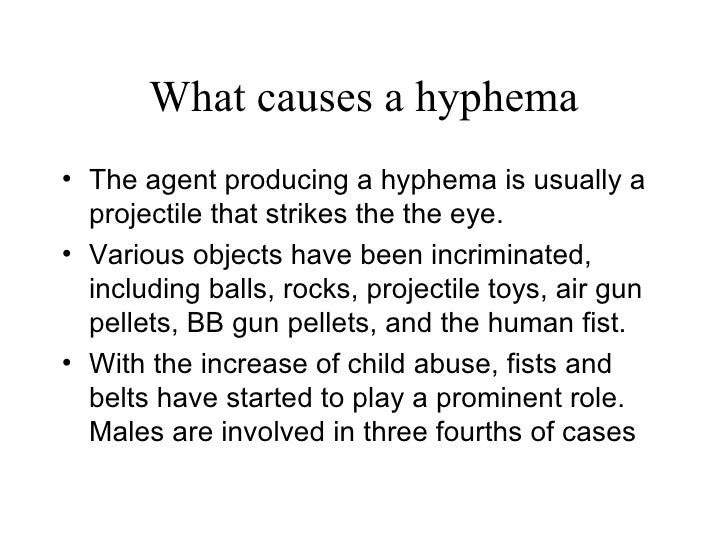
Blunt trauma is the most common cause of a hyphema. Compressive force to the globe can result in injury to the iris, ciliary body, trabecular meshwork, and their associated vasculature. The shearing forces from the injury can tear these vessels and result in the accumulation of red blood cells within the anterior chamber.
What is the best treatment for ciliary spasm?
Topical cycloplegic agents are also useful for patients with significant ciliary spasm or photophobia. In the setting of intraocular pressure elevation, topical aqueous suppressants are first line agents for pressure management (beta-blockers and alpha-agonists).
How to treat hyphema?
Treatment of a hyphema involves encouraging the blood to clear, treating any elevation in intraocular pressure, and trying to prevent additional bleeding. A period (often of several days) of limited activity or bed rest is recommended. The head is kept in an elevated position even during sleep, and the eye is protected with a shield.
What are the symptoms of hyphema?
What are the symptoms of a hyphema? Typical symptoms include eye pain, blurring or loss of vision, and photophobia or light sensitivity. Sometimes the accumulation of blood is visible to the naked eye.
What is hyphema in the eye?
Rarely, a hyphema occurs as a consequence of medical problems that can affect the eye such as juvenile xanthogranuloma and cancer. Fig. 1: A hyphemia is an accumulation of blood in the space between the cornea and the iris.
Where is hyphema located?
A hyphema is an accumulation of blood in the anterior chamber of the eye. This is the space between the cornea (front clear surface of the eye) and iris (colored part of the eye).
What to do if blood does not clear?
If the blood does not clear after a suitable period of time and conservative medical treatment, or if there is an uncontrollable rise in intraocular pressure, surgery may be performed to remove the blood .
Can you take ibuprofen for hyphema?
Steroid eye drops are often prescribed to limit inflammation and dilating drops can help alleviate pain. Patients with hyphemas should not take any products containing aspirin or ibuprofen.
Can hyphema cause glaucoma?
The blood from a hyphema can clog the drainage canals of the eye causing a rise in intraocular pressure. Prolonged elevated intraocular pressure can lead to glaucoma and irreversible optic nerve damage. This can be more common in those patients with sickle cell anemia.
Medicines
Cycloplegics: This medicine relaxes your eye muscles and decreases your pain so your eye can heal.
Follow up with your ophthalmologist in 1 day
Write down your questions so you remember to ask them during your visits.
Self-care
Rest: Rest when you feel it is needed. Raise the head of your bed, or rest in a recliner. This will help decrease the pressure in your eye.
Further information
Always consult your healthcare provider to ensure the information displayed on this page applies to your personal circumstances.
What is the best treatment for IOP?
Systemic Agents. Systemic carbonic anhydrase inhibitors are quite effective. Acetazolamide or methazolamide can be used in both children and adults.
Can prostaglandins be used for hyphema?
The use of prostaglandin analogs in cases of traumatic hyphema has not been well studied. At the present time, there appears to be no absolute contraindication, although increased intraocular inflammation may occur and would be undesirable in eyes with traumatic hyphemas.
Can antifibrinolytics be minimized?
It is important that the patient understand that certain side effects with antifibrinolytics are to be expected , but can be minimized . It is also important for the patient to understand that the concern is more likely due to the magnitude of a rebleed and not merely its presence.

Diagnosis
Clinical Diagnosis
Management
Acknowledgements
Specialist to consult
Additional Resources
See Also
- Laboratory test
All African American patients with a hyphema should be screened for sickle cell trait or disease with a sickle cell prep. A greater risk of trabecular meshwork obstruction with red blood cells exists while in the sickled state. Resultant elevated intraocular pressure levels put the patient at … - Differential diagnosis
1. Traumatic hyphema: Blunt trauma to the eye may result in injury to the iris, pupillary sphincter, angle structures, lens, zonules, retina, vitreous, optic nerve, and other intraocular structures. Blunt trauma to the eye is associated with a rapid, marked elevation in intraocular pressure with sudd…