What does it mean when your esophagus is thickening?
Barrett's esophagus is a condition in which the flat pink lining of the swallowing tube that connects the mouth to the stomach (esophagus) becomes damaged by acid reflux, which causes the lining to thicken and become red.
Can esophageal stricture be cured?
Various treatment methods can treat benign esophageal strictures effectively. However, esophageal strictures can reoccur, and people may need to have repeat dilations to reopen the esophagus. According to one source, 30 percent of people who have an esophageal dilation will require another dilation within a year.
How do you treat an esophageal stricture at home?
Lifestyle and home remediesAvoid foods that may increase reflux. ... Use good pill-taking habits. ... Lose weight. ... If you smoke, quit. ... Avoid certain medications. ... Avoid stooping or bending, especially soon after eating.Avoid lying down after eating. ... Raise the head of your bed.
Can esophagus repair itself?
The esophagus is a complex organ comprising nonredundant tissue that does not have the ability to regenerate. Hence, surgical repair and/or replacement of the esophagus are the only feasible treatment options upon extensive structural damage.
What foods help heal the esophagus?
Fiberfresh, frozen, and dried fruit.fresh and frozen vegetables.whole-grain breads and pasta.brown rice.beans.lentils.oats.couscous.More items...
How do you fix esophagus problems?
How are esophageal disorders treated?Antacids, proton pump inhibitors and histamine receptor (H2) blockers to reduce stomach acid.Endoscopic dilation to open a narrowed esophagus or relax a sphincter muscle.Botulinum toxin (Botox®) injections to temporarily stop esophageal spasms or relax the sphincter muscle.More items...•
How long does it take for esophagus to heal?
Untreated esophagitis can lead to ulcers, scarring, and severe narrowing of the esophagus, which can be a medical emergency. Your treatment options and outlook depend on the cause of your condition. Most healthy people improve within two to four weeks with proper treatment.
How long does it take for omeprazole to heal esophagitis?
The more severe the grade of esophagitis, the stronger the acid suppression and the longer the duration of therapy required to heal the mucosal lesions. uniformly found that omeprazole 40 mg/day will successfully heal nearly 90% of these patients within 12 weeks.
How to Treat Inflammation of the Esophagus Naturally
Esophagitis: Types, Symptom, and Risk Factors - Healthline
What is esophageal stricture?
What is an esophageal stricture? An esophageal stricture is an abnormal tightening or narrowing of the esophagus. Your esophagus is a muscular tube that connects the throat to the stomach, carrying food and liquid. A stricture narrows the esophagus, making it more difficult for food to travel down the tube. In severe cases, even drinking liquid can ...
What is the most common treatment for strictures?
Esophageal dilation is the most common treatment for strictures. Your provider uses a balloon or dilator (a long plastic or rubber cylinder) to widen the narrow area of the esophagus.
What is a stricture in the neck called?
This type of stricture is called a peptic stricture. Radiation therapy: Treatment for cancer in the head, neck or chest can cause strictures up to a year and a half later. Surgery: A procedure in the esophagus can leave inflammation and scarring, causing a stricture. Other causes: Ulcers, some medications (for example, ...
What is the name of the tumor that can cause strictures?
Esophageal cancer: When abnormal cells divide or grow out of control in esophageal tissue, the tumor can cause strictures.
What is complex stricture?
Complex strictures are longer and leave a narrower opening. They are not straight or symmetrical and have uneven surfaces and margins.
What to do if you have stricture?
If you have a stricture, see a healthcare provider. They can determine how narrow your esophagus is and treat any underlying conditions, such as GERD.
How does endoscope work for GERD?
Your provider will also numb your throat. If you have GERD, you may receive medication that makes your body produce less acid . Then your provider inserts an endoscope down your throat and into your esophagus.
Can a CT chest report show esophageal thickening?
Since mural thickening is not easily diagnosed by esophagram or by endoscopy, it is important that it be included in the CT chest report. Accurate description of the esophageal mural thickening will encourage referring physicians to consider infection, inflammation, and neoplasm – rather than fibrotic stricture or abnormal motility – as ...
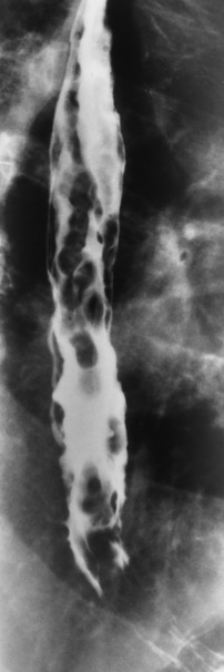
Is esophageal thickening a CT finding?
Imaging description Esophageal mural thickening is a nonspecific finding by CT chest. Mural thickening may be diffuse, segmental, or focal. It may occur in any segment of the esophagus, although it is more common distally. Intravenous contrast material administration is helpful in the CT evaluation of esophageal mural thickening. Esophagitis is more likely than esophageal carcinoma when uniform, circumferential mural thickening involves a long segment of the esophagus (Figure 41.1) [1]. Esophageal carcinoma is more likely when irregular, asymmetric mural thickening involves a short segment of the esophagus (Figure 41.2). Lymphadenopathy supports the diagnosis of esophageal cancer. Cancers of the mid and upper esophagus typically metastasize to paratracheal lymph nodes; cancers of the lower esophagus typically spread to gastrohepatic ligament lymph nodes [2]. Importance Esophageal mural thickening is never a normal finding. Since mural thickening is not easily diagnosed by esophagram or by endoscopy, it is important that it be included in the CT chest report. Accurate description of the esophageal mural thickening will encourage referring physicians to consider infection, inflammation, and neoplasm – rather than fibrotic stricture or abnormal motility – as the cause of any dysphagia reported by the patient. Over the last decade, eosinophilic esophagitis has gained greater recognition as a cause of esophagitis (Figure 41.3) [3].
Is esophageal carcinoma more likely to be asymmetric?
Esophageal carcinoma is more likely when irregular, asymmetric mural thickening involves a short segment of the esophagus (Figure 41.2). Lymphadenopathy supports the diagnosis of esophageal cancer. Cancers of the mid and upper esophagus typically metastasize to paratracheal lymph nodes; cancers of the lower esophagus typically spread ...
How to treat esophageal obstruction?
If your esophageal cancer has narrowed your esophagus, a surgeon may use an endoscope and special tools to place a metal tube (stent) to hold the esophagus open.
How does a surgeon remove the esophagus?
Surgery to remove a portion of the esophagus (esophagectomy). During esophagectomy, the surgeon removes the portion of your esophagus that contains the cancer, along with a portion of the upper part of your stomach, and nearby lymph nodes. The remaining esophagus is reconnected to your stomach. Usually this is done by pulling the stomach up to meet the remaining esophagus.
How does esophageal cancer surgery work?
During esophagectomy, your surgeon removes the portion of your esophagus that contains the tumor, along with a portion of the upper part of your stomach, and nearby lymph nodes. The remaining esophagus is reconnected to your stomach. Usually this is done by pulling the stomach up to meet the remaining esophagus.
What is the procedure that involves inserting a long, flexible tube (endoscope) down your throat and into?
Endoscopy . An endoscopy procedure involves inserting a long, flexible tube (endoscope) down your throat and into your esophagus. A tiny camera on the end of the endoscope lets your doctor examine your esophagus, stomach and the beginning of your small intestine (duodenum). Tests and procedures used to diagnose esophageal cancer include:
What is the purpose of a scope in an endoscopy?
Using a scope to examine your esophagus (endoscopy). During endoscopy, your doctor passes a flexible tube equipped with a video lens (videoendoscope) down your throat and into your esophagus. Using the endoscope, your doctor examines your esophagus, looking for cancer or areas of irritation.
Why do you need a feeding tube?
Providing nutrition. Your doctor may recommend a feeding tube if you're having trouble swallowing or if you're having esophagus surgery. A feeding tube allows nutrition to be delivered directly to your stomach or small intestine, giving your esophagus time to heal after cancer treatment.
What tests are done to determine if esophageal cancer is spread?
Tests may include: Bronchoscopy. Endoscopic ultrasound (EUS)
Why is my esophagus thick?
Follow Us: Possible causes of thickening of the esophagus include postirradiation scarring, reflux and monilial esophagitis, esophageal varices and esophageal carcinoma, according to the National Center for Biotechnology Information. A CT scan is sometimes used to detect thickened esophageal walls.
Why does the esophagus become narrower?
This cycle of inflammation, scarring and healing eventually causes the walls of the esophagus to become thicker, and the opening of the organ eventually becomes narrower. This type of constriction sometimes leads to a condition called dysphasia, in which a person has difficulty swallowing and develops a speech disorder due to lack of muscle control.
What is a CT scan of the esophagus?
A CT scan is sometimes used to detect thickened esophageal walls. Wake Gastroenterology explains that there are many causes for the thickening, or inflammation, of the walls in the esophagus.
What happens when acid is pushed into the esophagus?
When the gastroesophageal, or peptic, acid is repeatedly pushed into the esophagus, the walls of the organ become inflamed . The inflammation leads to scarring, which after healing, sits under newer layers of inflamed tissue.
How to identify candida esophagus?
The next step is to identify the source of these white plaques. The gold standard for the diagnosis of candida esophagus is by histological examination. Biopsy or brushing of the esophageal mucosa is taken during endoscopy, and staining by using hematoxylin and eosin is done. Candida yeast is almost always shown as pseudohyphae, which is an important basis for the diagnosis of esophageal candidiasis. The mucous membrane involved may present as desquamated parakeratosis, characterized by a group of squamous cells that have detached or are in the process of separating from the main squamous epithelium [11].
What is esophageal candidiasis?
Esophageal candidiasis (EC) is the most common type of infectious esophagitis. In the gastrointestinal tract, the esophagus is the second most susceptible to candida infection, only after the oropharynx. Immunocompromised patients are most at risk, including patients with HIV/AIDS, leukemia, diabetics, and those who are receiving corticosteroids, radiation, and chemotherapy. Another group includes those who used antibiotics frequently and those who have esophageal motility disorder (cardiac achalasia and scleroderma). Patients complained of pain on swallowing, difficulty swallowing, and pain behind the sternum. On physical examination, there is a plaque that often occurs together with oral thrush. Endoscopic examination is the best approach to diagnose this disease by directly observing the white mucosal plaque-like lesions and exudates adherent to the mucosa. These adherent lesions cannot be washed off with water from irrigation. This disease is confirmed histologically by taking the biopsy or brushings of yeast and pseudohyphae invading mucosal cells. The treatment is by systemic antifungal drugs given orally in a defined course. It is important to differentiate esophageal candidiasis from other forms of infectious esophagitis such as cytomegalovirus, herpes simplex virus, gastroesophageal reflux disease, medication-induced esophagitis, radiation-induced esophageal injury, and inflammatory conditions such as eosinophilic esophagitis. Except for a few complications such as necrotizing esophageal candidiasis, fistula, and sepsis, the prognosis of esophageal candidiasis has been good.
How old is the average person with esophageal candidiasis?
Worldwide, the median age of patients with esophageal candidiasis is 55.5 years. In the recent study, Kliemann et al. reported that the age range of esophageal candida disease patients was 21–88 years old (average 57.4 years old; standard deviation 16.7 years) [2]. However, other factors, such as the use of medications, can also contribute to changes in the average age at which the disease occurs. Therefore, the disease may occur at early ages or late. The average age of the patients at the time of diagnosis was 39.8 years [10].
Can esophageal candidiasis be treated with fluconazole?
Suspected cases of esophageal candidiasis should be treated with short-term fluconazole antifungal therapy. Esophageal candidiasis can be diagnosed when symptoms recover after fluconazole treatment. In these cases, no further investigation is required. If the infection persists, further investigation may be required and the patient will then conduct the following investigation.
Does esophageal candidiasis affect all patients?
Esophageal candidiasis affects all patients irrespective of gender. For example, a study conducted by Nassar et al. on individuals with this disease who were immunocompetent showed that there was no difference in terms of gender [10].
Is esophageal candidiasis a form of esophagitis?
Usually, esophageal candidiasis occurs in the form of superficial esophagitis. Few cases of transmural necrosis candidiasis have been reported and are associated with serious immunosuppression and neutropenia [34]or other comorbid conditions such as patients on hemodialysis [35]. The recovery of these patients is a critical concern because the mortality rate is high.
What is the treatment for Barrett's esophagus?
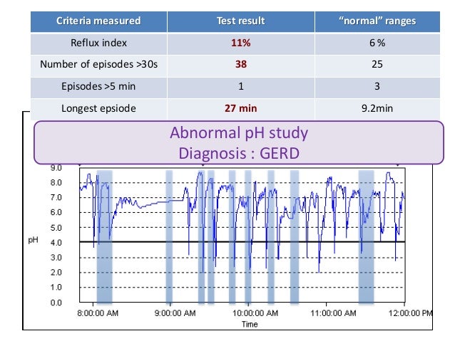
If you have Barrett’s esophagus and gastroesophageal reflux disease (GERD), your doctor will treat you with acid-suppressing medicines called proton pump inhibitors (PPIs). These medicines can prevent further damage to your esophagus and, in some cases, heal existing damage.
How long does it take to recover from esophageal surgery?
The surgery is performed at a hospital. You’ll receive general anesthesia, and you’ll stay in the hospital for 7 to 14 days after the surgery to recover.
How often should you do endoscopy surveillance?
Experts aren’t sure how often doctors should perform surveillance endoscopies. Talk with your doctor about what level of surveillance is best for you. Your doctor may recommend endoscopies more frequently if you have high-grade dysplasia rather than low-grade or no dysplasia. Read whether people with Barrett’s esophagus are more likely to develop cancer.
What is the alternative to endoscopic surgery?
Surgery called esophagectomy is an alternative to endoscopic therapies. Many doctors prefer endoscopic therapies because these procedures have fewer complications.
What is endoscopic ablative therapy?
Endoscopic ablative therapies use different techniques to destroy the dysplasia in your esophagus. After the therapies, your body should begin making normal esophageal cells. A doctor, usually a gastroenterologist or surgeon, performs these procedures at certain hospitals and outpatient centers.
How does Barrett's mucosal resection work?
In endoscopic mucosal resection, your doctor lifts the Barrett’s tissue, injects a solution underneath or applies suction to the tissue, and then cuts the tissue off. The doctor then removes the tissue with an endoscope. Gastroenterologists perform this procedure at certain hospitals and outpatient centers. You will receive local anesthesia to numb your throat and a sedative to help you relax and stay comfortable.
What is the procedure to numb your throat?
You will receive local anesthesia to numb your throat and a sedative to help you relax and stay comfortable. Before performing an endoscopic mucosal resection for cancer, your doctor will do an endoscopic ultrasound. Complications can include bleeding or tearing of your esophagus.
What is eosinophilic esophagitis?
Eosinophilic esophagitis (EoE) is a Th2, antigen driven disease in which chronic, eosinophil rich inflammation causes symptoms of esophageal dysfunction.2Esophageal symptoms due to EoE can manifest in multiple ways including heartburn/regurgitation, vomiting, dysphagia, food impactions, and even abdominal pain.
What is EoE diagnosis?
The differential diagnosis for EoE is broad and can include gastroesophageal reflux disease (GERD), parasitic and fungal infections, inflammatory bowel disease, allergic vasculitis, connective tissue disease, and other disorders associated with esophageal eosinophilia.
What are the symptoms of EoE in children?
Age-related differences in clinical presentation have been identified in children and adults.16, 17The most common presenting symptoms in adults are dysphagia, food impaction, heartburn , and chest pain with as many as 50% of adult patients initially presenting with food impaction having a final diagnosis EoE.7In contrast, children present more commonly with vomiting, heartburn, regurgitation, emesis, and abdominal pain. While younger children rarely present with dysphagia and food impaction typical of adult complaints, these presentations are commonly seen in older individuals over the age of 12 years.18Several validated tools are now available to gauge symptoms in adults and children.19–22A multicenter study demonstrates that the pediatric symptom scoring tool PEESSv2.0 can correlate with histologic changes including eosinophilia.15The Eosinophilic Esophagitis Activity Index (EEsAI) adult metric has good correlations between symptoms, histology and patient reported outcomes.21Despite this, symptoms do not provide adequate EoE diagnostic or management capacity and EoE patient care requires repeated biopsy to assess for esophageal inflammation.
How long does an empiric elimination diet last?
Typical duration for empiric elimination diets is 6–8 weeks followed by a repeat endoscopy. In patients demonstrating histologic response, eliminated food groups are sequentially reintroduced while monitoring for disease recurrence using endoscopic biopsies. While there is no standardized approach to food reintroduction and follow up endoscopy after an empiric elimination diet, typically a repeat endoscopy is performed after the introduction of 1–2 foods.65The current requirement for repeated endoscopies during the reintroduction is a considerable drawback to this approach particularly in pediatric patients who are exposed to general anesthesia.1Practically, the elimination diet can be onerous due to concerns with dietary contamination, psychosocial impact of restricted diets, and costs of allergen free food products.66Incorporation of a dietician and allergist to provide patient education and dietary monitoring likely improves the success of the elimination diet approach. The less invasive methods for esophageal sampling may make the process of diet reintroduction more palatable.32, 67
How much does PPI respond to EoE?
The reported response rates to PPI therapy in the EoE population can vary widely from 30–70%.2This is likely due to distinct clinical scenarios but there are currently no clinical features that clearly discern a patient who will respond to PPI monotherapy. Since high dose PPI is now considered an EoE directed therapy but the natural history of PPI responsive EoE is unclear, it is imperative to continue to follow patients. Differences in the pathophysiology between PPI responsive and resistant EoE remain to be determined in depth. Molecular transcriptomics demonstrate that expression of transcript for the potassium channel, Kir2.1 (gene KCNJ2) is lower in PPI responsive patients. If validated, this could provide a potential screen for personalized therapeutics.13Patients with allergic rhinitis and CYP2C19 rapid metabolizers are at higher risk for loss of EoE control despite continued PPI therapy.13
Is EoE a clinicopathological disease?
Recent consensus recommendations based on a systematic review of the literature and expert opinion led to the diagnostic criteria that EoE is a clinicopathological disease characterized by (a) esophageal symptoms including but not limited to dysphagia and food impaction in adults and feeding intolerance and gastroesophageal reflux disease (GERD) symptoms in children and (b) eosinophil predominant inflammation of ≥15 eosinophils per high power field in the esophageal tissue after exclusion of other disorders associated with similar clinical, histologic, or endoscopic features.2
Does esophageal dilation help luminal patency?
Effective treatment canreverse tissue fibrosis in some patients as well as decrease the rate of food impactions. Esophageal dilation may be required to increase luminal patency. The chronic nature of EoE necessitates long-term therapy in order to avoid disease recurrence and complications.
How to dilate the esophagus?
Mechanical dilatation of the esophagus via an endoscope with a small balloon attached to the end of it, via a dilator which is a long thin tube, or via inserting an esophageal stent to open the blocked esophagus.
How to treat esophageal dysphagia?
Treatment approaches for esophageal dysphagia may include: Try esophageal dilation with an endoscope with a special balloon attached to expand your esophagus, or use a tube to give your esophagus a stretch. Use surgery to remove a tumor, pharyngeal diverticula or other things that block the esophagus. Take medications.
What is benign esophageal stricture?
Benign esophageal stricture is defined as narrowing or contraction of the esophagus. Benign means that it is localized and has not spread into distant areas. Causes: Benign esophageal stricture characteristically occurs when stomach acid refluxes and causes deterioration of the lining of the esophagus over a period of time.
Why does my esophagus narrow?
If you are wondering why esophagus knowing happens and how to deal with it, here are the great answers for you. 1. Heartburn and GERD. Heartburn gives you a burning pain in the chest, which is just behind the breastbone and worsens when you try to bending over or lying down.
What is the term for the disease where cells that line the esophagus change, mutate and?
Esophageal cancer is defined as the disease where cells that line the esophagus change, mutate and become malignant cells. These cells multiply uncontrollably and form a tumor causing narrowing of the esophagus. There are two main types of esophageal cancer :
What causes scar tissue in the esophagus?
The esophagus links the throat to the stomach. Reflux of stomach acid, unwillingly swallowed chemicals, and other irritants may cause damage to the esophageal lining, producing signs of inflammation and the development of scar tissue. This may progressively lead to a narrowing esophagus, stopping food and fluids from getting into stomach.
What is the procedure to remove esophageal cancer?
Endoscopy: If the esophageal cancer is restricted within the walls of the esophagus and has not metastasized to other areas in the body, doctors may use endoscopic procedure to surgically remove the tumor.
Diagnosis

Treatment
- Treatments for esophagitis are intended to lessen symptoms, manage complications and treat underlying causes of the disorder. Treatment strategies vary primarily based on the cause of the disorder.
Clinical Trials
- Explore Mayo Clinic studiestesting new treatments, interventions and tests as a means to prevent, detect, treat or manage this condition.
Alternative Medicine
- No alternative medicine therapies have been proved to treat esophagitis. Still, some complementary and alternative therapies may provide some relief from heartburn or reflux symptoms when combined with your doctor's care. Talk to your doctor about what alternative treatments may be safe for you. Options may include: 1. Herbal remedies.Herbal remedies some…
Preparing For Your Appointment
- If you're experiencing severe chest pain that lasts more than a few minutes or if you suspect you have food lodged in your esophagus or are unable to swallow, get emergency medical care. If you have other signs or symptoms of esophagitis, you'll likely start by seeing your primary care doctor. For some diagnostic tests, your doctor may refer you to a specialist in digestive system disorder…
Diagnosis
Treatment
Clinical Trials
Alternative Medicine
Coping and Support
Preparing For Your Appointment
- If your family doctor suspects you have esophageal cancer, you may be referred to a number of doctors who will help evaluate your condition. Your health care team may include doctors who: 1. Evaluate the esophagus (gastroenterologists) 2. Treat cancer with chemotherapy and other medications (oncologists) 3. Perform surgery (surgeons) 4. Use radiati...