Sometimes the calcification will not show in the x-ray, but will appear on ultrasound or MRI
Magnetic resonance imaging
Magnetic resonance imaging is a medical imaging technique used in radiology to form pictures of the anatomy and the physiological processes of the body. MRI scanners use strong magnetic fields, magnetic field gradients, and radio waves to generate images of the organs in the body. MRI does not involve X-rays or the use of ionizing radiation, which distinguishes it from CT or CAT scans and PET sca…
What causes calcific tendinitis?
Calcific tendonitis is caused by calcium buildup in your tendons. These calcium deposits can accumulate in one area or may occur in more than one location. If the deposits grow bigger or become irritated, they can cause severe pain. Calcific tendonitis most often affects the shoulder — or rotator cuff — though it can occur anywhere in the body.
How to heal tendinitis in knee?
How To Treat Tendonitis Yourself
- Rest: try to avoid moving the tendon for 2 to 3 days.
- Ice: put an ice pack on the tendon for up to 20 minutes every 2 to 3 hours.
- Support: wrap an elastic bandage around the area, use a tube bandage, or use a soft brace. You can buy these from pharmacies. It should be snug, not tight.
Can the calf muscles prevent anterior knee pain?
Tid Bits of Info. Nearly 20% of all running injuries can be placed in the category of anterior knee pain. The soleus constitutes greater than 50% of the muscle mass in the calf. Isolating the soleus for stretching and strengthening requires the knee to be flexed. Increased stride rate and reduced stride length can help to prevent anterior knee pain in runners.
Can a cut tendon heal?
Tendons Can't Heal Themselves Though ligaments can sometimes heal on their own and bones are capable of healing without surgical intervention (so long as they are not displaced fractures), tendons cannot. A severed rope has about an equal chance of rejoining its ends when held together as a severed tendon does.
How long does it take to recover from calcific tendonitis?
What age is the most likely to get calcific tendonitis?
Where are calcific deposits located?
Can calcific tendonitis be prevented?
Can calcific tendonitis cause swelling?
What happens during the precalcific phase?
Is calcific tendonitis a type of arthritis?
See more
About this website
How do you get rid of calcification in the knee?
TreatmentResting the joint.Range of motion exercises.Medication.Arthroscopic surgery (for most severe cases)Lavage, which is when your doctor inserts two needles into your tendon and rinses the area out with a solution of saltwater.
How do you get rid of calcium calcifications?
laser therapy, the use of light energy to dissolve the calcium deposits. iontophoresis, the use of low levels of electric current to dissolve the calcium deposits by delivering medication — such as cortisone — directly to the affected areas. surgery to remove the calcium deposits.
What causes tendons to calcify?
Severe wear and tear, aging, or a combination of the two are involved in degenerative calcification. Some researchers think calcium deposits form because there is not enough oxygen to the tendon tissues. Others feel pressure on the tendons can damage them, causing the calcium deposits to form.
What does calcification of a tendon mean?
Calcific tendonitis (or tendinitis) occurs when calcium deposits build up in your muscles or tendons. Although this can happen anywhere in the body, it usually occurs in the rotator cuff.
What is the best treatment for calcific tendonitis?
What is the treatment for calcific tendonitis? Most cases of calcific tendonitis can be treated with steroid injections, physical therapy and non-steroidal anti-inflammatory drugs (NSAIDs).
What does calcification in the knee mean?
Objective. Pathologic calcification of articular cartilage in human knees is often associated with advanced age and conditions of osteoarthritis (OA). Coincidently, most studies that have characterized calcification in joint cartilage have examined populations that are aged and presenting with clinical symptoms.
Does exercise help calcific tendonitis?
Due to the different types of calcific tendonitis and because of the progressive stages of reactive calcific tendonitis, your pain level may vary. Physical Therapy can be very effective in decreasing the pain as well as the inflammation caused by this injury.
Is calcific tendonitis serious?
Summary: Calcific tendinitis of the shoulder, typically characterized by calcium deposits on the rotator cuff, is an extremely painful condition that can severely impair movement and life quality.
How long does it take to recover from calcific tendonitis?
Calcium usually disappears spontaneously with time. Complete resolution of symptoms can take 12 to 18 months. If symptoms are severe or resolution slow, then surgery is considered.
Is ice or heat better for calcific tendonitis?
Hot and Cold Compression: The application of moist heat is especially therapeutic in the relief of pain due to calcific tendonitis. While a warm washcloth can provide soothing warmth to the shoulder, an ice pack can help to reduce both pain and inflammation.
Does calcific tendonitis require surgery?
Treatment for Calcific Tendinitis. It merely decreases pain and local inflammation from the calcium deposit. Surgery is usually recommended only after pills, shots, and physical therapy fail to control the pain. If surgery is recommended, it is usually performed arthroscopically.
Can Massage Help calcific tendonitis?
For people suffering from tendonitis, it can help with pain relief and speed up the recovery process. Since tendonitis can take weeks to heal, using a massage therapy program to both relax and strengthen the inflamed tendon can give the sufferer a better chance of a full and speedy recovery.
I have been told I have calcium deposits in my shoulder… what is that?
I was diagnosed with calcific tendinitis through mri and exray. I tried a cortisone shot, it lasted 2 months. Im in alot of pain now. My dr told me he could break the calcium up by injection, but that hed probably have to do a rotar repair because he said when they go in it can tear the rotar cuff, leaving me out of work for 3 months.
Treatment Options for Calcific Tendonitis of the Shoulder
Physical therapy/exercises: Exercises and stretching can help prevent a stiff shoulder. One of the most difficult problems associated with calcific tendonitis is the development of a frozen shoulder because of pain. Specific exercises can help to improve the mechanics of the shoulder and decrease the burden on the tendons specifically affected by the problems.
Calcific tendonitis shoulder: what should you do it about it?
Surgery: In general, calcific tendonitis shoulder surgery is reserved for cases that fail other treatments.Most doctors agree that surgery should only be considered after six months. Surgery involves removing the calcium deposits and opening the space between the shoulder tendons and bone.
What causes calcific tendonitis?
Causes and risks. Diagnosis. Treatment. Recovery. Outlook. Calcific tendonitis is a condition caused by calcium deposits building up in a person’s muscles or tendons. If calcium builds up in an area, a person may feel pain and discomfort there. Although this condition can occur in other parts of the body, the most common.
Why are some people more prone to calcific tendonitis than others?
Doctors cannot say for sure why some people are more prone than others to calcific tendonitis.
What tests can be done to check for calcific tendonitis?
A doctor who suspects calcific tendonitis will usually request imaging tests, which will reveal any calcium deposits or other abnormalities in the joint.
What is the most painful part of the calcium buildup process?
The calcific stage. Calcium releases from the cells and begins to build up. During this stage, the body reabsorbs the calcium buildup, which is the most painful part of the process.
How to remove calcium deposits from skin?
There are two types of surgery for removing calcium deposits. Open surgery involves a doctor making an incision in the skin with a scalpel. They can then manually remove the deposit through the incision. Arthroscopic surgery involves a doctor making an incision where they will insert a tiny camera.
What are the stages of calcium build up?
The three stages are known as: Pre-calcification. The body undergoes cellular changes in the areas where the calcium will eventually build up. The calcific stage.
Where does calcific tendonitis occur?
Although this condition can occur in other parts of the body, the most common area for calcific tendonitis to develop is the rotator cuff. This is the group of muscles and tendons that provide strength and stability to the upper arm and shoulder.
How to get calcium out of a tendon?
Another possible treatment is called “barbotage,” or “fine needling.”. In this procedure, your doctor uses needles to suck the calcium deposits out of the tendon. Ultrasound and shockwave therapy are other ways to make the calcium deposits smaller or break them up. If the pain continues, you might need surgery.
How to treat a tendonitis?
Your doctor may suggest a procedure called “lava ge.”. This involves inserting two needles into the tendon and rinsing the area with a saltwater solution. Lavage can break the calcium particles loose and ease the pain. Another possible treatment is called “barbotage,” or “fine needling.”.
Why does it hurt to lift your arm?
That’s because it can take months or years for calcium deposits to form. Over time, calcific tendinitis can also make movement painful (especially in the morning) and can limit your range of movement. If it’s in your shoulder, it might hurt to lift your arm. The pain might also make it hard for you to sleep.
Where does calcium form in the shoulder?
The calcium deposits usually form in the rotator cuff -- a group of muscles and tendons that surround the shoulder joint. It keeps the top of your upper arm bone locked within the socket of your shoulder. Calcific tendonitis can also happen on the Achilles tendon. This connects your calf muscle to your heel bone.
Can calcific tendonitis cause thyroid problems?
It typically happens around age 30. And research shows there’s a link between calcium deposits in tendons and diabetes and thyroid disorders. Often, calcific tendonitis doesn’t cause problems.
Can you have surgery for calcific tendonitis?
If the pain continues, you might need surgery. In fact, if you have calcific tendinitis in your shoulder, there’s a 1 in 10 chance you will need it.
Can you have a calcium deposit removed?
In rare cases, you may need open surgery to remove the calcium deposit. Your surgeon will make a large cut to get to the calcium deposit. Whether you have surgery or not, you’ll likely need physical therapy. These are special exercises to stretch and strengthen the area affected by calcium deposits.
How to stop tendinitis pain in knees?
At the first sign of trouble: limit activities that put stress on your knees. apply ice. use over-the-counter pain relievers, ideally aspirin or another nonsteroidal anti-inflammatory like ibuprofen or naproxen . use a knee support.
What are the symptoms of tendinitis in the knee?
Symptoms of tendinitis of the knee include: pain above or below the kneecap. swelling. pain that recurs with particular activities and eases with rest. in severe cases, pain becomes constant (in spite of resting the joint) and can even disrupt sleep.
What is tendon inflammation?
Tendons are the bands of fibrous tissue that attach muscle to bone. Tendinitis — tendon inflammation — is often a repetitive strain injury. You get it by repeating the same motion over and over, which irritates the tendon. Joints commonly affected by tendinitis include the elbow, heel, and wrist.
How long does it take for tendinitis to go away?
Typically, tendinitis goes away in a few weeks or months. Your doctor may recommend extra treatments for particularly stubborn cases.
Can being overweight cause tendinitis?
Weekend warriors (folks who engage in high-intensity activities such as running or basketball on the weekend but do little to maintain conditioning during the week) often develop tendinitis in the knees. Simply being overweight can also contribute to knee tendinitis. Age is another risk factor. Over time, tendons become less flexible and the involved muscles lose strength, both of which further stress the tendons. Inflexible hamstring and quadricep muscles make you more susceptible as well.
How to treat calcific tendonitis?
The first step is commonly pain control with oral medications, such as anti-inflammatory medications, followed by physical therapy.
Why does calcification occur inside the tendon?
The key reason is an interruption of the normal repair process, leading to the forming of crystals inside the tendon. This affects proper function and causes pain.
Where is calcific tendonitis found?
It can be found at multiple locations, the most common being the rotator cuff in the shoulder (supraspinatus tendon), ...
How do you know if you have a patellar tendon?
The most common symptoms are pain related to activity, loss of range of motion, and tenderness to palpation at the involved tendon. This will affect the shoulder when elevating the arm above shoulder height and pain with high-impact activities such as running and jumping when it involves patellar or Achilles tendon.
What is the assessment of pain in sports medicine?
When you see your sports medicine specialist, you will have the painful structure evaluated. Generally, that will be an assessment of the location of pain, range of motion, and strength
Can you play with calcific tendon?
In order to prevent calcific tendinopathy, athletes should not repeat a painful repetitive motion, play through pain, or play while taking anti-inflammatory (NSAIDs) medications. If the injured tendon continues to be used, there can be worsening of symptoms with additional tendon tearing and prolonged recovery.
What is calcific tendonitis?
Calcific tendonitis symptoms and treatments. Calcific tendonitis is the unwanted buildup of calcium deposits in your muscles or tendons. Although this can happen anywhere in the body, it’s most common in the rotator cuff of your shoulder. This condition may also be described as calcium deposits in the shoulder.
Where does calcium calcification occur?
The calcification can occur in the glands (lobules) and ducts where milk is produced and carried to the nipple. Calcium deposits in the lobules are almost always benign. But deposits in the ducts can occasionally be a sign of ductal carcinoma in situ (DCIS), a form of breast cancer.
What is the name of the condition where the heel is pricked to draw blood?
Iatrogenic calcinosis is the name for calcium deposits that result from a medical procedure such as calcium injections or repeated heel sticks (pricking the heel to draw blood) with newborns. Idiopathic calcinosis is the name given when there’s no known cause for the condition. It’s usually localized in one area.
How to remove calcium from shoulder?
If surgery is needed, there are two choices: 1 In open surgery, your doctor uses a scalpel to manually remove the calcium deposit in the shoulder. 2 In arthroscopic surgery, your doctor makes a tiny incision and inserts a camera. The camera helps to guide a small surgical tool to remove the deposit.
Where does calcinosis cutis occur?
Calcinosis cutis is the deposit of calcium under the skin. This can happen anywhere on the body. One rare form#N#Trusted Source#N#of it can occur on the face or upper body after a case of acne.
How much calcium is in kidney stones?
Kidney stones are usually made up primarily of calcium. Your kidneys filter about 10 grams of calcium every day. When the body tries to remove a kidney stone by passing it through to the bladder and out during urination, it can be very painful.
Is calcium in your arteries a cause for concern?
Presence of calcium in your arteries isn’t necessarily a cause for concern. A heart specialist can discuss with you your total heart attack risk, whether you should consider a coronary artery scan for calcium, and what treatment is best for you. There is some evidence. Trusted Source.
What can I take for calcific tendonitis pain?
Anti-inflammatory medications: Anti-inflammatory medications can help decrease the pain associated with the calcific tendonitis. 2 No studies have shown a significant change in the time course of symptoms with these medications, but patients certainly have lessened symptoms. Before beginning any new medication be sure to confirm with your healthcare provider the medication is safe for you to take.
What is calcific tendinitis?
A Word From Verywell. Calcific tendinitis is a potential source of pain and difficulty moving the shoulder joint. Effective treatment can help to decrease the pain, improve shoulder function, and lessen the time with which you have to manage symptoms. Typically treatment starts with simple, noninvasive steps.
How long does it take to heal calcific tendinitis?
While treatment often takes 3 to 6 months, there are typically improvements without having to undergo a surgical procedure. 1 . Often the greatest challenge in the treatment of calcific tendinitis is having faith that simple efforts to alleviate symptoms will, in time, lead to improvement.
How long does it take to recover from rotator cuff surgery?
Full recovery for surgical treatment can be as quick as 6 weeks but is more commonly around 3 months. If the rotator cuff requires surgical repair the recovery may be up to 6 months in duration.
Why do surgeons remove bone from rotator cuff tendon?
One result of the removal of the calcium deposit can be a hole or defect in the rotator cuff tendon. Because the calcium deposit was inside the tendon, removing it can leave a gap.
How to treat a tendon in shoulder?
Surgical treatment is usually performed as arthroscopic shoulder surgery, although open surgical treatment can also be considered as an option. 1 The usual approach is to attempt to remove some, if not all, of the calcium deposit, and clean up the inflammation surrounding the tendon. In addition, some surgeons recommend removing some bone to create more space for the healing tendon, called subacromial decompression.
Do you need a surgical procedure for calcific deposits?
The next steps in treatment are considered minimally invasive, in that they do not require a surgical procedure, but they may require the use of a needle or specialized instruments to help address the calcific deposit. 3
How to treat calcific tendonitis?
If it doesn't, treatment options include painkillers, physical therapy, shock-wave therapy to break down the calcium buildup, a lavage treatment to "flush out" the deposits and, in extremely severe cases, surgery.
What causes calcium buildup in joints?
If you're worried about joint calcification, chat with your doctor. Some common causes of calcium buildup are injury, inflammation or another type of physical stress.
What Are Calcium Deposits?
Synovial fluid is a viscous substance that helps lubricate certain joints in your body. Harvard Health explains that both synovial fluid and the cartilage that lines your joints contain calcium and that calcium can crystallize into shards. These shards, Harvard says, can erode the surfaces of your joints and trigger the breakdown of cartilage.
What is the condition where calcium builds up in the bones?
Another form of calcium buildup is calcific tendonitis. This happens when calcium builds up on your tendons (the cords of tissue that connect muscles to bones), sometimes prompted by an injury or overuse of certain tendons, such as in the shoulders of those who frequently play racquet sports.
Why is calcium buildup so common?
Some common causes of calcium buildup are injury, inflammation or another type of physical stress. One potential way to lower your risk of joint calcification is to ensure you're not overdoing it during sports or workouts — doing your best to avoid overuse injuries. Advertisement.
Where does calcium build up occur?
Calcium buildup can occur in various places throughout the body, including in soft tissue, tendons and joints. There's not much you can do to prevent certain types of deposit. Calcium buildup is generally not linked to dietary calcium intake.
Does calcium help with calcification?
As such, loading your diet with calcium may actually prevent calcium buildup in your arteries.
How to treat patellar tendonitis?
Therapy. A variety of physical therapy techniques can help reduce the symptoms associated with patellar tendinitis, including: Stretching exercises. Regular, steady stretching exercises can reduce muscle spasm and help lengthen the muscle-tendon unit. Don't bounce during your stretch.
How to strengthen patellar tendon?
Strengthening exercises. Weak thigh muscles contribute to the strain on your patellar tendon. Exercises that involve lowering your leg very slowly after extending it can be particularly helpful, as can exercises that strengthen all of the leg muscles in combination, such as a leg press. Patellar tendon strap.
What to do if patellar tendon ruptures?
If conservative treatments don't help, your doctor may suggest other therapies, such as: Corticosteroid injection. An ultrasound-guided corticosteroid injection into the sheath around the patellar tendon may help relieve pain. But these types of drugs can also weaken tendons and make them more likely to rupture.
What injections help patellar tendon?
Platelet-rich plasma injection. This type of injection has been tried in some people with chronic patellar tendon problems. Studies are ongoing. It is hoped the injections might promote new tissue formation and help heal tendon damage.
What is the best way to diagnose knee pain?
Imaging tests. Your doctor may suggest one or more of the following imaging tests: X-rays. X-rays help to exclude other bone problems that can cause knee pain. Ultrasound. This test uses sound waves to create an image of your knee, revealing tears in your patellar tendon. Magnetic resonance imaging (MRI).
What to do when your knee hurts?
If your knee hurts, consider the following: Pain relievers. Over-the-counter medications such as ibuprofen and naproxen sodium may provide short-term pain relief. Avoid activity that causes pain. You may need to practice your sport less often or temporarily switch to a lower impact sport.
Where is patellar tendinitis pain?
Usually, pain from patellar tendinitis is on the front part of your knee, just below your kneecap.
How long does it take to recover from calcific tendonitis?
In most cases, recovery after calcific tendonitis surgery takes about six weeks. You may need to wear a sling to keep your shoulder from moving too much.
What age is the most likely to get calcific tendonitis?
People between the ages of 40 and 60 have a higher risk for calcific tendonitis. Women are also slightly more likely to be affected than men. Calcific tendonitis can happen to anyone and is not associated with any particular activity.
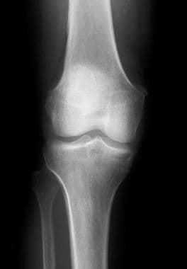
Where are calcific deposits located?
Calcific deposits located within the tendon can be seen in this x-ray.
Can calcific tendonitis be prevented?
Though calcific tendonitis can’t be prevented altogether, there are steps you can take that may reduce your risk. For example, if you develop pain in your shoulder, don’t participate in any strenuous activities until you have it checked out by your healthcare provider.
Can calcific tendonitis cause swelling?
Side effects depend on the treatment that is performed. Most non-surgical options have minimal side effects that may include temporary discomfort and swelling. Patients who undergo surgery for calcific tendonitis have a small risk of:
What happens during the precalcific phase?
Pre-calcific: During this beginning phase, movement causes pain and range of motion becomes limited. The area changes at a cellular level.
Is calcific tendonitis a type of arthritis?
No. Calcific tendonitis is inflammation of the tendons while arthritis is inflammation and damage of the joint itself. Calcific tendonitis may be confused with calcium pyrophosphate dihydrate deposition disease (CPPD, or pseudogout) — a type of arthritis in which calcium phosphate crystals form in the joints.
