Procedures
The appropriate moment for retinal surgery in these patients is when the vascular activity has subsided, and a retinal detachment is present. 40 Treating an eye with high vascular activity can lead to worse postoperative inflammation and vitreous hemorrhage.
Nutrition
Visual outcomes in pediatric patients after RD repair are generally poorer, even when anatomic success has been achieved, due to associated hereditary disorders with systemic comorbidities, and the presence of chronic detachments, macular involvement, and presence of PVR.
When is it appropriate to have retinal surgery for retinal detachment?
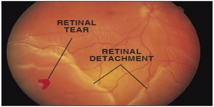
Coccius in 1853 followed by von Graefe in 1854, who also portrayed the course of retinal detachment, observed the first retinal tear [5,6]. The history of retinal detachment surgery can be divided into pre- (before 1920) and post-Jules Gonin's era (after
What is the prognosis of pediatric retinal detachment repair?
Options for surgical repair of pediatric rhegmatogenous retinal detachment are basically external reapposition of the retina versus intraocular subretinal fluid drainage and reattachment.
What is the history of retinal detachment?
What are the surgical options for pediatric rhegmatogenous retinal detachment?
When was the first retinal detachment surgery?
In 1929, at the International Congress of Ophthalmology in Amsterdam, Gonin (along with his disciples Arruga, Weve, and Amsler) conclusively proved to his audience that retinal breaks were the cause of retinal detachment and that closure of retinal breaks caused the retina to reattach [42, 43].
How quickly must a detached retina be treated?
If the macula detaches, it is too late to restore normal vision. Surgery can still be done to prevent total blindness. In these cases, eye doctors can wait a week to 10 days to schedule surgery.
When was the first vitrectomy performed?
David Kasner first described vitrectomy, or removal of the vitreous body, using an open-sky technique in 1969.
How long can a detached retina go untreated?
A retinal detachment may cause permanent blindness over a matter of days and should be considered an eye emergency until evaluated by a retina specialist. Most retinal detachments occur suddenly and can threaten the central vision within hours or days.
How urgent is a detached retina?
Retinal detachment is a potential medical emergency that can be corrected if it is caught early. However, if medical treatment is delayed too long, then it could lead to permanent damage that affects your sight or even causes blindness in the affected eye.
What is the success rate of retinal detachment surgery?
In most specialist centres around nine out of ten retinal detachments are successfully repaired with a single operation. In the remaining cases, the retina re-detaches and needs another operation. The final success rate is over 95 per cent.
Is vitrectomy a low risk procedure?
Although the procedure carries a low overall risk, there is still a risk of severe complications. These can compromise vision or even cause blindness.
What is the success rate of vitrectomy surgery?
The success rate for vitrectomy is around 90 percent, even if you're over 60.
Who invented vitrectomy?
Robert Machemer, the inventor of modern vitrectomy more than two decades ago, might scarcely recognize the procedure he invented.
How fast does a retinal detachment progress?
The rate of progression of a retinal detachment can vary from days to weeks depending on many factors such as patient age as well as the size and the number of retinal tears. Gradual loss of peripheral vision in the form of a shadow, curtain, or cloud (this corresponds to the retina detaching.)
What happens if you don't repair a detached retina?
If the retinal detachment isn't treated right away, more of the retina can detach — which increases the risk of permanent vision loss or blindness.
How can I repair my retina naturally?
How to Improve the Health of the RetinaHealthy and balanced diet. ... Avoiding unhealthy foods and drinks. ... Drinking plenty of water. ... Regular exercise. ... Wearing sunglass when out in the sun. ... Quitting smoking. ... Wearing eye protection. ... Regular eye check-up.
What is retinal detachment?
Retinal detachment is an eye problem that happens when your retina (a light-sensitive layer of tissue in the back of your eye) is pulled away from its normal position at the back of your eye.
What are the different types of retinal detachment?
There are 3 types of retinal detachment: rhegmatogenous, tractional, and exudative. Each type happens because of a different problem that causes your retina to move away from the back of your eye. Learn more about what causes each type of retinal detachment.
How to move retina back into place?
Surgery. If a larger part of your retina is detached from the back of your eye, you may need surgery to move your retina back into place. You may need to get these surgeries in a hospital. Treatment for retinal detachment works well, especially if the detachment is caught early.
How to prevent permanent vision loss?
Early treatment can help prevent permanent vision loss. It’s also important to get comprehensive dilated eye exams regularly. A dilated eye exam can help your eye doctor find a small retinal tear or detachment early, before it starts to affect your vision.
What to expect after a dilated eye exam?
Learn what to expect from a dilated eye exam. If your eye doctor still needs more information after a dilated eye exam, you may get an ultrasound or an optical coherence tomography (OCT) scan of your eye. Both of these tests are painless and can help your eye doctor see the exact position of your retina.
What is the term for the condition where the blood vessels in the retina are affected?
Diabetic retinopathy (a condition in people with diabetes that affects blood vessels in the retina) Extreme nearsightedness (myopia), especially a severe type called degenerative myopia. Posterior vitreous detachment (when the gel-like fluid in the center of the eye pulls away from the retina)
What is the procedure to repair a tear in the retina?
Freeze treatment (cryopexy) or laser surgery. If you have a small hole or tear in your retina, your doctor can use a freezing probe or a medical laser to seal any tears or breaks in your retina. You can usually get these treatments in the eye doctor’s office. Learn more about laser surgery and freezing treatment.
How to prevent retinal detachment?
When a retinal tear or hole hasn't yet progressed to detachment, your eye surgeon may suggest one of the following procedures to prevent retinal detachment and preserve vision. Laser surgery (photocoagulation). The surgeon directs a laser beam into the eye through the pupil. The laser makes burns around the retinal tear, ...
How to repair a detached retina?
The type of surgery your surgeon recommends will depend on several factors, including how severe the detachment is. Injecting air or gas into your eye.
What is the procedure to freeze a retinal tear?
Freezing (cryopexy). After giving you a local anesthetic to numb your eye, the surgeon applies a freezing probe to the outer surface of the eye directly over the tear.
What is the procedure called to remove the vitreous?
Draining and replacing the fluid in the eye. In this procedure, called vitrectomy (vih-TREK-tuh-me), the surgeon removes the vitreous along with any tissue that is tugging on the retina. Air, gas or silicone oil is then injected into the vitreous space to help flatten the retina.
What is the procedure called when you indent your eye?
Indenting the surface of your eye. This procedure, called scleral (SKLAIR-ul) buckling, involves the surgeon sewing (suturing) a piece of silicone material to the white of your eye (sclera) over the affected area.
What type of eye exam is used to see the retina?
This type of device provides a highly detailed view of your whole eye, allowing the doctor to see any retinal holes, tears or detachments. Ultrasound imaging.
Can a doctor examine both eyes?
Your doctor will likely examine both eyes even if you have symptoms in just one. If a tear is not identified at this visit, your doctor may ask you to return within a few weeks to confirm that your eye has not developed a delayed tear as a result of the same vitreous separation.
What is a pediatric retinal detachment?
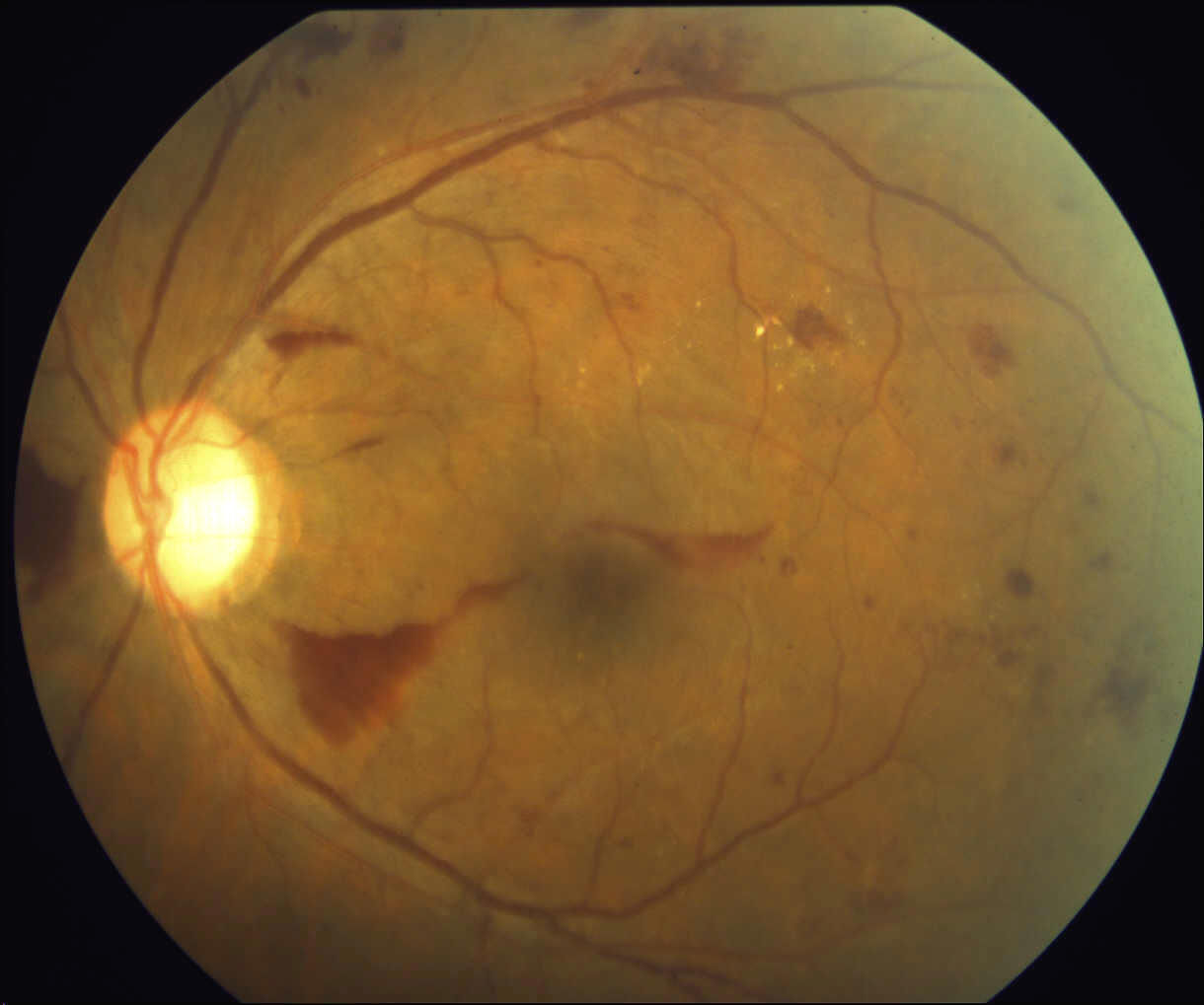
Pediatric retinal detachment (RD) is a rare and complicated disease, with a previously reported incidence that ranges from 3% to 13% . 1 Unlike RDs in adults, pediatric RDs tend to have a chronic duration , worse presenting visual acuity , macula involvement, and proliferative vitreoretinopathy (PVR) development at presentation. 1,2 Surgical repair of RD in pediatric patients is a challenge for the vitreoretinal surgeon, especially due to the associated systemic comorbidities usually present in this young population. 1,3
What is the tendency for late diagnosis in pediatric RDs?
Pediatric RDs have features that make them unique. The tendency for late diagnosis is an important characteristic that leads to macular involvement in 60%-85% of the cases and PVR grade C or worse in 20%-60%; these findings might be related to a delay in diagnosis due to delayed reporting of issues and higher degree of intraocular cellular activity and proliferation in the active immune system of young patients. 1,3-11
What percentage of RRDs are pediatric?
Pediatric RRDs account for 0.5%-8% of all patients suffering from RRD. 4-6,8,10,11,13,14 Predisposing factors include trauma (21.1% to 53%), myopia (11.5%-41.44%), previous intraocular surgery (30%-51%) and congenital-developmental anomalies (17%-65%). 3-7,10
Can a pediatric retinal exam be done in the operating room?
Therefore, in the operating room the eye to be treated should be examined with careful identification of any retinal breaks and extent of the subretinal fluid. As well, this is an excellent opportunity to examine the fellow eye and identify any retinal ...
Can anti-VEGF therapy cause vitrectomy?
Anti-VEGF therapy has been used to reduce vascular activity, as previously published by Cernichiaro-Espinosa and coauthors, but one must always be aware that anti-VEGF therapy can increase contraction of fibrovascular membranes and result in the need for vitrectomy. 44,48.
Can a scleral buckle be combined with vitrectomy?
Scleral buckle alone is not indicated in this pathology, due to the presence of foveal schisis, but can be combined with vitrectomy when RRD develops due to peripheral retinal breaks. 2,4. Pediatric patients with choroidal coloboma can also develop RRD, due to retinal breaks that develop at the borders of the coloboma.
How long does it take to reattach a detached retina?
If you have a retinal detachment, you may need surgery to reattach your retina to the back of your eye within a few days. After surgery, you may need to stay in the hospital for a short time — and it might take a few weeks before your vision starts getting better. There are 3 types of surgery that doctors can do to fix a detached retina: ...
What type of surgery is needed to fix a detached retina?
There are 3 types of surgery that doctors can do to fix a detached retina: Pneumatic retinopexy (“noo-mat-ick RET-ih-no-pek-see”) Scleral buckle. Vitrectomy. The type of surgery you need will depend on several things, including how much of your retina is detached and where in your eye it detached.
How to heal a swollen eye?
After the surgery, you’ll need to: 1 Hold your head in a certain position for several days to keep the air bubble in the right spot 2 Avoid some activities — like flying in an airplane, intense exercise, and heavy lifting — while your eye heals 3 Have a follow-up visit with your doctor to make sure your eye is healing
How to fix a hole in your retina?
Put numbing medicine in your eye. Insert a tiny needle into your eye and remove a small amount of fluid . Inject a small amount of air into your eye. Use laser or freeze treatment to repair any holes or tears in your retina. You’ll be able to see the air bubble in your peripheral (side) vision after the surgery.
Can you have more than one retinal surgery?
Some people may need more than one type of surgery at once . During the surgery, your doctor may also use laser or freeze treatments to repair tears or holes in your retina and help hold your retina in place after surgery. Learn more about laser surgery and freeze treatment.
Can you see bubbles in your eyes after a retinal surgery?
You can usually get this surgery in your doctor’s office. You’ll be able to see the air bubble in your peripheral (side) vision after the surgery.
Can you go home after eye surgery?
You won’t feel anything or remember the surgery. Most people can go home the same day, but you’ll need someone to drive you home. After the surgery, your eye may feel a little sore. You’ll need to: Wear a patch over your eye for about a day.
How to treat retinal tear?
Ophthalmologists occasionally perform cryotherapy if the location of the tear makes it difficult to perform laser photocoagulation. Laser photocoagulation and cryotherapy can also be used to treat a retinal detachment and prevent it from becoming bigger.
What is the procedure to tear the retina?
With retinal tears, the procedure prevents fluid from traveling underneath the retina, where it can cause detachment. After this relatively painless procedure, your surgeon may administer a topical steroid to prevent inflammation.
What is the procedure to remove vitreous gel?
Vitrectomy. During a vitrectomy, your doctor makes an incision in the sclera of the eye and inserts an instrument to remove the vitreous gel. After the vitreous is removed, your doctor may treat the retina with photocoagulation or cryotherapy to seal the tear.
How long does a scleral buckle last?
Surgery usually lasts two hours.
What happens after retinal surgery?
Some people become more prone to developing a cataract after retinal surgery because surgery can trigger changes in the lens of the eye.
How long does a numbing eye procedure last?
Your doctor then injects a small amount of intraocular gas into the vitreous. The gas lasts for several days and gently pushes the retina against the back of the eye.
Does NYU Langone have cataract surgery?
NYU Langone offers three surgical outpatient procedures to treat retinal detachment. Doctors determine the type of surgery needed based on several factors, including the location and size of the detachment and whether the person has had cataract surgery.
