
How long does it take for neurocysticercosis to develop?
Still, many people with neurocysticercosis have no symptoms. Other people may take months or years to start developing symptoms, according to 2017 treatment guidelines. Cysts, called cysticerci, can develop in the muscles, eyes, brain, and spinal cord.
Are there guidelines for the clinical management of neurocysticercosis?
Keywords: Taenia solium, cysticercosis, neurocysticersosis, taeniasis Guidelines for the clinical management of patients with neurocysticercosis (NCC) were prepared by a panel of the Infectious Diseases Society of America (IDSA) and the American Society of Tropical Medicine and Hygiene (ASTMH).
Is there a cure for cysticercosis?
Not all cases of cysticercosis need treatment. Even if you don’t need treatment to kill the parasite, you may need treatment for the symptoms caused by the infection, such as medication to reduce the number of seizures you have. Can cysticercosis be spread from person to person?
What are the symptoms of neurocysticercosis?
Still, many people with neurocysticercosis have no symptoms. Other people may take months or years to start developing symptoms, according to 2017 treatment guidelines. Cysts, called cysticerci, can develop in the muscles, eyes, brain, and spinal cord. The person’s symptoms can also depend on the location, size, number, and stage of these cysts.
How long does it take to treat neurocysticercosis?
Drugs. Several studies suggest that albendazole (conventional dosage 15 mg/kg/day in 2 divided doses for 15 days) may be superior to praziquantel (50 mg/kg/day for 15 days) for the treatment of neurocysticercosis.
Can neurocysticercosis be cured completely?
Most importantly, neurocysticercosis is one of the few diseases that can be eradicated, an accomplishment that would prevent millions of cases of epilepsy.
How do you get rid of neurocysticercosis?
For intraventricular cysts, endoscopic surgery is the procedure of choice as it is minimally invasive. For incompletely excised cysts and cysts or granulomas in locations such as the spinal cord, medical treatment with steroids and albendazole is recommended.
Can you recover from cysticercosis?
Since then, an alternative opinion has been voiced that the acute, severe brain inflammation resulting from their use is unnecessary because parenchymal brain cysticercosis follows a benign course and cysts will degenerate and heal by natural evolution of the disease (17, 18, 64-66).
Is neurocysticercosis serious?
Neurocysticercosis, which affects the brain and is the most severe form of the disease, can be fatal. Neurocysticercosis is considered a Neglected Parasitic Infection, one of a group of diseases that results in significant illness among those who are infected and is often poorly understood by health care providers.
Which drug is used in the treatment of neurocysticercosis?
Praziquantel (an isoquinolone) and albendazole (an imidazole) are anticysticercal drugs that are currently being used for the treatment of neurocysticercosis. Both have been reported to eliminate or markedly reduce the number and size of cysticerci.
Is cysticercosis serious?
Symptoms include severe headache, blindness, convulsions and epileptic seizures and can be fatal. Neurocysticercosis is the most frequent preventable cause of epilepsy worldwide and is estimated to cause 30% of all epilepsy cases in countries where the parasite is endemic.
What is cysticercosis How is it treated?
Cysticercosis located outside of the nervous system usually does not require specific treatment. The treatment for cysticercosis located within the nervous system (neurocysticercosis) consists of antiparasitic therapy, corticosteroids, antiepileptic drugs, and/or surgery.
What is calcified neurocysticercosis?
Neurocysticercosis is responsible for increased rates of seizures and epilepsy in endemic regions. The most common form of the disease, chronic calcific neurocysticercosis, is the end result of the host's inflammatory response to the larval cysticercus of Taenia solium.
Can neurocysticercosis reoccur?
Conclusion: Recurrent SCGs are not uncommon in NCC and recurrence is more likely to occur at the site of initial infection and therefore may be mistaken for persistent infection or other granulomatous lesions. Key Message: Recurrent NCC is not rare and is most likely to occur at the site of initial infection.
In which stage of neurocysticercosis is there no edema?
Vesicular stage: Cysts follow the CSF signal; T2 hyperintense scolex may be seen, with no edema and usually no enhancement.
How do you get rid of tape worm?
Tapeworms are usually treated with a medicine taken by mouth. The most commonly used medicine for tapeworms is praziquantel (Biltricide). These medications paralyze the tapeworms, which let go of the intestine, dissolve, and pass from your body with bowel movements.
Abstract
Taenia solium neurocysticercosis is a common cause of epileptic seizures and other neurological morbidity in most developing countries. It is also an increasingly common diagnosis in industrialized countries because of immigration from areas where it is endemic.
INTRODUCTION
Cysticercosis, the infection caused by the larval stage of the tapeworm Taenia solium, is the most common parasitic disease of the nervous system in humans and the single most common cause of acquired epileptic seizures in the developing world, where prevalence rates of active epilepsy are twice those in developed countries ( 41, 53, 56, 91, 107 ).
DISCUSSION GUIDELINES AND METHODS
When the panel began to discuss the preferred treatment for neurocysticercosis, it immediately became clear that neurocysticercosis is not a single disease for which one therapy can be recommended. There are marked differences in clinical presentation, pathogenesis, natural history, and treatment options for the different forms.
OVERVIEW OF PARENCHYMAL BRAIN CYSTICERCOSIS
There was agreement that albendazole and praziquantel are effective antiparasitic agents, destroying most viable cysts.
OVERVIEW OF EXTRAPARENCHYMAL CYSTICERCOSIS
Whenever hydrocephalus or intracranial hypertension is present, its management should be the first priority. It can generally be managed by means of a ventricular shunt. Since extraparenchymal neurocysticercosis is associated with a worse prognosis, there was a consensus towards aggressive management.
COMMENTS
Cysticercosis should no longer be considered an “exotic” disease.
REFERENCES
1. Agapejev, S., M. D. Da Silva, and A. K. Ueda. 1996. Severe forms of neurocysticercosis: treatment with albendazole. Arq. Neuropsiquiatr. 54:82-93. [ PubMed] [ Google Scholar]
What is neurocysticercosis?
Neurocysticercosis is a neurological disease that occurs when a particular type of parasitic tapeworm invades the central nervous system. In this article, we explore neurocysticercosis, including its symptoms, diagnosis, treatment, and more.
How many people have neurocysticercosis?
Reports show that neurocysticercosis affects around 50 million people worldwide, and according to a 2017 entry in the International Encyclopedia of Public Health, the disease is a major cause of epilepsy and seizures across the globe.
What are the symptoms of cysticerci?
Common symptoms and signs include: seizures. chronic headaches. increased pressure in the brain. Seizures are the most common.
How to treat parasites?
Treatments focus on removing the parasite and managing symptoms#N#Trusted Source#N#, by controlling seizures, swelling, and intracranial hypertension, for example.#N#The right course of treatment will also depend on the position, size, abundance, and maturity of the parasites.#N#After assessing the person’s symptoms and the extent of the infection, a doctor may use antiparasitic and anti-inflammatory therapy. In some cases, surgery is necessary to remove cysts.
Can tapeworms cause cysticercosis?
However, if a person consumes the parasite’s larvae, they are at risk of cysticercosis.
Do people with neurocysticercosis have symptoms?
from 2014, symptoms of neurocysticercosis depend on where the lesions form, the extent of the infection, and the person’s immune response. Still, many people with neurocysticercosis have no symptoms.
How long does a parenchymal NCC last?
The natural history of parenchymal NCC includes an asymptomatic period that typically lasts several years, followed by gradual degeneration over a period of at least a year. While the exact proportion is unknown, many patients with parenchymal NCC go on to develop calcified lesions, a risk factor for chronic epilepsy.
How many seizures are caused by NCC?
Epidemiologic studies suggest that NCC is the cause of approximately 29% of seizures in endemic areas and about 2% of patients presenting with seizures presenting to US emergency rooms [2–5, 8–10]. The seizures can be focal, focal with generalization, or generalized.
How big is a cyst in the gyri?
Most are between 5 and 20 mm in diameter, but they can be larger, especially if located in the gyri and fissures. Parenchymal lesions with cystic areas diameter >20 mm, with irregular borders, or accompanied by midline shift are more likely to have other causes [12]. Midline shift is usually limited to larger cysts.
How to treat neurocysticercosis?
Although the heterogeneity of the clinical picture of neurocysticercosis requires individual tailoring of treatment and management, several general principles apply: 1 Anthelminthic therapy is generally indicated for symptomatic patients with multiple, live (noncalcified) cysticerci. 2 Anthelminthic treatment will not benefit patients with dead worms (calcified cysts). 3 Concomitant administration of steroids (e.g. dexamethasone) is often indicated to suppress the inflammatory response induced by destruction of live cysticerci. 4 Conventional anticonvulsant therapy is the mainstay of management of neurocysticercosis-associated seizure disorders. 5 Intraventricular cysts should usually be treated by surgical removal (endoscopic if possible). Anthelminthics are relatively contraindicated, because the resulting inflammatory response could precipitate obstructive hydrocephalus. 6 Although our understanding of subarachnoid neurocysticercosis is evolving, treatment with both anthelminthics and corticosteroids is usually required. Ventricular shunting is often necessary as well.
Where do cysticerci occur?
In humans, cysticerci (encysted larvae) often occur in skeletal muscles. However, the manifestations that most frequently lead patients to visit health care providers are caused by cysts in the central nervous system (CNS), known as neurocysticercosis. Less frequently, cysticerci may localize in the eyes, skin, or heart.
How many cysticercus are there in the host?
The number of cysticerci in the host can vary from one to more than 1,000. In the absence of massive numbers of cysticerci, the initial host tissue reaction is usually minimal. The developing cysticercus affects the surrounding tissue as a slowly growing mass that may cause pressure atrophy.
What is the diagnosis of CNS disease?
Diagnosis. Diagnosis typically requires both CNS imaging and serological testing. A careful history should be taken, including questions regarding residence or extended travel in developing countries, and consumption of food prepared by someone who has lived in a high-risk area.
What is the inflammatory reaction in the CNS?
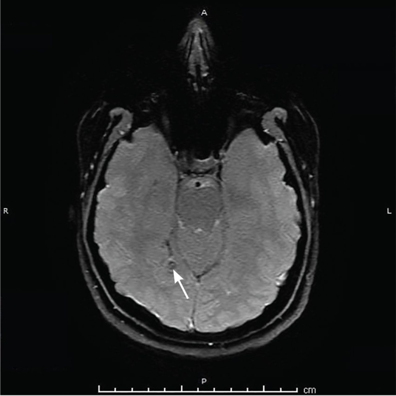
In the CNS, the inflammatory reaction and resultant edema appear as a contrast-enhancing ring around the cyst on imaging. There may be CSF pleocytosis as well. Necrotic larvae are completely or partially resorbed, but may become calcified, resulting in focal scarring that may provide a focus for seizures.
Is cysticercosis a reportable disease?
For confirmatory testing in cases where EITB is not available, contact the CDC directly. Cysticercosis is a reportable disease in several states. Health care providers should check with their state health department to determine if they require notification of patients testing positive for cysticercosis. Back To Top.
Can cysticerci be seen on imaging?
In this instance, serological results may be negative, but the lesions may be visible on imaging. A patient may have cysticerci in locations other than the brain.
